
Electrochemical Biosensor

a biosensor and electrochemical technology, applied in the field of electrochemical biosensors, can solve the problems of ineffective assays, many current assay systems suffer from interference, and the time and labor required for assays is so grea

Inactive Publication Date: 2009-10-01
SITDIKOV RAVIL +4
View PDF1 Cites 51 Cited by
  • Summary
  • Abstract
  • Description
  • Claims
  • Application Information

AI Technical Summary

Benefits of technology

[0010]Various embodiments of the present disclosure provide a simple, fast, selective and highly sensitive electrochemical method assay and disposable device for detection of viruses, bacteria, proteins, DNA, and / or organic / inorganic compounds. The sensor of the invention is of a multi-layered construction, with each successive layer performing a different function. Miniaturization allows packing of numerous microscopic electrode transducers onto a small footprint of a biochip device, and hence the design of high-density arrays.
[0011]The present disclosure improves upon previous target analyte assays by requiring fewer steps, detecting specific targets at lower concentrations, and needing less time to complete. The inventions of the present disclosure can be used in field conditions, outside of a well-equipped laboratory setting. Complex instrumentation is not required because the probes and sensors may be used with an inexpensive, hand-held meter. This unique assay approach greatly reduces unwanted background signal, enabling the rapid identification of captured biomolecules with high sensitivity and specificity with little or no sample processing.
[0012]Accordingly, in one embodiment, the present disclosure provides an improved biosensor and method for the simultaneous conduct of a multiplicity of binding reactions on a substrate.

Problems solved by technology

However, many current assay systems suffer from interference caused by background sample material.
The time and labor required for assay is so great that the assays are not useful and less sensitive means of analysis are employed.
However, this test required about 4 h to perform and required a fluorescence microscope and a senior technician experienced in the reading of immunofluorescence slides.
These assays are difficult to conduct in the non-laboratory conditions typically encountered in a field setting or a remote location.
Both direct and indirect immunofluorescence antibody tests can produce a high frequency of false negative results caused by the low sensitivities of the tests compared with viral culture.
The accuracy of the clinical diagnosis of influenza is limited, even during peak influenza activity, because other co-circulating respiratory viruses (such as adenoviruses, parainfluenza viruses, respiratory syncytial virus, rhinoviruses, human metapneumovirus), or other organisms (such as Streptococcus pneumoniae, Chlamydia pneumoniae and Mycoplasma pneumoniae) can cause symptoms similar to those of influenza viruses.
Existing diagnostic ELISA tests are also not sensitive enough and detect proteins at levels corresponding to advanced stages of the disease.
Thus, these kinds of diagnostic tools have several shortcomings that must be overcome: 1) they are slow to recognize the presence of a target virus; 2) they lack adequate sensitivity; 3) the bioanalytical systems are often transportable rather than portable and require highly trained personnel to properly operate them.
Other limitations are the complexity of the instrumentation, and the multi-step and time consuming procedures that are always required.
In addition, running these instruments is both expensive and labor-intensive.
Rapid detection also may decrease the use of antibiotics in patients with respiratory tract infections.

Method used


Image

  • Electrochemical Biosensor
  • Electrochemical Biosensor
  • Electrochemical Biosensor

Examples


example 1

Detection of Influenza Viruses: Label-Free Detection

[0089]Virus Samples. Different concentration virus solutions are prepared from sticks by dilution on 0.01 M phosphate buffer (pH 6.0) containing 0.01 M KCl (assay buffer); influenza type A at different concentrations.

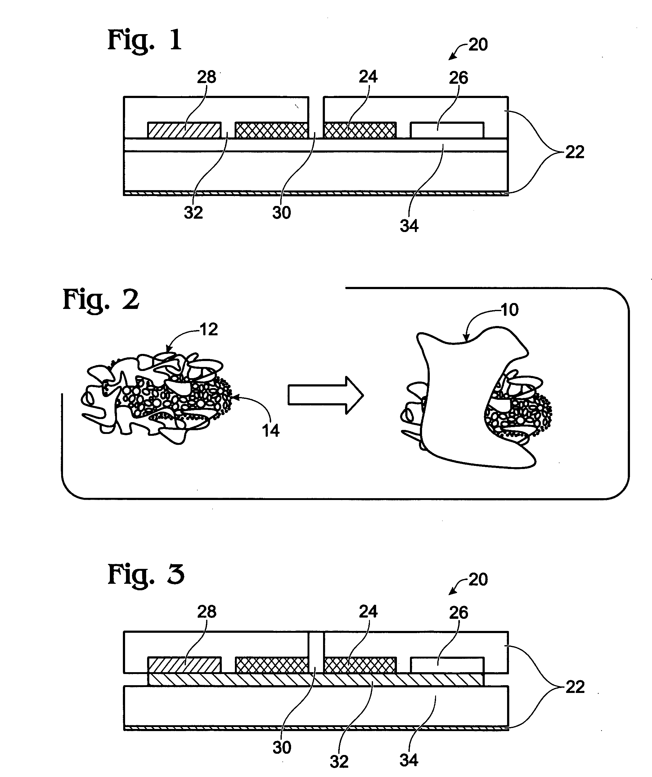
[0090]Electrical Measurements. The three-dimensional multi-channel electrochemical flow-thru cells were used for direct electrical detection of influenza viruses. The basic building blocks of biosensor device are biorecognition and transducing elements and the readout modality. The biosensor assembly is comprised of flow-through electrochemical cell, the AndCare 800 8-Well Sensor Strip Reader, which operates by using Intermittent Pulse Amperometry (IPA) and a special software program. The electrode assembly is located perpendicular to the flow of sample through the device. The microchannels of the working electrode contained carbon nanotubes with immobilized antibodies against influenza A virus, on which the immunochem...

example 2

Enzyme Immunoassay of Influenza A
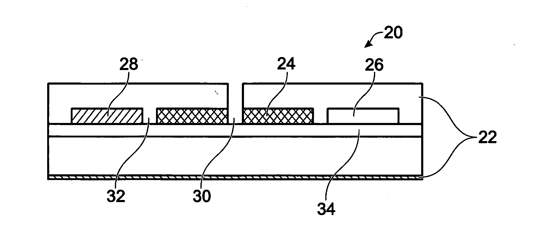
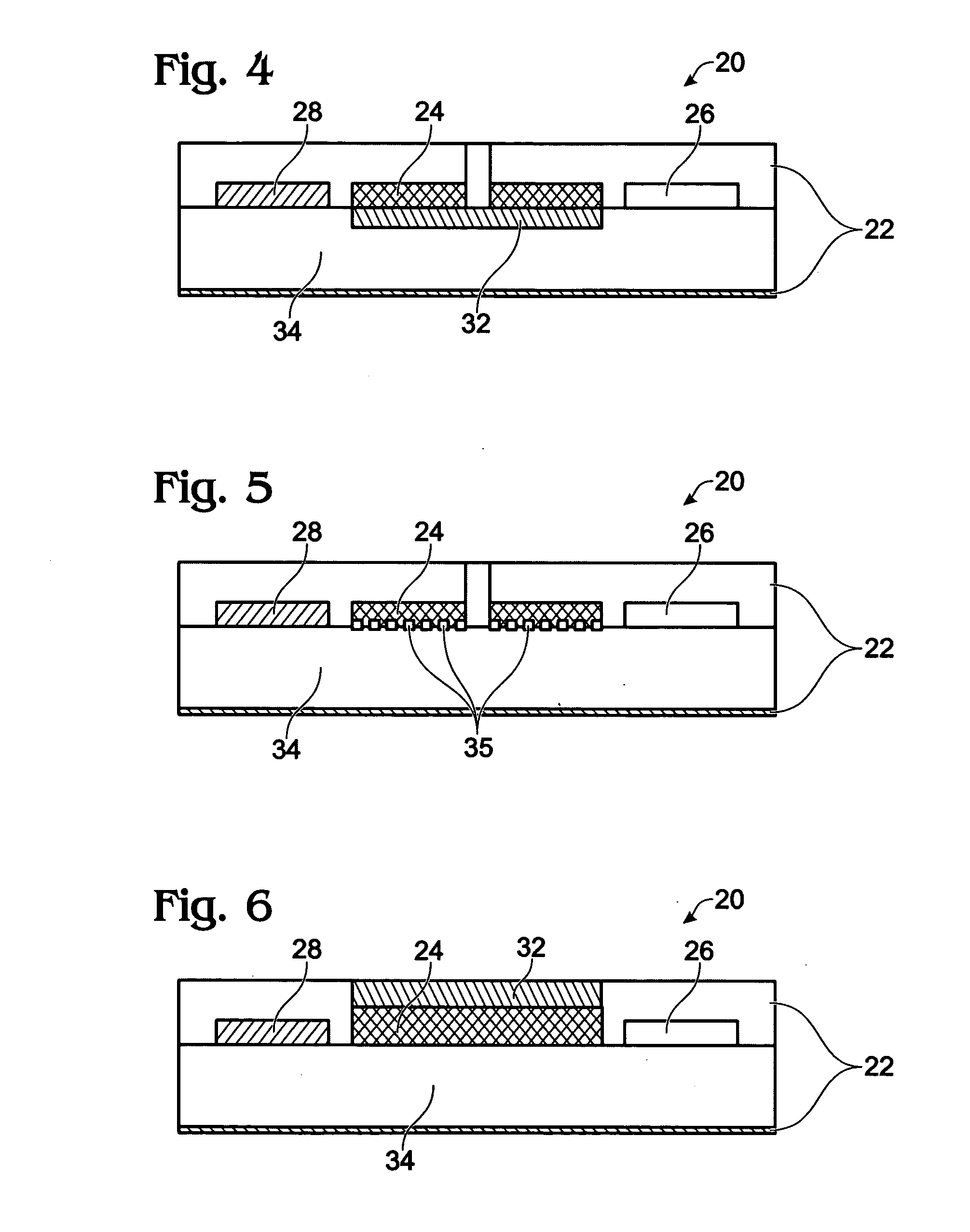
Device Layout

[0099]The flow-thru biosensor is composed of an array of microporous working microelectrodes. Each working electrode includes microscopic channels and arrays of probes (Abs, Enzymes, DNA, cells, or receptors), which are deposited on the side surface of microchannels. Microporous working electrodes are used both as a matrix for probe immobilization and as a transducer. Sample flows through the microchannels of the working electrodes and biological recognition reactions occur within the microchannels with electrochemical detection following.

[0100]Eight well Carbon Sensor Strips (Alderson Biosciences, Inc. Beaufort, N.C.) form arrays of 8 three-electrode independent electrochemical sensor elements made on one plastic support using screen printing technology. Each electrochemical cell (a volume 300 ul) consists of carbon working, counter, and silver reference electrodes. The 8-well sensor strip is used as disposable 8-channel probes for a mu...


PUM

PropertyMeasurementUnit
Concentrationaaaaaaaaaa
Densityaaaaaaaaaa
Electrical conductoraaaaaaaaaa

Abstract

A simple, fast, selective and highly sensitive electrochemical method assay and disposable device for detection of viruses, bacteria, proteins, DNA, and / or organic / inorganic compounds. The sensor has a multi-layered construction, with each successive layer performing a different function. The design further allows for the packing of numerous microscopic electrode transducers onto the small footprint of a biochip device, allowing for a high-density array of sensors.

Description

CROSS-REFERENCE TO RELATED APPLICATIONS[0001]The following application claims benefit of U.S. Provisional Patent Application No. 61 / 010,227, filed Jan. 7, 2008, which is hereby incorporated by reference in its entirety.STATEMENT REGARDING GOVERNMENT SPONSORED RESEARCH[0002]This invention was made with Government support under Grants Nos. DMR-0611616 and CTS-0332315, both awarded by the National Science Foundation. The U.S. Government has certain rights in this invention.BACKGROUND[0003]1. Field of the Invention[0004]The invention relates to the fields of electrochemical flow-through sensor technology and molecular diagnostics. More particularly, the invention relates to three dimensional, flow-through, disposable sensors for detecting and quantifying viruses, bacteria, proteins, DNA, or compounds such as pollutants, illegal drugs, etc. in air, water, food and complex biological mediums such as blood or saliva in real-time.[0005]2. Description of the Related Art[0006]The need to impr...

Claims


Application Information

Patent Timeline
IPC IPC(8): G01N27/26
CPCB82Y5/00G01N2333/11G01N33/557
Inventor SITDIKOV, RAVILIVNITSKI, DMITRILOPEZ, GABRIELRAMIREZ, BRIANAATANASSOV, PLAMEN
Owner SITDIKOV RAVIL
Learn More