
Multi-Layered Antiadhesion Barrier

Inactive Publication Date: 2008-10-16
BIORANE
View PDF7 Cites 66 Cited by
  • Summary
  • Abstract
  • Description
  • Claims
  • Application Information

AI Technical Summary

Benefits of technology

[0070]The multi-layered anti-adhesion barrier of the present invention can solve the problems of conventional gel, solution, sponge, film or nonwoven type anti-adhesion systems, including adhesion to tissues or organs, flexibility, physical strength, ease of handling (ease of folding and bending), etc., offers improved user convenience and a method for the preparing the same. With a nanofibrous structure, the multi-layered anti-adhesion barrier of the present invention effectively blocks the infiltration or migration of blood and cells and promotes the healing of wound. It is not torn or broken when folded or rolled and can be easily handled using small surgical instruments. Thus, it can minimize foreign body reaction when used in various surgical operations.

Problems solved by technology

Post-surgical adhesion is a very critical medical situation, which may result in pains, ileus, infertility, etc.
Sometimes, it causes malfunction of organs or tissues, leading to another surgery or possibly loss of life.
But, the anti-adhesion barriers in the form of a gel, fluid, foam, etc. are not accurately fixed at the wound site; they move downward because of gravity and, thus, are less effective in healing wounds and reducing adhesion.
However, carboxymethylcellulose is less biocompatible than bio-originated materials.
However, since materials having a small molecular weight are absorbed quickly, the role of the anti-adhesion barrier cannot be sustained sufficiently.
But, this gel type anti-adhesion barrier is also problematic in accurately fixing it at such wound sites as the abdominal internal organs or tissues which are constantly moving.
However, it is degraded quickly, with a half life of only 1 to 3 days, and is problematic when used as anti-adhesion barrier.
Since the crosslinked hyaluronic acid is a water-soluble polymer, its mechanical strength weakens when in contact with water because it absorbs a lot of water.
There also remains the problem of removing the residuals of the crosslinking agent used to chemically crosslink hyaluronic acid in order to delay its degradation.
However, these patents are also not without the problems of quick degradation and use of non-bio-originated material.
But, since celluloses are sensitive to pH, there is a difficulty in processing them.
Also, although they are natural polymers, celluloses are not a constituent of the human body and are known to have the potential to cause a foreign body reaction.
But, as mentioned earlier, ORC is a non-bio-oriented material and has poor biocompatibility.
Also, because of a very large pore size, cells or blood proteins may easily penetrate the barrier, and the anti-adhesion barrier is deformed by external force during handling.
However, it tends to roll when in contact with water and be brittle when it is dry.
Especially, Seprafilm is restricted to use in laparoscopic surgery.
Films made of PLA or poly(glycolic acid) (PGA) are easy to roll to one side, but they do not adhere well to the three-dimensionally, highly irregular surfaces of organs or tissues.
Also, since these materials are hydrophobic, they do not absorb moisture well.
Therefore, they do not adhere well to the wet surface of organs or tissues.
Besides, when hydrolyzed in the body, they give acidic degradation products, which may cause inflammation and adhesion.
However, it has relatively weak physical strength and, because of excessive moisture absorption, tends to be too heavy to handle or transport to another site.
Additionally, because a material derived from an animal source is used, there is a possibility of immune rejection or exposure to animal pathogens or viruses.
However, since synthetic polymers contact tissues, a foreign body reaction or inflammation may occur.
In addition, they are not effective in preventing adhesion caused by infiltration of blood or cells, because of the inability to control the pore size.
However, the intermediate cell layer may be the cause of increased adhesion because of growth and proliferation of cells in the layer.
However, because the tissue engineering devices have a large pore size for easier transportation of nutrients and oxygen, they may increase adhesion caused by infiltration, attachment and proliferation of cells.
But, since the hydrophilic polymer or the weakly hydrophobic polymer loses mechanical strength when swollen by water, the fiber may be deformed or torn during handling.
The foregoing techniques, in which biodegradable synthetic polymers are used, are problematic in that inflammation cannot be avoided when the polymers directly contact tissues or blood, because they are bio-originated materials.
Despite the superior flexibility of nanofibers, non-hydrophilic materials do not adhere well to wet tissues, and thus are not easily fixed at a specific site.
To conclude, the conventional techniques have the problem that, since synthetic polymers are used, and although they are biodegradable, inflammation cannot be avoided when the polymers directly contact tissues or blood, because they are bio-originated materials.
Also, despite the superior flexibility of nanofibers, non-hydrophilic materials do not adhere well to wet tissues, and thus are not easily fixed at a specific site.
Further, the small diameter and porosity designed to improve transportation of drugs and cells or to cover the wound are not appropriate in an anti-adhesion barrier for internal organs.

Method used


Image

  • Multi-Layered Antiadhesion Barrier
  • Multi-Layered Antiadhesion Barrier
  • Multi-Layered Antiadhesion Barrier

Examples


examples 10 to 18

Preparation of Multi-Layered Anti-Adhesion Barriers

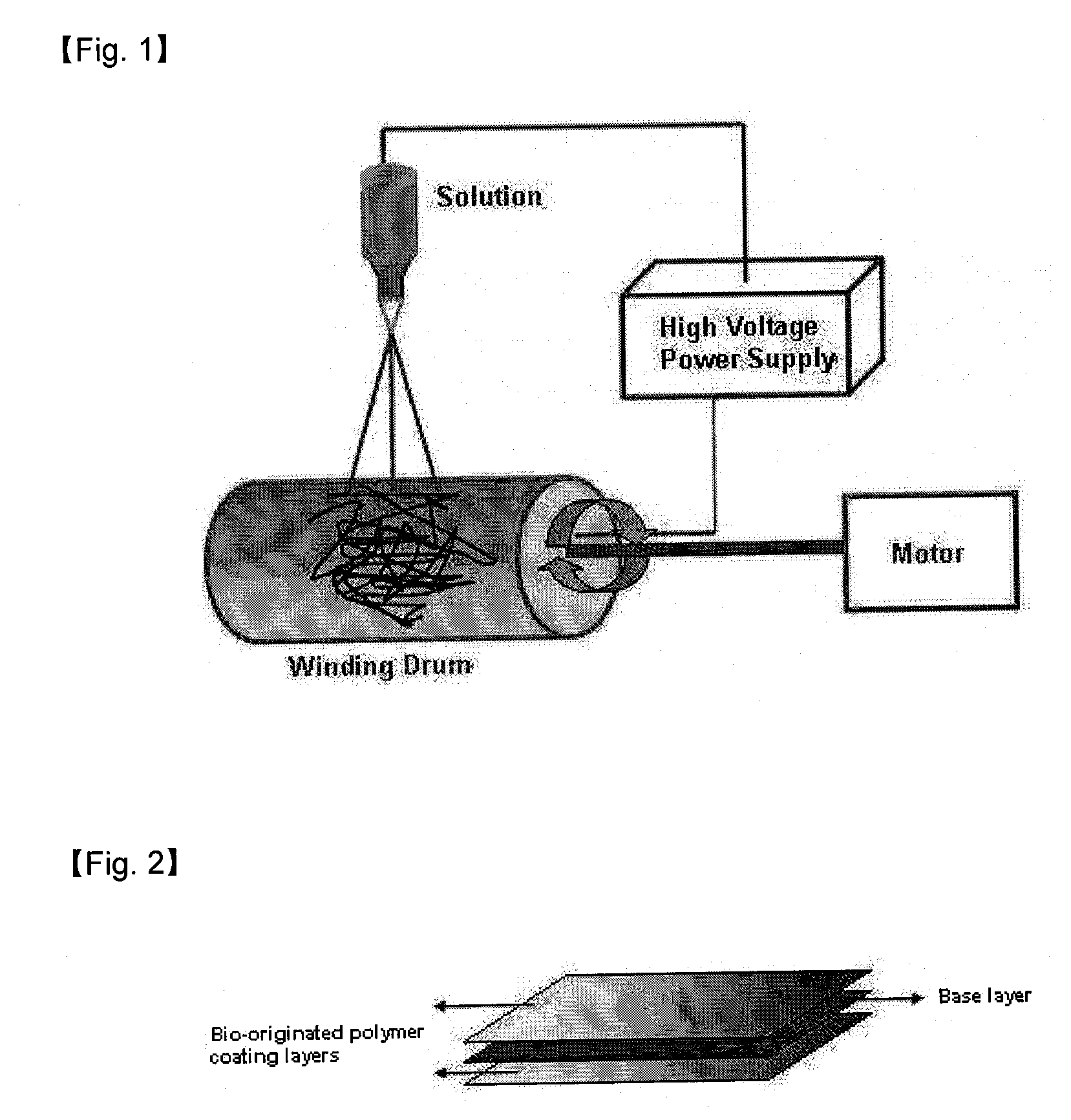
[0079]Multi-layered anti-adhesion barriers were prepared by coating a bio-originated polymer selected from polylactide-co-glycolide, poly ε-caprolactone, polylactide and hyaluronic acid on the nanofibrous structured base layers prepared in Examples 1 to 9 with different coating methods (see Table 2 below). Electrospinning was carried out using the electrospinning apparatus illustrated in FIG. 2 and a spinning solution in which the bio-originated polymer was dissolved at a voltage of 10 to 40 kV. Dip coating was carried out by dip coating the bio-originated polymer solution and drying the anti-adhesion barrier in an oven of 70° C. Casting was carried out by coating the bio-originated polymer solution on the base layer, casting it into a film and drying the anti-adhesion barrier. Spray coating was carried out by spraying the bio-originated polymer solution on the base layer and drying the anti-adhesion barrier in an oven of 70° C. for...

example 19

[0081]Poly(lactide-co-glycolide) (PLGA) having a lactide / glycolide ratio of 70:30 was dissolved in chloroform to 2 wt % and electrospun to form a nano structured base layer having a thickness of 60 μm. Subsequently, hyaluronic acid (HA) was dissolved in distilled water to 1 wt %, adjusted to pH 1.5 with 1 N HCl, uniformly coated on the nano structured base layer by casting to form a polymer layer having a thickness of 50 μm thickness. The procedure of freezing at −20° C. for 22 hours and thawing at 25° C. for 2 hours repeated twice. A multi-layered anti-adhesion barrier was obtained following neutralization with PBS, washing and freeze drying.

example 20

[0082]Dissolved HA was coated on the nano structured base layer of PLGA prepared in Example 19 and dried to prepare a PLGA / HA film. Subsequently, 1-ethyl-3-(3-dimethylaminopropyl)carbodiimide (EDAC), which is the crosslinking agent for HA, was added to a 90:10 (w / w) mixture of ethanol and water. The PLGA / HA film was immersed in the resultant solution and dried to obtain a multi-layered anti-adhesion barrier.


PUM

PropertyMeasurementUnit
Pore sizeaaaaaaaaaa
Pore sizeaaaaaaaaaa
Pore sizeaaaaaaaaaa

Abstract

The present invention relates to a multi-layered anti-adhesion barrier, particularly to a multi-layered anti-adhesion barrier comprising a nanofibrous structured base layer electrospun from a hydrophobic, biodegradable, biocompatible polymer and a polymer layer formed by coating a hydrophilic, biooriginated polymer on the base layer, and a method for the preparing the same. The multi-layered anti-adhesion barrier of the present invention can solve the problems of the conventional gel, solution, sponge, film or nonwoven type anti-adhesion systems, including adhesion to tissues or organs, flexibility, physical strength, ease of handling (ease of folding and bending), etc., offers improved user convenience. With a nanofibrous structure, the multi-layered anti-adhesion barrier of the present invention effectively blocks the infiltration or migration of blood and cells and promotes the healing of wounds. It is not torn or broken when folded or rolled and can be easily handled using small surgical instruments. Thus, it can minimize a foreign body reaction when used in various surgical operations.

Description

TECHNICAL FIELD[0001]The present invention relates to a multi-layered anti-adhesion barrier, and more particularly to a multi-layered anti-adhesion barrier having improved anti-adhesion properties by solving the problems of the conventional gel, solution, sponge, film or nonwoven type anti-adhesion systems, including adhesion to tissues or organs, flexibility, physical strength, ease of handling (ease of folding and bending), etc., offers improved user convenience, and a method for the preparing the same. With a nanofibrous structure, the multi-layered anti-adhesion barrier of the present invention effectively blocks the infiltration or migration of blood and cells and promotes the healing of wounds. It is not torn or broken when folded or rolled and can be easily handled using small surgical instruments. Thus, it can minimize a foreign body reaction when used in various surgical operations.BACKGROUND ART[0002]Adhesion occurs when blood flows out and is clotted during the healing of...

Claims


Application Information

Patent Timeline
IPC IPC(8): A61L31/04B32B5/02B32B5/18B05D3/00B05D1/00A61K38/48A61K38/57A61K31/727A61K38/49
CPCA61L31/10A61L2400/12D01D5/003Y10T428/249921Y10T428/249988A61K31/74A61L27/14B82Y5/00
Inventor LEE, YOUNG-WOOCHU, BO-YOUNG
Owner BIORANE
Learn More