Retinal dystrophin transgene and methods of use thereof

Inactive Publication Date: 2008-02-21
WHITE ROBERT L +2
View PDF8 Cites 11 Cited by
  • Summary
  • Abstract
  • Description
  • Claims
  • Application Information

AI Technical Summary

Benefits of technology

[0016] In another embodiment of the present invention, a Dp 260 transgene is inserted into an animal's genome by a microinjection process that includes freeing the transgene from its plasmid by restriction digest, and injecting it directly into the animal's oocytes. Animals that have incorporated the transgene into their genome are identified by appropriate conventional methods including sequencing and PCR reactions. Preferably, these animals express Dp260 in their muscle cells, a property that can be tested using conventional techniques such as PCR and western blotting. Animals benefitting from such an embodiment include humans, mice, dogs, and horses. In one example of this embodiment, the preferred human Dp260 transgene was inserted into the genome of double mutant (DM) mice by injecting the Dp260 transgene into DM mouse oocytes, followed by a series of crosses with mdx and utrophin knockout mice. Of course, mice could also be transfected through any conventional method including by the use of other vectors such as adenoviruses or lentiviruses, as well as electoporation of naked DNA. Untransformed DM mice exhibit physiological symptoms similar to muscular dystrophy in humans, and produce neither dystrophin, nor its murine analogue, utrophin. Additionally, DM mice show a severe phenotype, have short lifespans, have high levels of necrosis in their muscles, and exhibit an increasing incidence of Complex Repetitive Discharges (CRDs), a hallmark of muscular dystrophy, as they age. In contrast, DM mice expressing the Dp260 transgene (DM / Tg+) show symptoms of only a mild myopathy, and have normal lifespans. Additionally, DM / Tg+ mice do not have the severe spinal curvature (kyphosis) or limb muscle weakness seen in DM mice. They also show lower levels of necrosis and lower incidence of CRDs as they age. Due to the similarities between DM mice and human individuals that suffer from DMD, the DM mice appear to be an ideal model for the disease.
[0018] In still another embodiment of the present invention, methods of supplying Dp260 in animals through the use of gene therapy is provided. Preferably, the animals are mammals, and more preferably are selected from the group consisting of humans, mice, dogs, and horses. The goal of such therapy would be the alleviation of muscular dystrophy symptoms. In one preferred form of this embodiment, cells would be removed from the patient, and stably transfected with a transgene preferably containing a DNA sequence having at least 80%, more preferably at least 85%, still more preferably at least 90%, even more preferably at least 95%, still more preferably at least 97%, and most preferably 99-100% sequence identity with human Dp260. In some preferred forms of this method, such cells would be transfected with a DNA sequence containing a form of Dp260 that includes human dystrophin exon 71. Preferred transgenes of the present invention would also include the appropriate regulatory elements for stable expression of Dp260. Preferably, the cells transfected would be myoblast or bone marrow cells. Even more preferably, these cells would be side population bone marrow cells, as described above, with cell surface markers as described above, such cells being particularly likely to differentiate into muscle cells. Most preferably, these cells would be taken from the patient receiving therapy, transfected outside the body with the Dp260 transgene, and replaced in the same patient in an autologous transplant. Such autologous transplantation decreases the likelihood of generating an immune response, and may further eliminate the need for immunosuppression, as the transfected cells are the patient's own. Autologous bone marrow transplants of transfected cells could be used at a variety of points in time in the course of the disease. Bone marrow cells are more strongly attracted to more damaged cells, thus making this procedure appropriate for older patients who have suffered muscular dystrophy for long periods of time. Also, this process could occur several times throughout a patient's lifetime, because the effects of such autologous bone marrow transplants are additive, thereby increasing healthy, functional muscle mass.
[0019] Importantly, the present invention is advantageous in an immunological sense. In general terms, an obstacle to any type of gene therapy is the immunogenicity of the transgene product. Full length dystrophin can induce an immunogenic response which can result in failed expression of the transgene (1). The unique nature of the Dp260 transgene is that it expresses a naturally occurring isoform of human dystrophin. The Dp260 protein is expressed primarily in retina and in small amounts in other tissues. Therefore, retinal dystrophin is a natural isoform. The introduction of Dp260 from a transgene will not induce an immunogenic response especially in patients that have deletions upstream of exon 30 which do not affect the expression of Dp260. This is a distinct advantage over full length dystrophin transgenes as well as micro-dystrophin transgenes in which most of the spectrin domain coding region is removed or gutted. The microdystrophins will also potentially induce an immunogenic response since the protein can be considered a neoantigen (the microdystrophin protein contains sequences which are foreign to patients with Duchenne muscular dystrophy). The Dp260 transgene of the present invention overcomes this important barrier to successful gene therapy.

Problems solved by technology

Presently there are no treatments that can prolong life or significantly alter the clinical course of the disease.
Without dystrophin the membrane loses mechanical stability allowing an influx of calcium ions and ultimately leads to muscle fiber necrosis.

Method used


Image

  • Retinal dystrophin transgene and methods of use thereof
  • Retinal dystrophin transgene and methods of use thereof
  • Retinal dystrophin transgene and methods of use thereof

Examples


example 1

Preparation and Analysis of Human Dp260 Transgene Construct

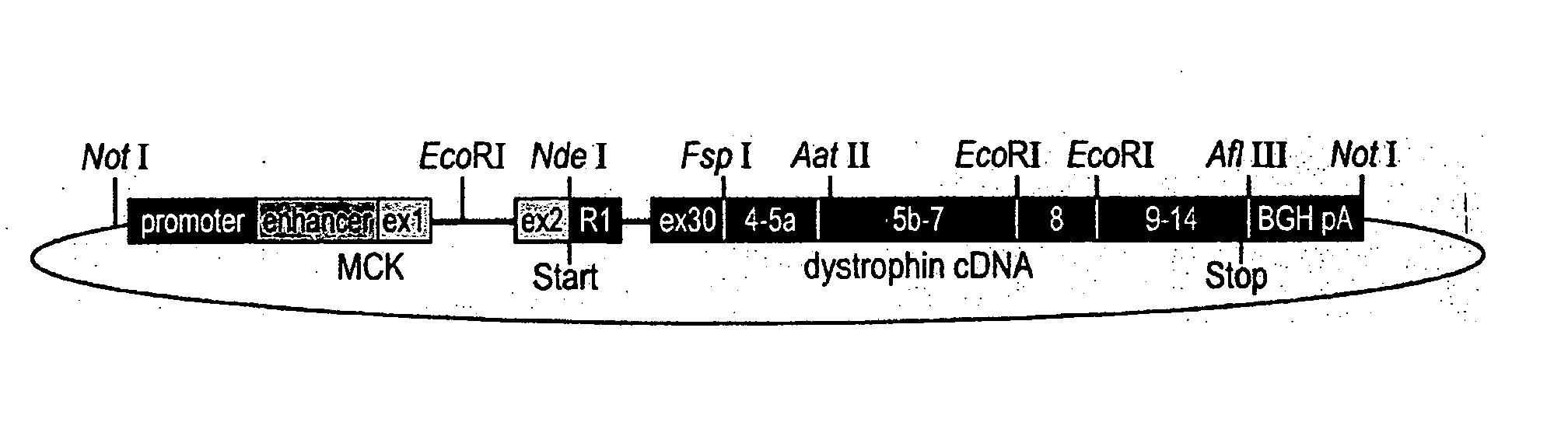
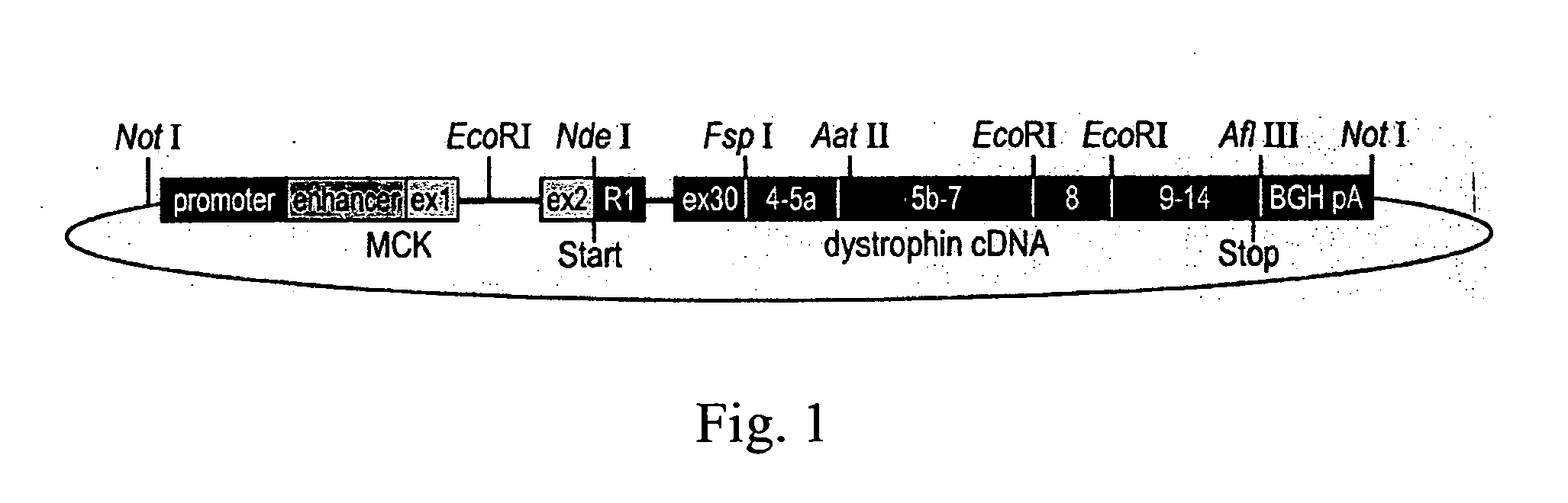
[0054] The mouse muscle creatine kinase (MCK) promoter and enhancer (SEQ ID NO: 1), along with MCK exon 1 (SEQ ID NO: 2), intron 1 (SEQ ID NO: 3), and a portion of exon 2 (SEQ ID NO: 4) comprising the 5′ untranslated portion of exon 2, were used to produce the final transgene. The regulatory elements of MCK with its first exon and part of its first intron (SEQ ID NO: 5) were cloned directly into a pBluescript II SK vector (Stratagene, La Jolla, Calif.). The first PCR amplicon, consisting of the remainder of MCK intron 1 and exon 2, up to the MCK ATG start codon (SEQ ID NO: 6), was amplified by PCR to generate an NdeI restriction site. This allowed ligation to the NdeI restriction site of a human genomic PCR amplicon. The second PCR amplicon started with the ATG start codon of the retinal dystrophin unique first exon R1, continued with intron R1, and ended in exon 30 (SEQ ID NO: 7), which was placed at the exact position whe...

example 2

Production of DM Human Dp260 Transgenic Mice

[0057] The human Dp260 transgene construct was extracted with the Endo Free Plasmid Kit (Quiagen, Valencia, Calif.) and was released from the plasmid vector by restriction digest with NotI prior to oocyte injection. The construct was injected into 200 eggs, which were then transplanted into psuedopregnant females, delivered, and weaned. Genotyping for the Dp260 transgene identified two mice that had incorporated the human Dp260 transgene. Genotyping was performed by PCR reactions using an MCK-specific forward primer (SEQ ID NO: 14) and a dystrophin human exon 30-specific reverse primer (SEQ ID NO: 15) which amplified a transgene-specific product of less than 400 bp (SEQ ID NO: 16). Both lines of mice showed strong expression of the transgene and may differ by the location of insertion into the genome, and the number of copies of the transgene inserted into the mouse's genome. The transgenic mice thusly identified as having the TgN(DMD 260...

example 3

Western Blotting

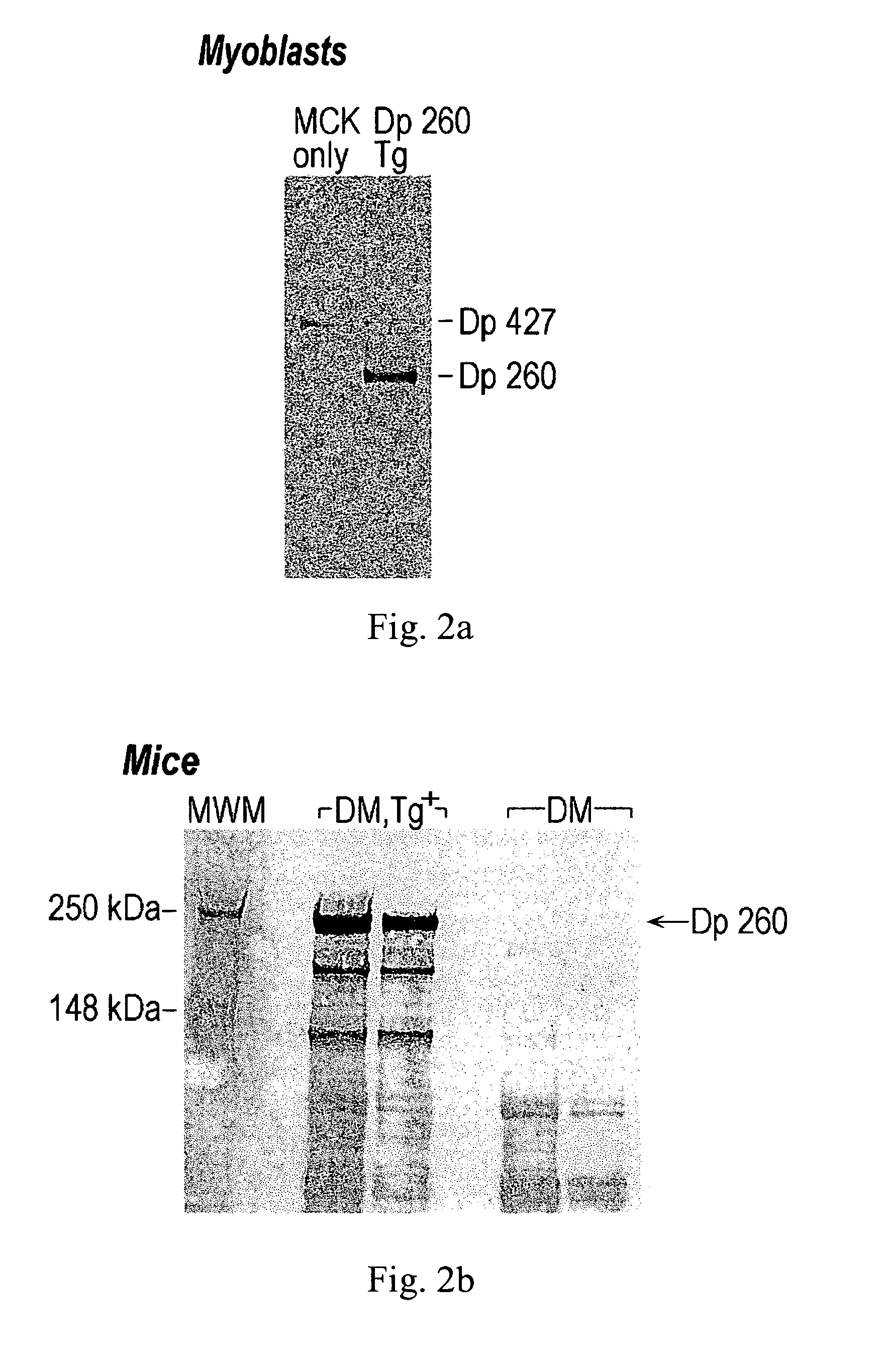
[0060] Differentiated MM14 myoblast cell cultures, stably transfected with either the human MCK / Dp260 Tg or the MCK plasmid alone, were harvested. Protein was extracted from 3 million cells by homogenizing in 1 mL of homogenization buffer (50 mM Tris pH 8, 150 mM NaCl, 1 mM EDTA, 0.04 mg / mL aprotinin, 0.0025 mg / mL pepstatin A, 0.025 leupeptin, 1 mM phenylmethyl sulfonylfluoride, 0.1% Triton X100) in a Dounce homogenizer. Muscle tissue was also harvested (100 mg) from the hind legs of DM / Tg+, and DM mice. The tissue was frozen and was homogenized in 1 mL homogenization buffer using a chilled mortar and pestle. The homogenates were centrifuged for 10 minutes at 13,000 rpm at 4° C. to sediment cell debris.

[0061] A 4× loading buffer (Invitrogen) was added to the supernatant, and the proteins were heat denatured at 70° C. for 10 minutes. Aliquots of 24 μL were analyzed on 4-8% acrylamide gels using a NuPAGE Tris-Acetate SDS Gel System (Invitrogen). Proteins were transfe...


PUM

PropertyMeasurementUnit
Fractionaaaaaaaaaa
Timeaaaaaaaaaa
Weightaaaaaaaaaa

Abstract

Duchenne muscular dystrophy (DMD) is a progressive muscle disease that is caused by severe defects in the dystrophin gene and results in the patient's death by the third decade. The present invention utilizes the Double Mutant mice (DM) as an appropriate human model for DMD as these mice are deficient for both dystrophin and utrophin (mdx / +, utrn − / −), die at 3 months of age and suffer from severe muscle weakness, pronounced growth retardation, kyphosis, weight loss, slack posture, and immobility. Expression from a transgene of novel human retinal dystrophin Dp260 was shown to prevent premature death and reduce the severe muscular dystrophy phenotype to a mild clinical myopathy. Electromyography, histology, radiography, magnetic resonance imaging, and behavior studies concluded that DM transgenic mice grew normally, had normal spinal curvature and mobility, and had reduced muscle pathology. EMG and histologic data from transgenic DM mice showed decreased abnormalities to levels typical of mild myopathy, while the DM mice exhibited severe abnormalities commonly seen in human dystrophinopathies. The transgenic DM mice also had measurable movement levels comparable to those of untreated mdx mice and controls.

Description

RELATED APPLICATIONS [0001] The following application claims the benefit of Provisional Application Serial Nos.: 60 / 588,700; Filed: Jul. 16, 2004; 60 / 608,252; Filed: Sep. 9, 2004; and 60 / 613,026; Filed: Sep. 24, 2004, the teachings and contents of which are hereby enclosed by reference.SEQUENCE LISTING [0002] The present application contains a sequence listing in both computer readable format and on paper. The computer readable format copies are labeled as 34444.txt Copy 1 and 34444.txt Copy 2. These copies are identical to one another and are identical to the paper copy of the sequence listing included herewith. Each of these sequence listings are expressly incorporated by reference into the present application. BACKGROUND OF THE INVENTION [0003] 1. Field of the Invention [0004] The present invention relates to Duchenne muscular dystrophy (DMD). More particularly, the present invention is concerned with a novel model for DMD as well as treatments for DMD. Still more particularly, t...

Claims


Application Information

Patent Timeline
IPC IPC(8): A61K31/70A01K67/00A61K35/00A61P43/00C07H21/04C12N15/00C12N5/06
CPCA01K67/0275A01K67/0276A01K2217/05A01K2217/075C12N2830/008A01K2267/0306C07K14/4708C12N15/8509A01K2227/105A61P21/00A61P21/04A61P25/00A61P43/00C12Q1/68
Inventor WHITE, ROBERT L.GAEDIGK, ROGERFITZGERALD-GUSTAFSON, KATHLEEN
Owner WHITE ROBERT L
Why Eureka
Try Eureka