
Ocular pressure regulation

a technology of ocular pressure and regulation, which is applied in the field of ocular pressure regulation, can solve the problems of glaucoma patients exposed to side effects, loss of field of vision, and tunnel vision and blindness

Active Publication Date: 2007-04-19
ALCON INC
View PDF99 Cites 61 Cited by
  • Summary
  • Abstract
  • Description
  • Claims
  • Application Information

AI Technical Summary

Benefits of technology

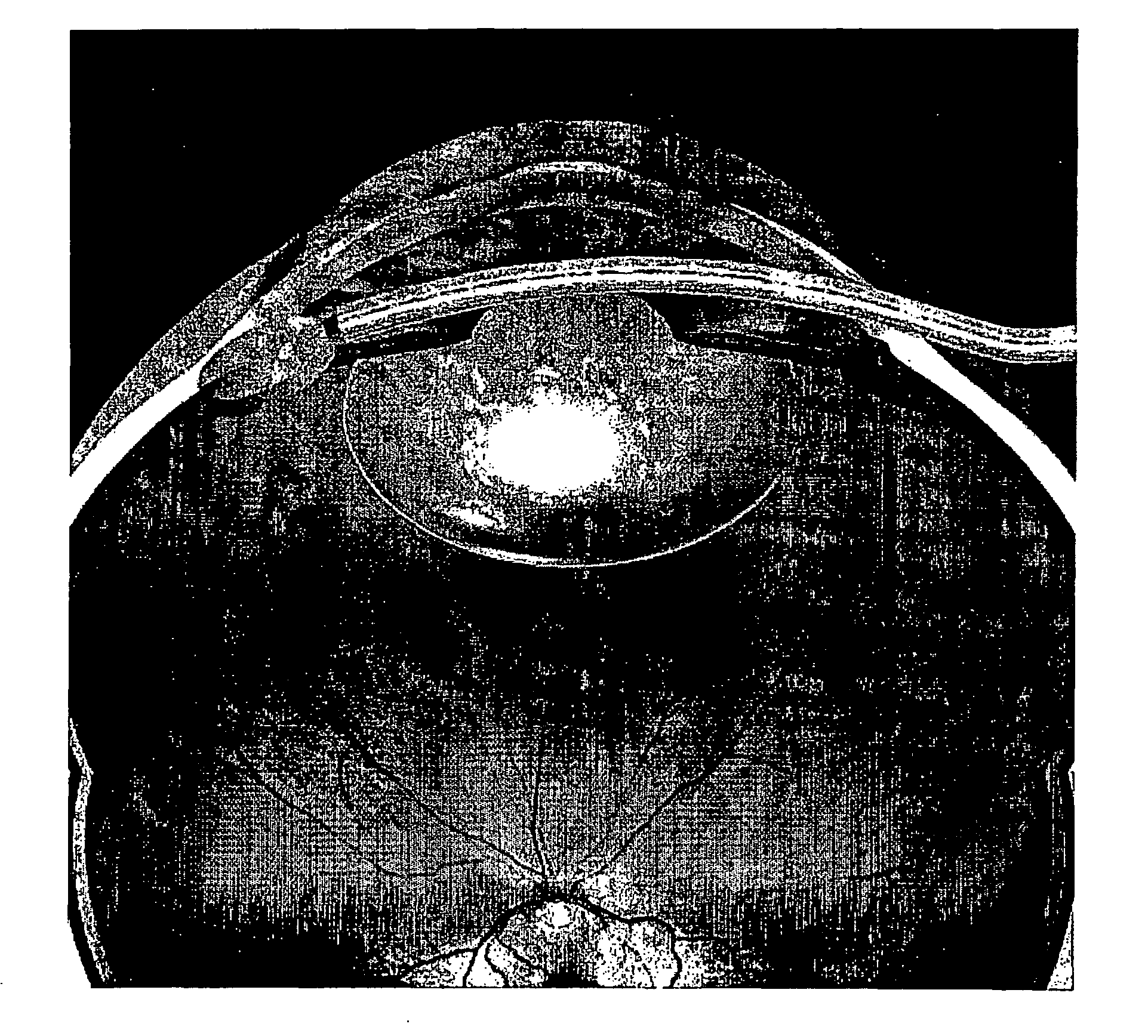
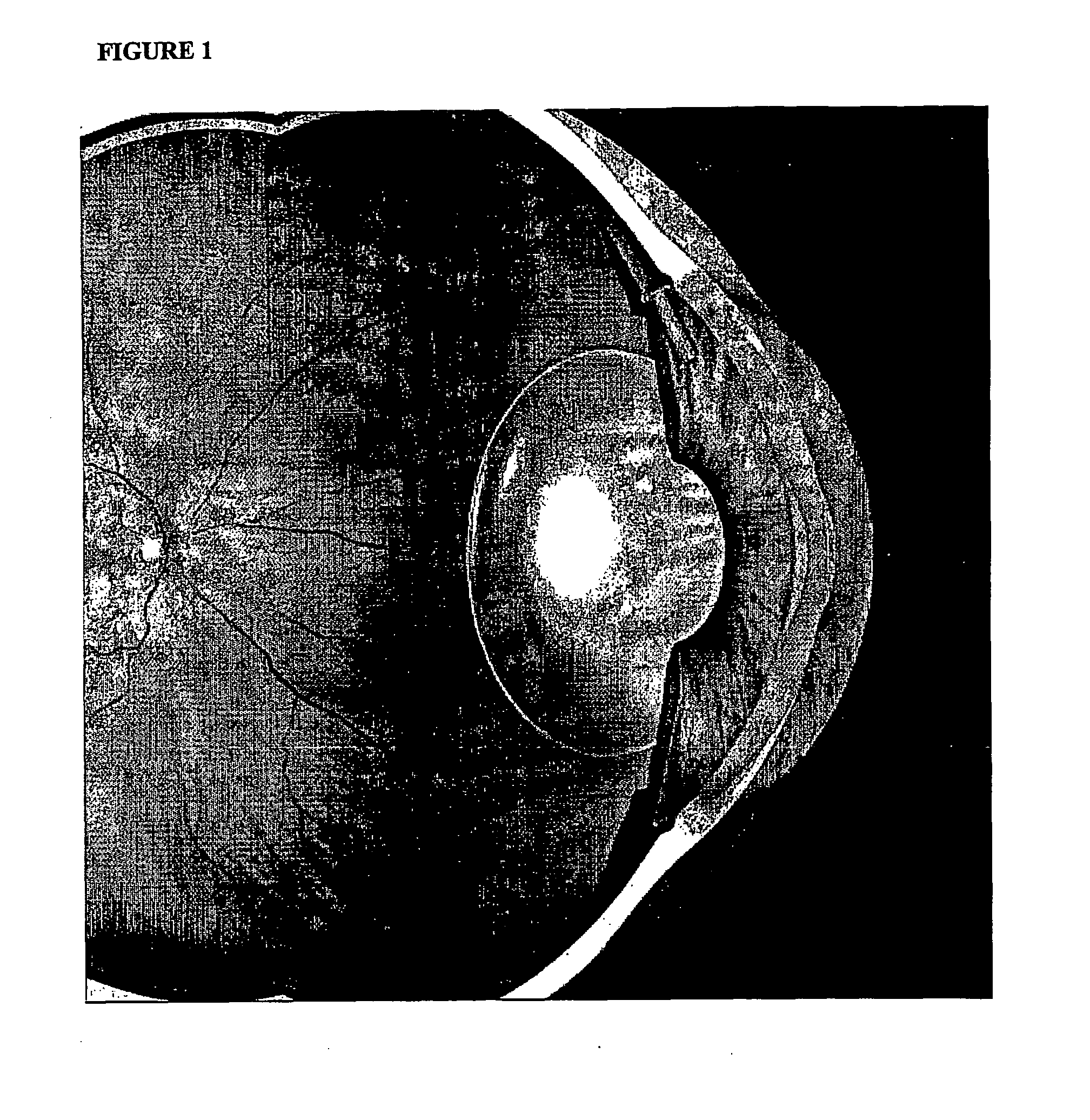
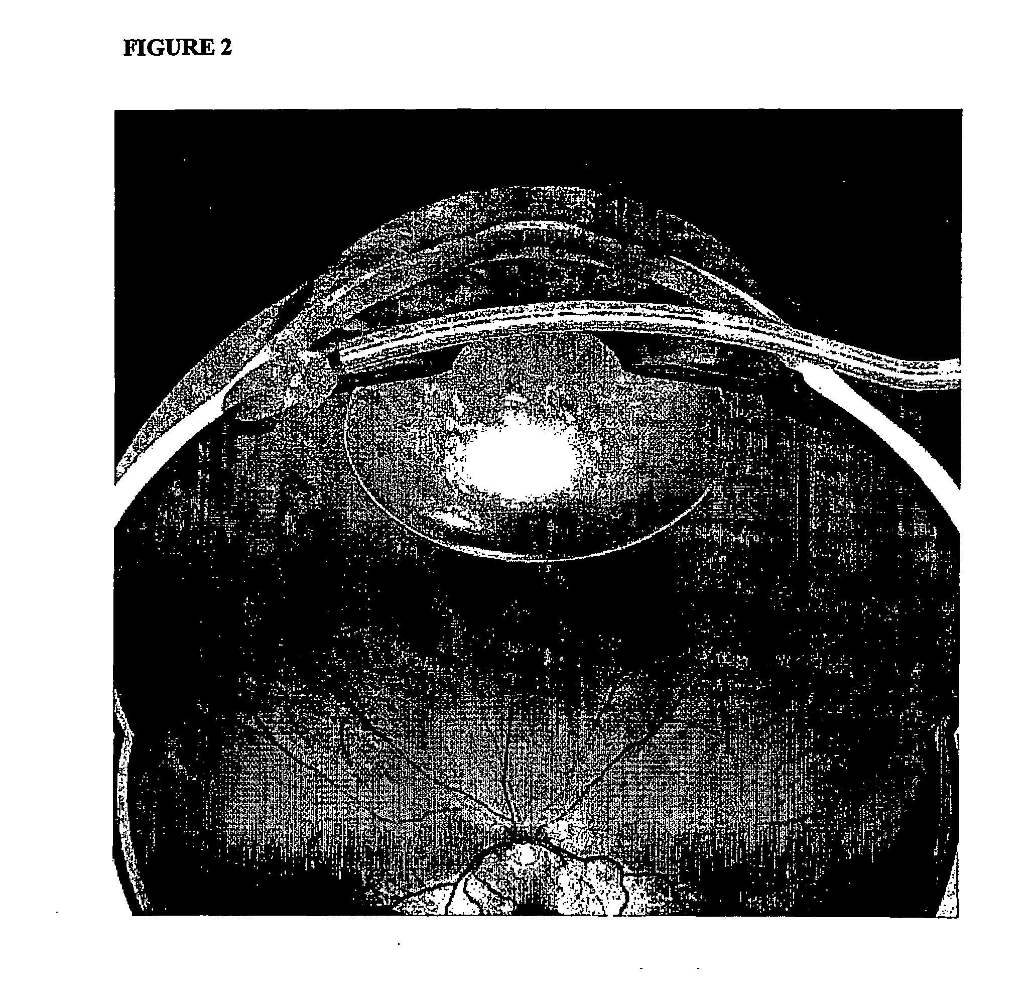
The present invention provides a flexible ocular device for implantation into the eye that can regulate aqueous pressure. The device is made of a biocompatible elastomeric material and has a fluid drainage tube with a valve to control pressure. The device can be implanted through a small incision in the eye and can also be used to prevent ocular pressure spikes after surgery. The technical effects of the invention include improved treatment and prevention of glaucoma and reduction of ocular pressure spikes.

Problems solved by technology

Elevated intraocular pressure results in the death of retinal ganglion cells (which convey retinal information to the brain) resulting in a characteristic pattern of loss of the field of vision, progressing to tunnel vision and blindness if left untreated.
Although pharmacological treatments of glaucoma have improved, they have important implications for the patient's quality of life, have compliance issues which are important in the elderly (in whom glaucoma is prevalent), expose the patient of glaucoma to side effects, and over a lifetime are costly.
These suffer from similar problems of scarring (Classen L, Kivela T, Tarkkanen “A Histopathologic and immunohistochemical analysis of the filtration bleb after unsuccessful glaucoma seton implantation”Am J Ophthalmol, 1996; 122:205-12) around the external opening of the tube devices in the subconjunctival space—the development of a large number of these devices is testament to the fact that many fail in the longer term.
Whereas cataract surgery has been revolutionized in the last two decades, improvements in glaucoma surgery have been slower.
Glaucoma shunts have had limited success in eyes that have “failed” multiple standard procedures.
However complications with malpositioned tubes, erosion & strabismus persist.
A considerable issue is the lack of reproducibility and predictability in achieving the desired target intraocular pressure (IOP).
Final IOP is largely determined by healing which can be unpredictable—in view of vast biological variations, it is impossible to predict which eyes will rapidly scar causing failure and which will fail to heal resulting in prolonged post-operative hypotony.
Scarring remains a significant problem in all these external drainage proposals, where aqueous drains into the conjunctiva, or surgical chambers in the sclera.
However the success rate of such approaches has been low (as low as 15%, Rosenburg & Krupin ibid and Gross R L, Feldman R M, Spaeth G L, et al “Surgical therapy of chronic glaucoma in aphalia and pseudophakia”Ophthalmology, 1988; 95:1195-201).
Failure was due to uncontrolled low pressure (hypotony) with consequentiqal macular edema, bleeding (hyphema) and inadequate pressure control.
Such pressure spikes can cause pain and may increase the risk of sight-threatening complications such as retinal vascular occlusion, increases loss of visual filed in advanced glaucoma and ischemic optic neuropathy—effects in otherwise healthy eyes are unknown (Hildebrand G D et al, ibid).
A number of prophylactic treatments are used with limited success—these include intacameral carbachol or acetylcholine, topical timolol, dorzolamide, aproclonidine, latanoprost and systemic acetazolamide (see Hildebrand G D et al, ibid).
This also exposes the patient to the risk of drug side effects, increased cost and it has been postulated that reducing the flow of aqueous humor post surgery prolongs the residence time of bacteria that frequently (46.3% of cases) contaminate the anterior chamber during surgery (Srinivasan R, Tiroumal S, Kanungo R, Natarajan M K.
This may increase the risk of endopthalmitis one of the most devastating sequeleae of intraocular surgery, since the bacteria are not being “flushed out” of the eye by the normal production of aqueous humour, the secretion of which has been suppressed by the drugs.
This requires surveillance and could increase the risk of infection.

Method used


Image

  • Ocular pressure regulation
  • Ocular pressure regulation
  • Ocular pressure regulation

Examples


example 1

[0038] Fresh whole porcine eyes were taken and mounted in a temperature controlled (37°) perfusion chamber. The eyes were perfused with Balanced Salt Solution via a 30 gauge needle inserted via a paracentesis into the anterior chamber. A peristaltic pump was used at a flow rate of 2 μl / min. Intraocular pressure was continuously monitored via a second paracentesis.

[0039] Typically intraocular pressures stabilized at 10-15 mm Hg and fell with time (the “washout effect”, as glycosan aminoglycans are washed out of the trabecular meshwork with time). Creation of a cyclodialysis (initially with a small spatula, then viscoelastic injection to enlarge the area of detachment of the ciliary body from the sclera) with or without insertion of the device in the cyclodialysis cleft (silicone tubing, length 3 mm, external diameter—1 mm, plate diameter 3 mm) resulted in lower intraocular pressures (below 10 mm Hg) on reperfusion at the same perfusion rate as control eyes.

example 2

[0040] Adequate anesthesia is provided to the eye of a glaucoma patient prepared for intraocular surgery. A paracentesis (opening into anterior chamber from without at the junction of the cornea and sclera—the limbus) is performed and the anterior chamber is filled with a viscoelastic substance. A surgical gonioscopy lens is placed on the cornea (or anterior segment endoscope is used) and a cyclodialysis instrument is introduced via the paracentesis—the paracentesis is carried out 180° away from the planned implant insertion site. The cyclodialysis instrument tip is advanced into the angle and pushed into the space between the ciliary body and sclera creating a cycodialysis—this is carried out with direct visualization via the gonioscopy lens viewed through an operating microscope. In order to minimize bleeding, the area in the angle (anterior ciliary body face and overlying trabecular meshwork) can be lasered either preoperatively or at the time of surgery to abalate surface blood ...

example 3

[0042] Fresh whole porcine eyes were taken and mounted in a temperature controlled (37°) perfusion chamber as in Example 1. The eyes were perfused with Balanced Salt Solution via a 30 gauge needle inserted via a paracentesis into the anterior chamber. A peristaltic pump was used at a flow rate of 2 μl / min. Intraocular pressure was continuously monitored via a second paracentesis.

[0043] Typically intraocular pressures stabilized at 10-15 mmmHg and fell with time (the “washout effect, as glycoaminoglycans are washed out of the trabecular meshwork with time). Silicone tubing, length 3 mm, external diameter 1 mm was introduced into one paracentesis port. One end of the port (outer end) was flush with the cornea and the inner end of the port extended slightly into the anterior chamber. Intraocular pressure did not exceed 10 mm Hg.


PUM


Abstract

This invention comprises a flexible ocular device for implantation into the eye formed of a biocompatible elastomeric material, foldable to a diameter of 1.5 mm or less, comprising a fluid drainage tube having at one end a foldable plate adapted to located the device on the inner surface of the sclera in a suprachoroidal space formed by cyclodialyis, said drainage tube opening on to the disc at one end and opening to the anterior chamber when implanted into the eye at its other end, so as to provide aqueous pressure regulation. Also provided are methods for the treatment of glaucoma utilizing the flexible ocular device, and an ocular pressure spike shunt.

Description

FIELD OF THE INVENTION [0001] This invention is directed to therapeutic methods and devices for the treatment of glaucoma. In particular, this invention is concerned with the use of a shunt or drain for the treatment of glaucoma. In another aspect this invention is concerned with ocular pressure spikes shunts and use of the same in ocular surgery. BACKGROUND OF THE INVENTION [0002] The glaucomas are a common group of blinding conditions usually associated with elevated intraocular pressure. This elevated pressure in the eye may be regarded as a disorder of the drainage system of the eye which gives rise to the glaucomas. [0003] Aqueous humor of the eye (“aqueous”) is a flowing liquid fluid (composed of sodium, chloride, bicarb, amino acids, glucose, ascorbic acid, and water) that is actively secreted by the ciliary body and flows out past the iris into the anterior chamber (are between the lens / iris and the cornea). The aqueous drains out through angle formed by the iris and the scl...

Claims


Application Information

Patent Timeline
Patent Type & Authority Applications(United States)
IPC IPC(8): A61M5/00A61F9/007
CPCA61F9/00781A61F2/148
Inventor CORONEO, MINAS THEODORE
Owner ALCON INC
Learn More