Eureka AIR delivers breakthrough ideas for toughest innovation challenges, trusted by R&D personnel around the world.

Methods for detection of mycobacterium tuberculosis

a technology of mycobacterium tuberculosis and detection methods, which is applied in the field of methods for detection of mycobacterium tuberculosis, can solve the problems of the inability to find the indian laboratory, and the inability to use conventional methods

Inactive Publication Date: 2007-03-29
ALL INDIA INST OF MEDICAL SCI & DEPT OF BIOTECH
View PDF0 Cites 3 Cited by
  • Summary
  • Abstract
  • Description
  • Claims
  • Application Information

AI Technical Summary

Benefits of technology

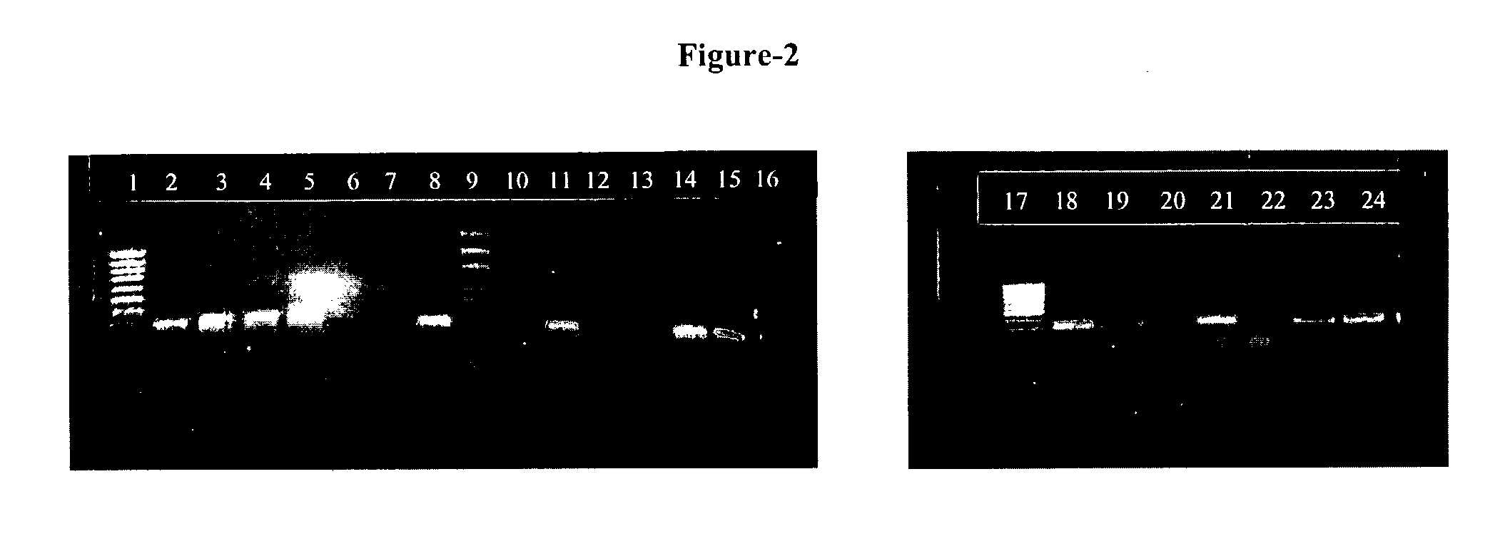
[0019] The present invention provides novel oligonucleotide primer pair having SEQ ID NO: 3 and SEQ ID NO: 4 for amplification of Early Secretory Antigenic Target (esat)-6-gene of Mycobacterium species. The invention also provides a method for detecting M. tuberculosis in a sample based on the amplification of esat-6 gene, comprising isolating DNA template from the sample, amplifying with the above oligonucleotide primer pair and subjecting the amplified DNA product to separation and staining to detect the presence of amplified DNA product for identifying Mycobacteri

Problems solved by technology

However, in India the diagnosis of Mycobacterial infections is established empirically on clinico-radiological basis or only by sputum smear examination, and the infections caused by non-tubercular Mycobacteria are under-diagnosed due to lack of diagnostic facilities.
The speciation of Mycobacteria using conventional methods is very slow, labor intensive, hazardous and not always reproducible (33) and hence left unattempted by most of the Indian Laboratories.
However, its further utility is restricted only for taxonomic purposes after sequencing the amplified gene product.
Therefore, the single PCR method can not answer the most important clinical question, whether the Mycobacterium is M. tuberculosis or other than M. tuberculosis.
The antigen- antibody based test methods have been a research topic but no antigen has been found to be satisfactory.
Even the M. tuberculosis specific antigen (ESAT-6 antigen in this case) has a fundamental drawback of poor predictive value to make an organ specific disease diagnosis.
Therefore, if a pre-exposed asymptomatic person (antibodies already positive) gets fresh TB brain infection (TB meningitis) and in another pre-exposed person there is no such fresh infection, the antibody detection assays will not be useful for the specific diagnostic use in such cases, as both these patients will be positive for these antibodies.
To explain it in other words, antibody detection assays, for diseases of high endemicity (e.g. Tuberculosis which is airborne) have very poor specificity and organ specific diagnosis can not be made at all, as the antibody detection methods are indirect evidences of infection.

Method used


Image

  • Methods for detection of mycobacterium tuberculosis
  • Methods for detection of mycobacterium tuberculosis
  • Methods for detection of mycobacterium tuberculosis

Examples


example 1

Clinical Samples:

[0053] Clinical samples were obtained from patients attending the out patient department (OPD), or hospitalized patients at the All India Institute of Medical Sciences (AIIMS), New Delhi, India. All the samples were processed in the Clinical Microbiology Division, Department of Laboratory Medicine. A total of 3072 patients who attended the general OPD of AIIMS, with complaints of fever, cough and expectoration were investigated to rule out tuberculosis, as a routine work-up. However, only 265 of these patients had clinico-radiological findings suggestive of tubercular etiology and were referred to specialized clinics. These referred patients fulfilled the inclusion criteria as per CDC guidelines and were included in this study. After informed consent of the patients, their routine investigations were carried out and their blood samples were also tested for human Immunodeficiency virus (HIV) infection. The clinical samples investigated for Mycobacterial isolation i...

example 2

Microscopy and Routine Culture:

[0054] The slide smears were air dried and heat fixed. One set of smears was stained with auramine-O (A-O) fluorochrome staining and examined under epifluorescence at 400× using Nikon®™ fluorescent microscope and the another set stained with Zeihl-Neelsen staining and examined under oil emersion (1000×). The results and quantification of acid fast bacilli (AFB) were reported as per revised WHO guidelines (33): scanty (1 or 2 AFB in 300 oil fields), 1+(3-9 AFB in 100 fields), 2+(1-10 AFB per oil field), or 3+(10 or more AFB per oil field).

[0055] Culture and identifications were made according to the standard method (12, 36) by inoculating on a Lowenstein-Jensen (L-J) slant with 0.2 to 0.5 ml of the decontaminated sputum sample. Cultures were incubated at 37° C. till the growth or for 8 weeks whatsoever was early. The cultured Mycobacteria were identified by staining and standard biochemical methods including Niacin, heat stable catalase, aryl sulphat...

example 3

Extraction of Genomic DNA:

[0056] The DNA was isolated directly from the Mycobacterium cultures by a procedure which the applicants have been following in the laboratory for the last several years. Briefly: 2-3 loopful (˜100 mg) of Mycobacterial cultures was transferred to an eppendorf tube containing 200 μl of sterile distilled water. The suspension was incubated at 80° C. in a water bath for 20 minutes. To the suspension 200 μl chloroform was added followed by vortexing. The suspension was again incubated at 65° C. for 10 minutes and centrifuged at 9000 rpm for 2 minutes. The clear supernatant containing Mycobacterial DNA was taken for assay.


PUM

PropertyMeasurementUnit
Lengthaaaaaaaaaa
Stabilityaaaaaaaaaa

Abstract

The present invention provides an oligonucleotide primer pair having SEQ ID NO: 3 and SEQ ID NO: 4 for amplification of Early Secretory Antigenic Target (esat)-6-gene of Mycobacterium species. The invention also provides a method for detecting M .tuberculosis in a sample based on the amplification of esat-6 gene, comprising isolating DNA template from the sample, amplifying with the above oligonucleotide primer pair and subjecting the amplified DNA product to separation and staining to detect the presence of amplified DNA product for identifying Mycobacterium tuberculosis in the sample. The invention further provides a diagnostic kit for detection of Mycobacterium tuberculosis. The invention also provides a method of detecting Mycobacterium tuberculosis from a sample by amplifying the 16s rRNA region from the isolated DNA template by conventional methods to detect Mycobacterium species and further amplifying the positive sample contains Mycobacterium species using primers positive for ESAT-6 region detection of Mycobacterium tuberculosis.

Description

TECHNICAL FIELD [0001] The invention provides novel oligonucleotide primers for the amplification of Early Secretory Antigenic Target (ESAT)-6 regions for detection of Mycobacterium species. The primers are used for differentiating the Mycobacterium tuberculosis from other species of Mycobacterium. Further, the invention provides a method for detection of Mycobacterium tuberculosis based on the DNA amplification of the ESAT-6 region. BACKGROUND AND PRIOR ART [0002] According to the WHO, tuberculosis still kills 3 million individuals per year, making it the leading infectious cause of death. It is believed that one in every three individuals on the planet harbor the causative microorganism belonging to the genus Mycobacterium (13, 24). This genus represents a complex phenotypic and genotypic diversity amongst its more than 100 odd species (4,9,10,14,15,17,27,28,30,34). Though, the most important human pathogenic species is Mycobacterium tuberculosis (M.tb), other species commonly kno...

Claims


Application Information

Patent Timeline
IPC IPC(8): C12Q1/68C07H21/04
CPCC12Q1/689C12Q1/686C12Q2600/16
Inventor SINGH, SARMANSHARMA, PAWAN
Owner ALL INDIA INST OF MEDICAL SCI & DEPT OF BIOTECH
Why Eureka
Eureka Blog
Learn More