Eureka AIR delivers breakthrough ideas for toughest innovation challenges, trusted by R&D personnel around the world.

Method for acquiring information of a biochip using time of flight secondary ion mass spectrometry and an apparatus for acquiring information for the application thereof

a biochip and secondary ion mass spectrometry technology, applied in the field of biochip secondary ion mass spectrometry acquisition information and the application of an apparatus, can solve the problems of complicated method, unclear determination of the position of the probe portion, and special facilities and special equipment, and achieve the effect of high resistivity and better quantitative ability

Inactive Publication Date: 2007-02-22
JACKSON BRAD
View PDF16 Cites 13 Cited by
  • Summary
  • Abstract
  • Description
  • Claims
  • Application Information

AI Technical Summary

Benefits of technology

[0023] The present invention provides a solution for the aforementioned problems. The present invention provides a measurement method, which enables one to obtain a two-dimensional image with better quantitative-ability by suppressing the influence of the charge accumulation when the two-dimensional secondary ion image is obtained for a biological material fixed on a substrate having high resistivity by utilizing a TOF-SIMS method over a wide area.

Problems solved by technology

In addition, as described later, if there is no physical address for indicating the expected position of a matrix on the substrate that is employed for forming chips, an additional problem may occur.
More specifically, when the biochip is formed by using a method of supplying fine droplets of a probe solution thereto via the ink jet method, for example, an absence of the physical address thereon may lead to an unclear determination of the position of the probe portion when the analysis is conducted on the biochip, depending on employed method.
However, the probe on the biochip exists principally as a monolayer or less, and in general, the analysis of the biological materials including the clear determination of the matrix position requires highly sensitive surface analysis techniques.
However, this method has various disadvantages from the viewpoint of general purpose usage.
Specifically the method requires complicated labeling, as well as special facilities and special equipment, because the employed isotope itself may be a source of a radioactive emission.
However, such a method also has various problems with respect to achieving higher quantification-ability, such as a problem of the chemical stability of the fluorescent dye used for labeling, a problem of the fluorescent quenching, a problem of the nonspecific adsorption of the fluorescent dye onto the substrate surface, or additionally the problem of the quantification-ability (i.e., stability, reproducibility) of the specific binding-ability (i.e., hybridization) Thus, there are a number of problems for quantitatively detecting the amount of the existing probe itself.
However, these methods do not involve sufficient sensitivity for the quantitative analysis of the probe on a biochip, i.e., a biological material, or imaging thereof.
In particular, when a general purpose glass is employed as a substrate for producing the biochip, these methods are not available, because the absorption due to the glass substrate itself adversely affects the analysis results when the FT-IR (ATR) method is employed, for example, or because a charge-up occurred on the glass, which is an electrically insulating material, adversely affecting the analysis when the XPS method is employed.
However, these methods have a technical limitation in that only a limited number of elements can be detected.
Thus, the amount of the information on the chemical structures obtained from the mass spectrum is not sufficient.
Thus, the method is not generally suitable, because the obtained information is not sufficient for the analysis of organic compounds such as, for example, nucleic acid-related materials having only four common bases.
Thus, information on the chemical structure obtained from the mass spectrum, e.g., mass range, is limited.
In turn, when the positive charge accumulates beyond a certain level (i.e., charge-up condition), the excessive positive charge may disturb the quantitative measurements.

Method used


Image

  • Method for acquiring information of a biochip using time of flight secondary ion mass spectrometry and an apparatus for acquiring information for the application thereof
  • Method for acquiring information of a biochip using time of flight secondary ion mass spectrometry and an apparatus for acquiring information for the application thereof
  • Method for acquiring information of a biochip using time of flight secondary ion mass spectrometry and an apparatus for acquiring information for the application thereof

Examples


example 1

Preparation of a Nucleic Acid Probe Chip by Using a dT40 Probe

[0068] A nucleic acid probe was prepared by using quartz glass, similarly as in the method described in the Japanese Patent Laid-Open No. H11-187,900 (1999).

(1) Washing of the Substrate

[0069] A 25.4 mm×25.4 mm synthesized quartz substrate mm was placed on a rack and immersed in a solution containing a detergent for ultrasonic washing (GPIII, commercially available from BRANSON) diluted to 10% with pure water for one night. Then, the substrate was ultrasonic-washed in the detergent solution for 20 minutes and then washed with water to remove the detergent. After being rinsed with pure water, the substrate was further ultrasonic-washed within a container containing pure water for 20 minutes. Next, the substrate was immersed in an aqueous solution of 1N sodium hydroxide that was pre-heated to 80° C. for 10 minutes. Sequentially, the substrate was washed with water and further washed with pure water, and the washed substr...

example 2

Imaging and Composition Analysis via TOF-SIMS

(1) Operations

[0076] The imaging and the composition analysis for the DNA chip prepared in the above-mentioned Example 1 were carried out by using a “TOF-SIMS IV” apparatus, which is commercially available from ION TOF CO. LTD.

[0077] The apparatus and conditions used in this operation are listed below.

[0078]

[0079] primary ion beam: 25 kV, Ga+, 0.6 pA (pulse current), random scan mode;

[0080] pulse frequency of the primary ion beam: 2.5 kHz (400 μsec. / shot);

[0081] pulse width of the primary ion beam: 1 ns; and

[0082] beam diameter of the primary ion beam: 5 μm.

[0083]

[0084] detection mode for secondary ion: negative;

[0085] area for the measurement: 300 μm×300 μm;

[0086] number of pixel in the secondary ion image: 128×128 pixels; and

[0087] number of integrating operation: 256.

[0088] (2) Measurement Results

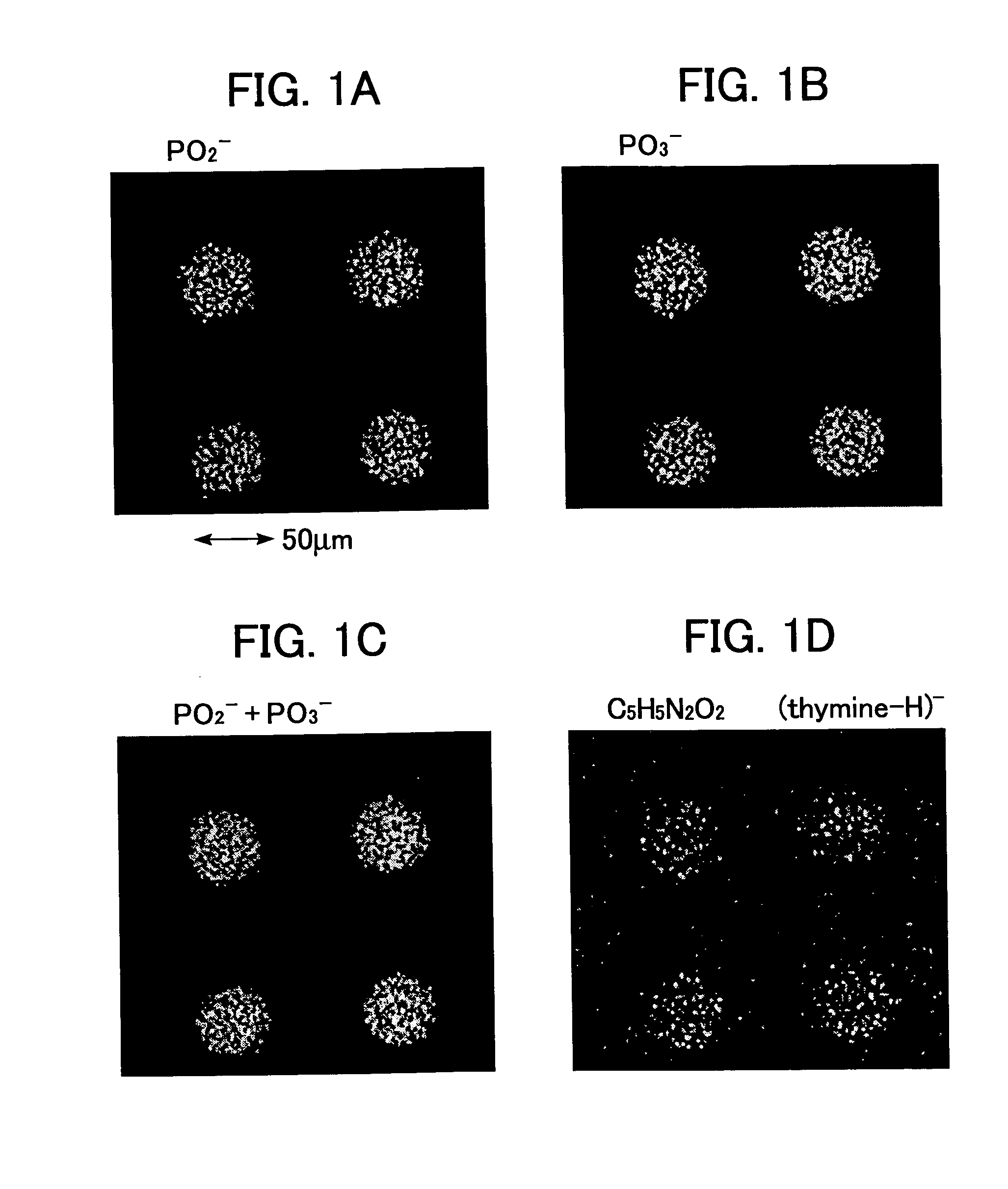
[0089]FIG. 1 shows the results of the imaging for the typical ion species from the data obtained by analyzing the DNA chip prep...

example 3

Preparation of a Nucleic Acid Probe Array by Employing 50mer Probe Containing Mixed Four Types of Nucleic Acid Bases, Imaging and Component Analysis Thereof

(1) Preparation of DNA chip

[0092] DNA chip was prepared with DNA of the following base sequence No. 2, in the procedure identical to the procedure described in Example 1.

Sequence No. 25′HS-(CH2)6-O-PO2-O-TGCAGGCATG CAAGCTTGGCACTGGCCGTC GTTTTACAAC GTCGTGACTG 3′

(2) Imaging and Composition Analysis Via TOF-SIMS

[0093] Imaging and composition analysis for the DNA chip comprising DNA of the above-identified sequence No. 2 were conducted via the method and conditions identical to these described in Example 2.

[0094] The results show that the imaging and the component analysis by the respective fragment ions of (adenine-H)−, (guanine-H)− and (cytosine-H)− can be conducted, as well as the imaging and the component analysis for the fragment ions for the phosphate backbone and the fragment ions, such as (thymine-H)− described in Exam...


PUM

PropertyMeasurementUnit
volumetric resistivityaaaaaaaaaa
diameteraaaaaaaaaa
voltageaaaaaaaaaa

Abstract

A measurement method is provided, which enables to obtain a two-dimensional image with better quantitative-ability by suppressing the influence of the charge-up, when the two-dimensional secondary ion image is obtained for a biological material fixed on a substrate having a high resistivity by utilizing a TOF-SIMS method in a certain wide area. A two-dimensional image having considerably high positioning resolution-ability can be obtained by the procedure in which the pulsed primary ion beam is irradiated at a spot, and the pulse-wise spot-applications of the primary ion beam and the simultaneous detection of the secondary ion generated from the irradiated primary ion beam proceed along with a discontinuous scanning pattern, and eventually the results of these secondary ion measurements are reconstructed into a two-dimensional image in line with the aforementioned discontinuous scanning pattern.

Description

BACKGROUND OF THE INVENTION [0001] 1. Field of the Invention [0002] The present invention relates to an imaging of a matrix disposed on a surface of a biochip, which includes a substrate and a plurality of biological materials disposed on a surface of the substrate in a matrix form, and also relates to an analysis of the components of the matrix. [0003] 2. Description of the Related Art [0004] A biochip, such as a DNA chip, protein chip and so on, which includes a substrate and various molecular probes disposed on a surface of the substrate in a matrix form, has been employed for the purposes of analyzing a genome or analyzing a generation of a gene. Further, it is expected that the result of the analysis employing biochips provides a critical index for diagnosis of cancers, genetic diseases, life style-related diseases, infectious diseases and the like, prediction for prognostics, or a decision on treatment policy and so on. [0005] Several methods for preparing biochips are known. ...

Claims


Application Information

Patent Timeline
Patent Type & Authority Applications(United States)
IPC IPC(8): G06F19/00G01N33/50G01N23/02G01N23/225G01N27/62G01N33/483G01N33/53G01N33/566H01J49/16H01J49/40
CPCG01N23/2258H01J49/16H01J49/40
Inventor OKAMOTO, TADASHITAKASE, HIROMITSUHASHIMOTO, HIROYUKI
Owner JACKSON BRAD
Why Eureka
Eureka Blog
Learn More