Method for obtaining dynamic and structural data pertaining to proteins and protein/ligand complexes
- Summary
- Abstract
- Description
- Claims
- Application Information
AI Technical Summary
Benefits of technology
Problems solved by technology
Method used
Image

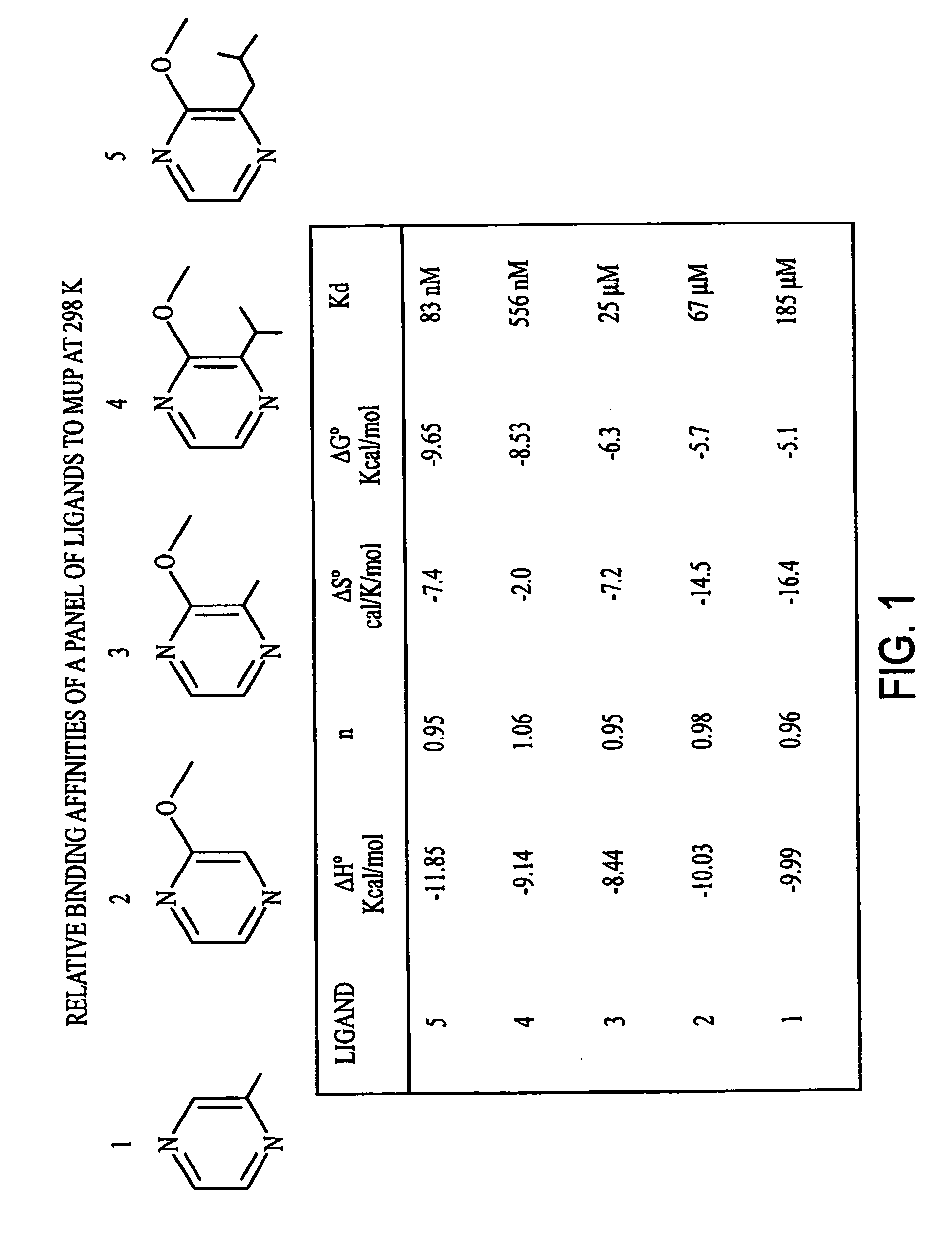
Examples
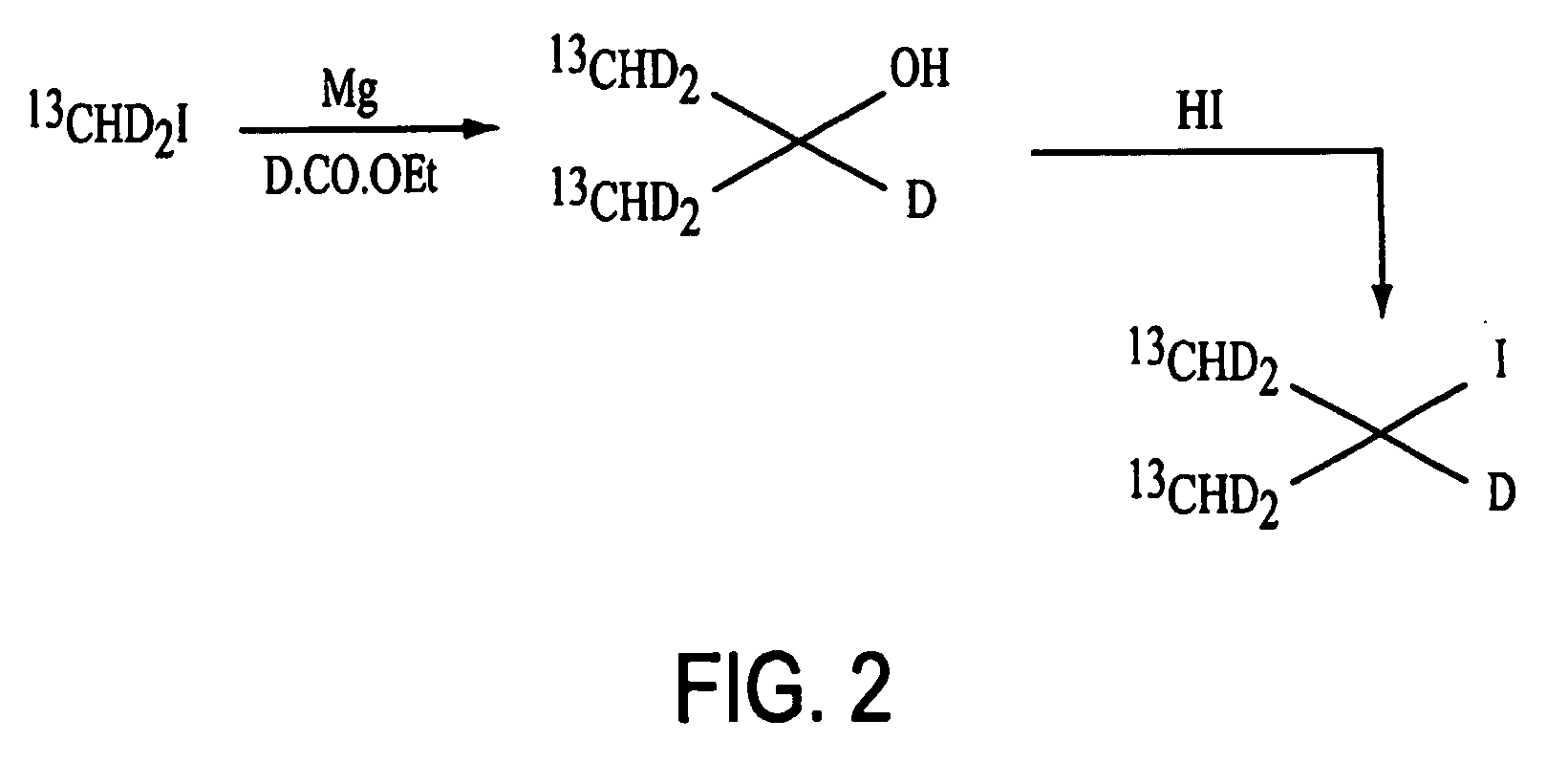
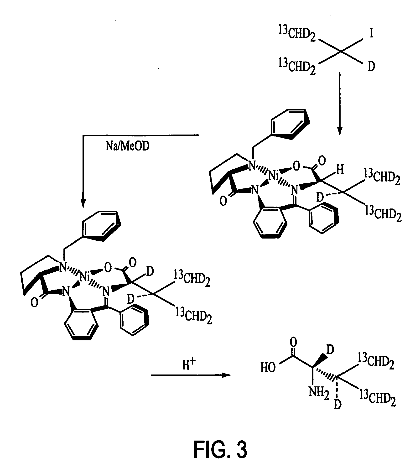
example 1
Synthesis of L-(13Cγ1γ2HD2)212Cα,βD-L-Valine
[0059] Magnesium turnings (6.08 g, 250.00 mmol, 2.50 equiv.) and anhydrous ether (100 mL) were added into a 3-neck 500 ml round bottom flask equipped with condenser, mechanical stirrer and heating mantle. The mixture was stirred and heated until under gentle reflux. 13CHD2-I (Cambridge Isotope Labs, 28.99 g, 200.00 mmol, 2.00 equiv.) in anhydrous ether (50 mL) was added dropwise into the Mg / ether mixture over 30 minutes and refluxing was continued for another 2 hours with the heating mantle to form a Grignard reagent. The reaction was then cooled in an ice bath.
[0060] D-CO—OCH3 (Cambridge Isotope Labs), 6.11 g, 100.00 mmol, 1.00 equiv.) in anhydrous ether (50 mL) was added slowly into the Grignard reagent over 15 minutes. The ice bath was removed and the reaction mixture was stirred for another 4 hours. The reaction mixture then was cooled again in an ice bath and saturated aqueous NH4Cl solution (35 mL) was added slowly over 15 minutes ...
example 2
Expression and Purification of Murine Urinary Protein (MUP) Containing L-valine-α-D-12CD(13CHD2)2
[0064] A 20 mL stock culture of M15 cells transformed with the vector pqe30 MUP was used to inoculate 1 L of medium containing 500 mg alanine, 400 mg arginine, 400 mg aspartic acid, 50 mg cysteine, 400 mg glutamine, 650 mg glutamic acid, 550 mg glycine, 100 mg histidine, 230 mg isoleucine, 230 mg leucine, 420 mg lysine HCl, 250 mg methionine, 130 mg phenylalanine, 100 mg proline, 2.1 g serine, 230 mg threonine, 170 mg tyrosine, 230 mg valine, 500 mg adenine, 650 mg guanosine, 200 mg thymine, 500 mg uracil, 200 mg cytosine, 1.5 g sodium acetate (anhydrous), 1.5 g succinic acid, 750 mg NH4Cl, 850 mg NaOH, 10.5 g K2HPO4 (anhydrous), 2 mg CaCl2 2H2O, 2 mg ZnSO4 7H2O, 2 mg MnSO4H2O, 50 mg tryptophan, 50 mg thiamine, 50 mg niacin, 1 mg biotin, 20 g glucose, 4 mL 1 M MgSO4, 1 mL 0.01 M FeCl3, 15 mg ampicillin, and 50 mg kanamycin.
[0065] When cell density had reached an OD of 1.2, the cells we...
example 3
NMR Analysis of Murine Urinary Protein (MUP) Containing L-valine-α-D-12CD(13CHD2)2
[0069] A 15 mg sample of L-valine-α-D-12CD(13CHD2)2 labeled MUP was dissolved in 650 μL phosphate buffered saline (10 mM potassium phosphate; 200 mM sodium chloride), to which was added 50 μL deuterium oxide. 13C Relaxation rates (R2 and R1) of 13CHD2 groups were determined using pulse sequences as described in Ishima et al., J. Am. Chem. Soc. 121:11589-11590 (1999). Spectral parameters were as follows: spectral width in the 13C dimension, 900 Hz; spectral width in the 1H dimension, 5200 Hz; number of real data points in the 13C dimension, 128; number of real points in the 1H dimension, 1408; number of transients per t1 increment, 8; probe temperature, 298 K. Prior to two-dimensional Fourier transformation, free induction decays were apodized with cosine-bell windowing functions according to known methods.
[0070] The NMR analysis was then repeated following addition of the small molecule ligand 1 μL o...
PUM
| Property | Measurement | Unit |
|---|---|---|
| Force | aaaaa | aaaaa |
| Force | aaaaa | aaaaa |
| Length | aaaaa | aaaaa |
Abstract
Description
Claims
Application Information
 - R&D
- Intellectual Property
- Life Sciences
- Materials
- Tech Scout
- Unparalleled Data Quality
- Higher Quality Content
- 60% Fewer Hallucinations
Browse by: Latest US Patents, China's latest patents, Technical Efficacy Thesaurus, Application Domain, Technology Topic, Popular Technical Reports.
© 2025 PatSnap. All rights reserved.Legal|Privacy policy|Modern Slavery Act Transparency Statement|Sitemap|About US| Contact US: help@patsnap.com