Endothelial cell growth factor methods of isolation and expression
a technology of growth factor and endothelial cells, applied in the field of new growth factor, can solve the problems of not being able to purify or characterize mitogenic activity, and no highly suitable mitogenic factor exists for this type of application, and achieve the effect of promoting re-endothelialization
- Summary
- Abstract
- Description
- Claims
- Application Information
AI Technical Summary
Benefits of technology
Problems solved by technology
Method used
Image


Examples
example a
[0235] Purpose of this Example was the determination of the molecular weight of the endothelial cell growth factor secreted in the medium by follicular cells.
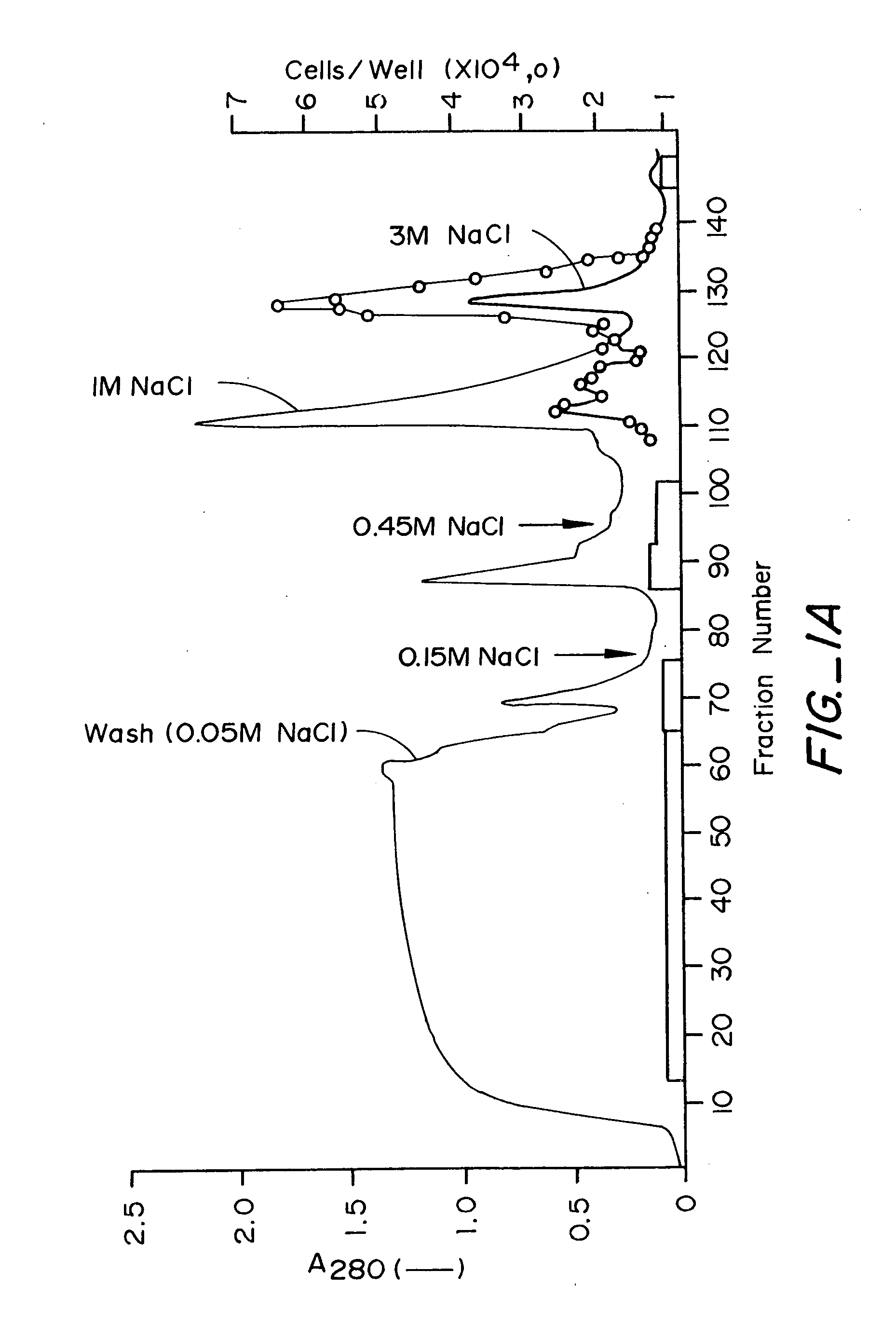
[0236] Confluent cultures of follicular cells were incubated for three days in a serum-free medium consisting of low glucose Dulbecco's modified Eagle's medium supplemented with transferrin (10 μl / ml), insulin (5 μg / ml), 2 mM glutamine and antibiotics. The conditioned medium (CM) (150 ml) was then collected centrifuged (10000×g, 15 min. 4° C.) in order to remove cell debris, and then applied to a Heparin-Sepharose column which had been preequilibrated with 10 mM Tris / Cl, pH 7.0. The column was then sequentially eluted with 10 mM Tris / Cl, pH 7.0 containing 0.6, 1 and 3 M NaCl. The flow rate was 21 ml / h. Fractions of 700 μl were collected and aliquots were tested for bioactivity on adrenal cortex-derived microvascular endothelial cells. The majority of the bioactivity was eluted in the present of 0.6 M NaCl. This chromatographic...
example 1
Culture of Follicular Cells and Media Collection
[0240] Primary cultures of bovine pituitary FC were established as previously described (20,41). In one embodiment in the culturing the 20% fetal bovine serum in reference 20 was reduced to 10%. Concentrations of 5 to 20% should be effective. Also no DNAase is used. All other components are the same. At confluency, cells were passaged into large scale tissue culture plates in the presence of low glucose Dulbecco's modified Eagle's medium (DMEM) supplemented with 10% fetal bovine serum, 2 mM glutamine and antibiotics. Shortly after reaching confluency the cultures were extensively washed with PBS in order to remove serum components. The cells were then incubated in a serum-free medium consisting of DMEM plus transferrin (10 μl / ml), insulin (5 μg / ml), selenium (10−8 M), 2 mM glutamine and antibiotics. After three or four days, the medium was collected and replaced with fresh serum free medium. The collected medium was centrifuged (1000 ...
example 2
Concentration of Conditioned Medium
[0241] Four to six liter batches of conditioned medium (CM) were subjected to ammonium sulfate precipitation. Ammonium sulfate (500 g / L) was added under constant stirring, until the salt was completely in solution. After 8-12 hours in the cold room, the material was centrifuged (20,000×g, 45 min at 4° C.). The supernatant was discarded and the pellet was resuspended with 10 mM Tris / Cl, pH 7.2, 50 mM NaCl and dialyzed at 4° C. against the same buffer for 8-12 h. The final volume was 50-60 fold less than the original.
[0242] Alternatively, the CM is concentrated using ultrafiltration using an Amecon stir cell (2.5 liter unit) using a membrane having a molecular weight cut off of 10,000 daltons with similar results.
PUM
 Abstract
Description
Claims
Application Information
 - R&D
- Intellectual Property
- Life Sciences
- Materials
- Tech Scout
- Unparalleled Data Quality
- Higher Quality Content
- 60% Fewer Hallucinations
Browse by: Latest US Patents, China's latest patents, Technical Efficacy Thesaurus, Application Domain, Technology Topic, Popular Technical Reports.
© 2025 PatSnap. All rights reserved.Legal|Privacy policy|Modern Slavery Act Transparency Statement|Sitemap|About US| Contact US: help@patsnap.com