Ocular pressure regulation
a technology of ocular pressure and regulation, which is applied in the field of ocular pressure regulation, can solve the problems of glaucoma patients exposed to side effects, loss of field of vision, and tunnel vision and blindness
- Summary
- Abstract
- Description
- Claims
- Application Information
AI Technical Summary
Benefits of technology
Problems solved by technology
Method used
Image
Examples
example 1
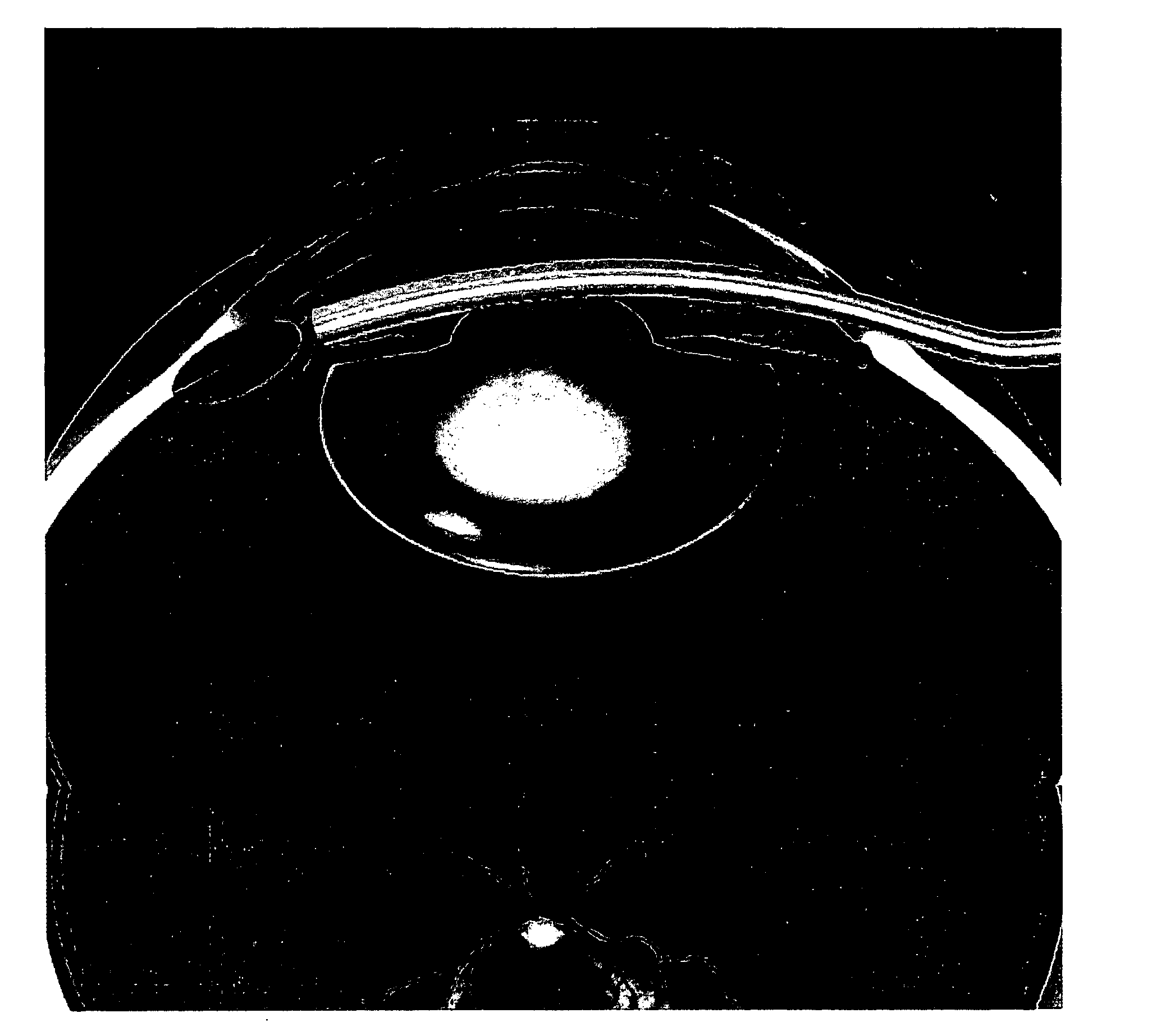
[0038] Fresh whole porcine eyes were taken and mounted in a temperature controlled (37°) perfusion chamber. The eyes were perfused with Balanced Salt Solution via a 30 gauge needle inserted via a paracentesis into the anterior chamber. A peristaltic pump was used at a flow rate of 2 μl / min. Intraocular pressure was continuously monitored via a second paracentesis.
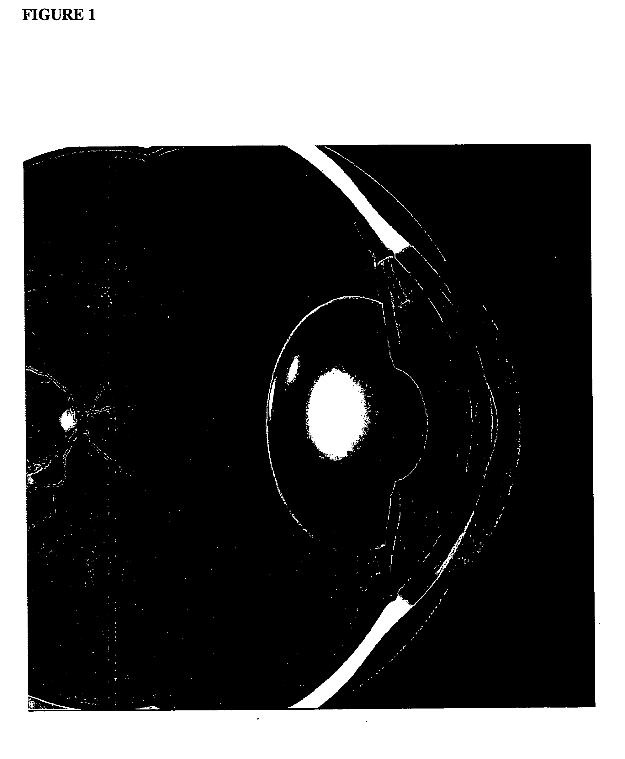
[0039] Typically intraocular pressures stabilized at 10-15 mm Hg and fell with time (the “washout effect”, as glycosan aminoglycans are washed out of the trabecular meshwork with time). Creation of a cyclodialysis (initially with a small spatula, then viscoelastic injection to enlarge the area of detachment of the ciliary body from the sclera) with or without insertion of the device in the cyclodialysis cleft (silicone tubing, length 3 mm, external diameter—1 mm, plate diameter 3 mm) resulted in lower intraocular pressures (below 10 mm Hg) on reperfusion at the same perfusion rate as control eyes.
example 2
[0040] Adequate anesthesia is provided to the eye of a glaucoma patient prepared for intraocular surgery. A paracentesis (opening into anterior chamber from without at the junction of the cornea and sclera—the limbus) is performed and the anterior chamber is filled with a viscoelastic substance. A surgical gonioscopy lens is placed on the cornea (or anterior segment endoscope is used) and a cyclodialysis instrument is introduced via the paracentesis—the paracentesis is carried out 180° away from the planned implant insertion site. The cyclodialysis instrument tip is advanced into the angle and pushed into the space between the ciliary body and sclera creating a cycodialysis—this is carried out with direct visualization via the gonioscopy lens viewed through an operating microscope. In order to minimize bleeding, the area in the angle (anterior ciliary body face and overlying trabecular meshwork) can be lasered either preoperatively or at the time of surgery to abalate surface blood ...
example 3
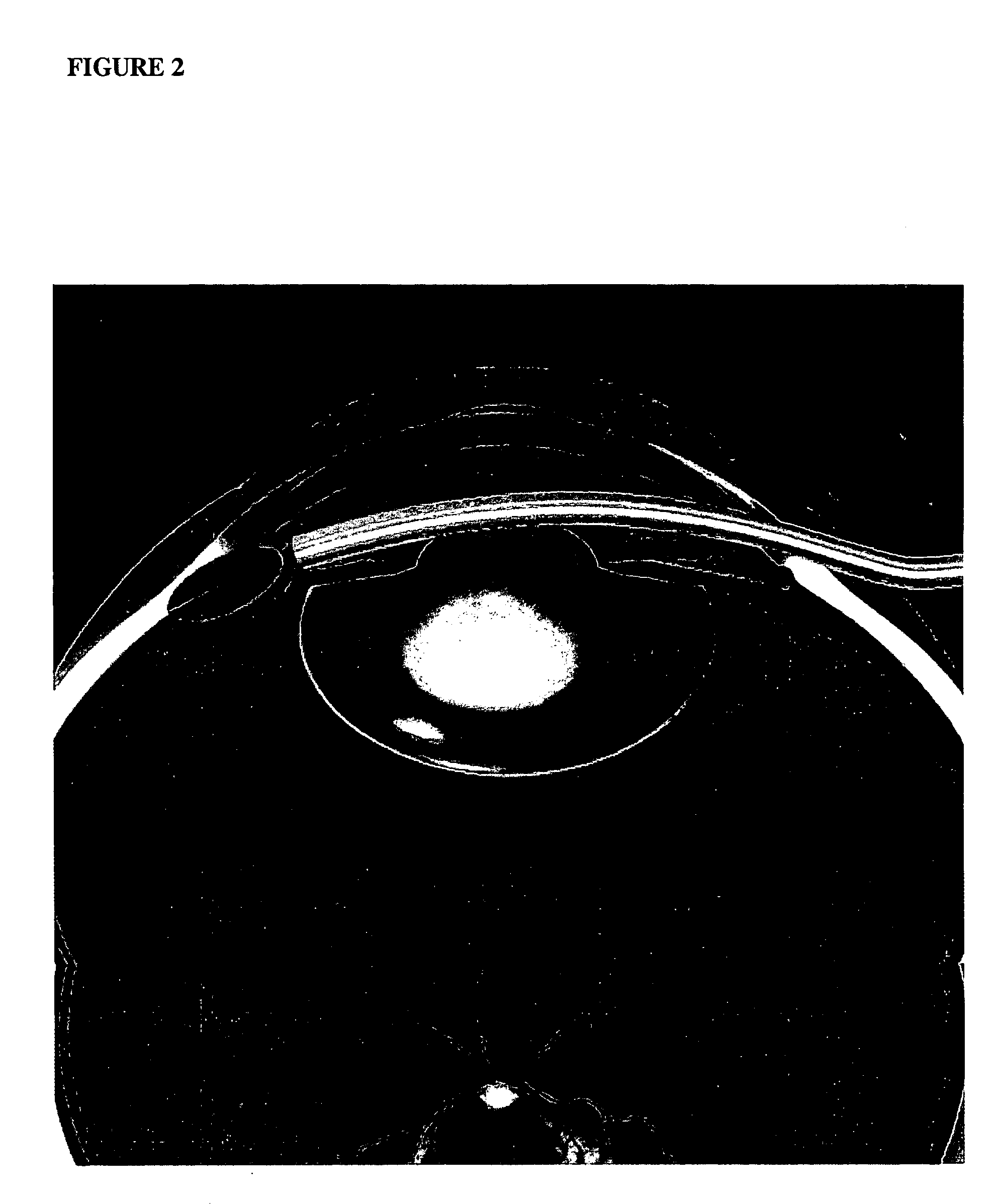
[0042] Fresh whole porcine eyes were taken and mounted in a temperature controlled (37°) perfusion chamber as in Example 1. The eyes were perfused with Balanced Salt Solution via a 30 gauge needle inserted via a paracentesis into the anterior chamber. A peristaltic pump was used at a flow rate of 2 μl / min. Intraocular pressure was continuously monitored via a second paracentesis.
[0043] Typically intraocular pressures stabilized at 10-15 mmmHg and fell with time (the “washout effect, as glycoaminoglycans are washed out of the trabecular meshwork with time). Silicone tubing, length 3 mm, external diameter 1 mm was introduced into one paracentesis port. One end of the port (outer end) was flush with the cornea and the inner end of the port extended slightly into the anterior chamber. Intraocular pressure did not exceed 10 mm Hg.
PUM
 Abstract
Description
Claims
Application Information
 - R&D
- Intellectual Property
- Life Sciences
- Materials
- Tech Scout
- Unparalleled Data Quality
- Higher Quality Content
- 60% Fewer Hallucinations
Browse by: Latest US Patents, China's latest patents, Technical Efficacy Thesaurus, Application Domain, Technology Topic, Popular Technical Reports.
© 2025 PatSnap. All rights reserved.Legal|Privacy policy|Modern Slavery Act Transparency Statement|Sitemap|About US| Contact US: help@patsnap.com


