
Intradermal delivery of vaccines and gene therapeutic agents via microcannula

a gene therapy and intradermal delivery technology, applied in the field of intradermal delivery of vaccines and gene therapy agents, can solve the problems of rarely targeted, unfavorable absorption, variable clinical results, etc., and achieve the effect of improving systemic immunogenic or therapeutic response, accurate targeting, and improving vaccine availability

Inactive Publication Date: 2004-07-08
BECTON DICKINSON & CO
View PDF98 Cites 11 Cited by
  • Summary
  • Abstract
  • Description
  • Claims
  • Application Information

AI Technical Summary

Benefits of technology

[0014] The present invention improves the clinical utility of ID delivery of vaccines and gene therapeutic agents to humans or animals. The methods employ devices to directly target the intradermal space and to deliver substances to the intradermal space as a bolus or by infusion. It has been discovered that the placement of the substance within the dermis provides for efficacious and / or improved responsiveness to vaccines and gene therapeutic agents. The device is so designed as to prevent leakage of the substance from the skin and improve adsorption or cellular uptake within the intradermal space. The immunological response to a vaccine delivered according to the methods of the invention has been found to be equivalent to or improved over conventional IM delivery of the vaccine, indicating that ID administration according to the methods of the invention will in many cases provide improved clinical results, in addition to the other advantages of ID delivery.
[0016] Accordingly, it is one object of the invention to provide a method to accurately target the ID tissue to deliver a vaccine or a medicament comprising genetic material to afford an immunogenic or therapeutic response.

Problems solved by technology

However, such delivery systems do not reproducibly breach the skin barriers or deliver the pharmaceutical substance to a given depth below the surface of the skin and consequently, clinical results can be variable.
The dermis, however, has rarely been targeted as a site for administration of substances, and this may be due, at least in part, to the difficulty of precise needle placement into the intradermal space.
Furthermore, even though the dermis, in particular, the papillary dermis has been known to have a high degree of vascularity, it has not heretofore been appreciated that one could take advantage of this high degree of vascularity to obtain an improved absorption profile for administered substances compared to subcutaneous administration.
On the other hand, large molecules such as proteins are typically not well absorbed through the capillary epithelium regardless of the degree of vascularity so that one would not have expected to achieve a significant absorption advantage over subcutaneous administration by the more difficult to achieve intradermal administration even for large molecules.
The technique is known to be quite difficult to perform and requires specialized training.
A degree of imprecision in placement of the injection results in a significant number of false negative test results.
Moreover, the test involves a localized injection to elicit a response at the site of injection and the Mantoux approach has not led to the use of intradermal injection for systemic administration of substances.
However, as discussed above, intradermal (ID) injection using standard needles and syringes is technically very difficult to perform and is painful.
The prior art contains several references to ID delivery of both DNA-based and conventional vaccines and therapeutic agents, however results have been conflicting, at least in part due to difficulties in accurately targeting the ID tissue with existing techniques.
A major factor that has precluded the widespread use of the ID delivery route and has contributed to the conflicting results described above is the lack of suitable devices to accomplish reproducible delivery to the epidermal and dermal skin layers.
Standard needles commonly used to inject vaccines are too large to accurately target these tissue layers when inserted into the skin.
This technique is difficult to perform, unreliable and painful to the subject.

Method used


Image

  • Intradermal delivery of vaccines and gene therapeutic agents via microcannula
  • Intradermal delivery of vaccines and gene therapeutic agents via microcannula
  • Intradermal delivery of vaccines and gene therapeutic agents via microcannula

Examples


example 1

ID Delivery and Expression of Model Genetic Therapeutic / Prophylactic Agents, Guinea Pig Model

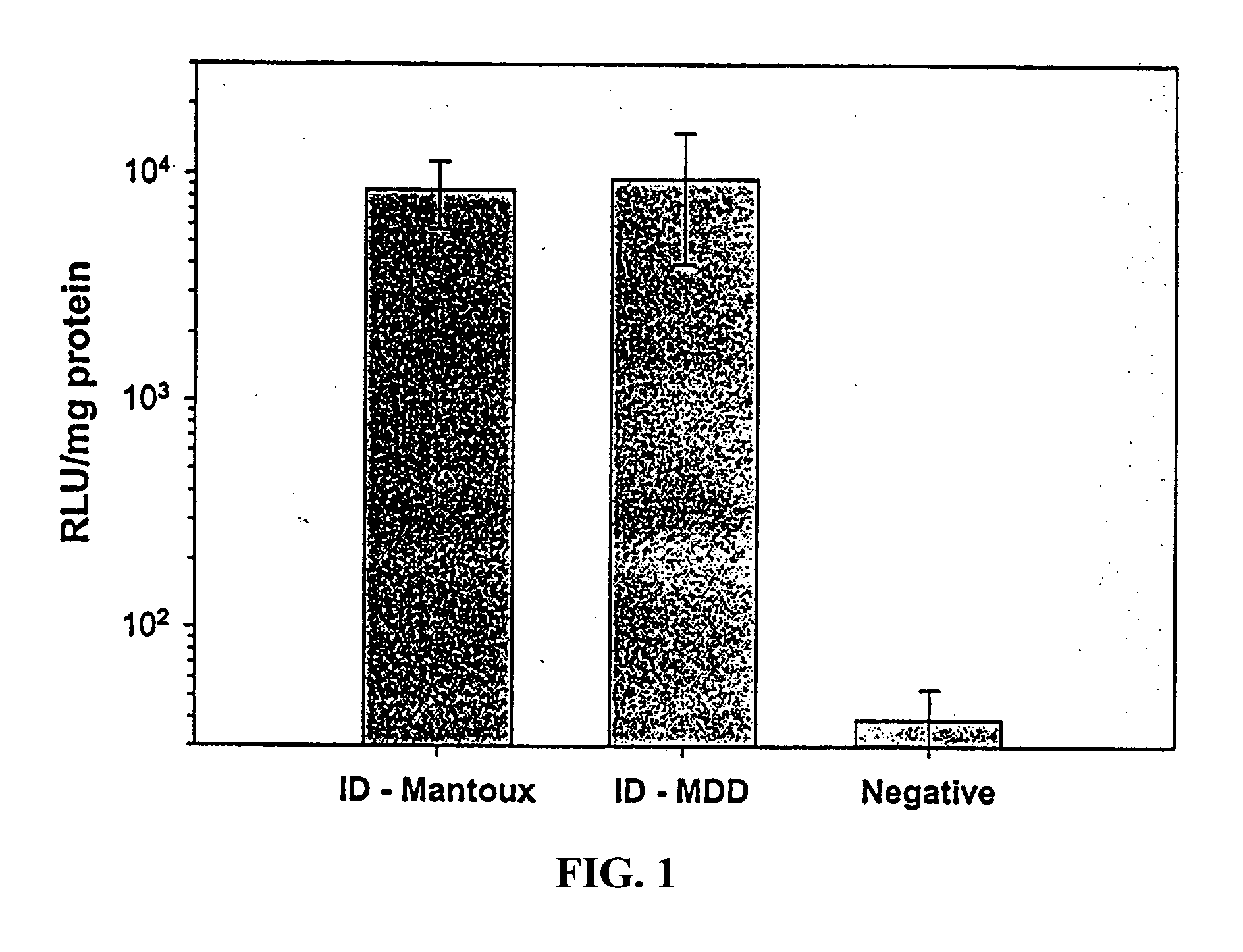
[0084] Uptake and expression of DNA by cells in vivo are critical to effective gene therapy and genetic immunization. Plasmid DNA encoding the reporter gene, firefly luciferase, was used as a model gene therapeutic agent (Aldevron, Fargo, N. Dak.). DNA was administered to Hartley guinea pigs (Charles River, Raleigh, N.C.) intradermally (ID) via the Mantoux (ID-Mantoux) technique using a standard 30 G needle or was delivered ID via MDD (ID-MDD) using a 34 G steel micro-cannula of 1 mm length (MDD device) inserted approximately perpendicular. Plasmid DNA was applied topically to shaved skin as a negative control (the size of the plasmid is too large to allow for passive uptake into the skin). Total dose was 100 .mu.g per animal in total volume of 40 .mu.l PBS delivered as a rapid bolus injection (<1 min) using a 1 cc syringe. Full thickness skin biopsies of the administration sites were collec...

example 2

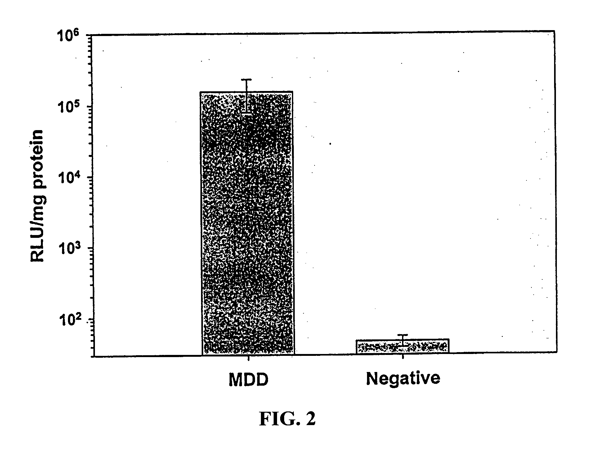
ID Delivery and Expression of Model Genetic Therapeutic / Prophylactic Agents, Rat Model

[0086] Experiments similar (without Mantoux control) to those described in Example 1 above were performed in Brown-Norway rats (Charles River, Raleigh, N.C.) to evaluate the utility of this platform across multiple species. The same protocol was used as in Example 1, except that the total plasmid DNA load was reduced to 50 .mu.g in 50 .mu.l volume of PBS. In addition, an unrelated plasmid DNA (encoding b-galactosidase) injected ID (using the MDD device) was used as negative control. (n=4 animals per group). Luciferase activity in skin was determined as described in Example 1 above.

[0087] The results, shown in FIG. 2, demonstrate very significant geneexpression following ID delivery via the MDD device. Luciferase activity in recovered skin sites was >3000-fold greater than in negative controls. These results further demonstrate the utility of the method of the present invention in delivering gene ba...

example 3

ID Delivery and Expression of Model Genetic Therapeutic / Prophylactic Agents, Pig Model

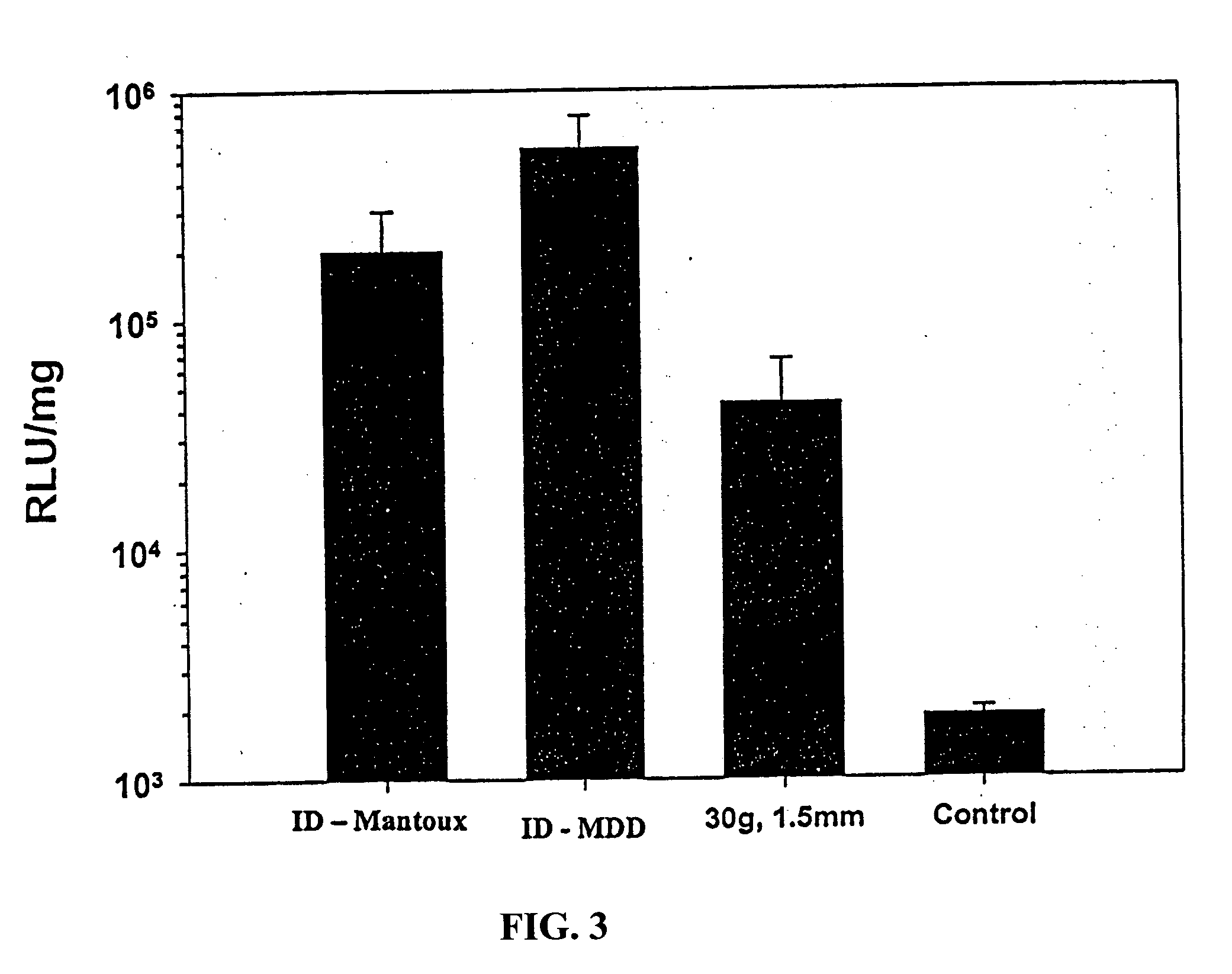
[0088] The pig has long been recognized as a preferred animal model for skin based delivery studies. Swine skin is more similar to human skin in total thickness and hair follicle density than is rodent skin. Thus, the pig model (Yorkshire swine; Archer Farms, Belcamp, Md.) was used as a means to predict the utility of this system in humans. Experiments were performed as above in Examples 1 and 2, except using a different reporter gene system, .beta.-galactosidase (Aldevron, Fargo, N. Dak.). Total delivery dose was 50 .mu.g in 50 .mu.l volume. DNA was injected using the following methods I) via Mantoux method using a 30 G needle and syringe, ii) by ID delivery via perpendicular insertion into skin using a 30 / 31 G needle equipped with a feature to limit the needle penetration depth to 1.5 mm, and iii) by ID delivery via perpendicular insertion into skin using a 34 G needle equipped with a feature to ...


PUM

PropertyMeasurementUnit
total thicknessaaaaaaaaaa
depthsaaaaaaaaaa

Abstract

The present invention relates to methods and devices for administration of vaccines and gene therapeutic agents into the intradermal layer of skin.

Description

[0001] This application is a continuation-in-part of application Ser. No. 10 / 185,717, filed Jul. 1, 2002, which is a continuation-in-part of application Ser. No. 10 / 044,504 filed Jan. 10, 2002, which is a continuation-in-part of application Ser. Nos. 09 / 834,438 and 09 / 835,243, filed Apr. 13, 2001, which are continuations-in-part of application Ser. No. 09 / 417,671, filed Oct. 10, 1999 and claims priority to U.S. provisional application No. 60 / 301,476, filed Jun. 29, 2001, each of which is incorporated herein by reference in its entirety.1. BACKGROUND OF THE INVENTION[0002] 1.1 Field of the Invention[0003] The present invention relates to methods and devices for administration of vaccines and gene therapeutic agents into the intradermal layer of skin.[0004] 1.2 Background Information[0005] The importance of efficiently and safely administering pharmaceutical substances for the purpose of prophylaxis, diagnosis or treatment has long been recognized. The use of conventional needles has ...

Claims


Application Information

Patent Timeline
Patent Type & Authority Applications(United States)
IPC IPC(8): C12N15/09A61B17/20A61D7/00A61K39/00A61K39/02A61K39/12A61K39/145A61K47/36A61K47/46A61K48/00A61M5/158A61M5/28A61M5/32A61M5/46A61M37/00A61P31/04A61P31/12A61P31/16A61P37/06
CPCA61B17/205C12N2760/16134A61K39/00A61K39/145A61K2039/53A61K2039/54A61M5/158A61M5/46A61M37/00A61M37/0015A61M2037/0061A61M2202/0445A61K2039/5252A61K2039/545A61K2039/55566A61K2039/55572A61D7/00A61K39/12A61P31/04A61P31/12A61P31/16A61P37/06Y02A50/30
Inventor MIKSZTA, JOHN A.ALARCON, JASON B.DEAN, CHERYLWATERSTON, ANDREA
Owner BECTON DICKINSON & CO
Learn More