
Apparatus and method for purification and assay of neurites

a technology of neurite and apparatus, applied in the field of cell biology, can solve the problems of affecting the study of neurites and their growth, severely affecting the understanding of the role of these neurite organelles in development, and the means to achieve the correct separation of neurites from neuronal cell bodies are by no means obvious, so as to facilitate rapid contact with neural cells and enhance the possibility of new drug discovery

Inactive Publication Date: 2004-04-08
INNOVATIV CELL SYST
View PDF0 Cites 16 Cited by
  • Summary
  • Abstract
  • Description
  • Claims
  • Application Information

AI Technical Summary

Benefits of technology

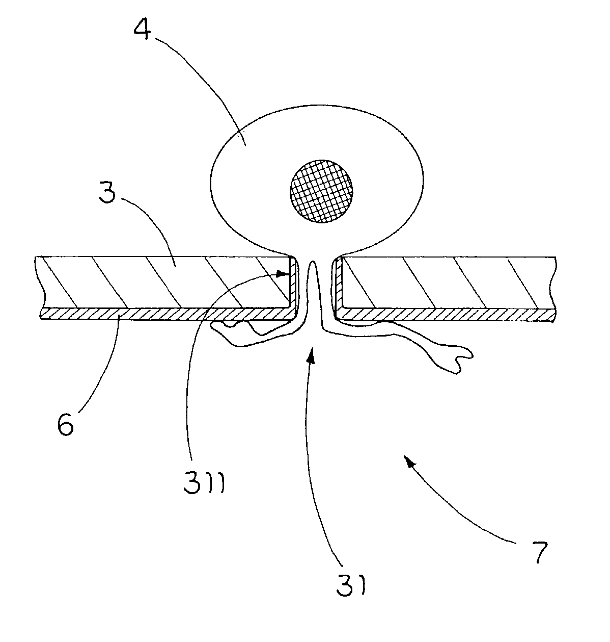
[0015] It is a main objective of the present invention to provide an apparatus and method for purification and assay of neurites to rapidly and conveniently contact neural cells and neurites with diverse panels of organic and inorganic substances, substantially enhancing the possibility of new drug discovery. The present invention further provides neurites useful in all aspects of their physical, physiological, biological and biochemical characterization.
[0034] In view of the above preferred embodiment, the present invention provides means to rapidly and conveniently contact neuronal cells and neurites with diverse panels of organic and inorganic substances and / or macromolecules, substantially enhancing the possibility of new drug discovery. Practically, neurite culture devices can be provided in any suitable form, for instance, as membranes, tubes, microtiter wells, columns, hollow fibers, roller bottles, plates, dishes, and solid, hollow, or porous beads. The filter membrane of the neurite culture device is coated at one side the cell adhesion factor for the outgrown neurite to be attracted and attached. Accordingly, the present invention is capable of providing neurites useful in all aspects of their physical, physiological, biological and biochemical characterization.
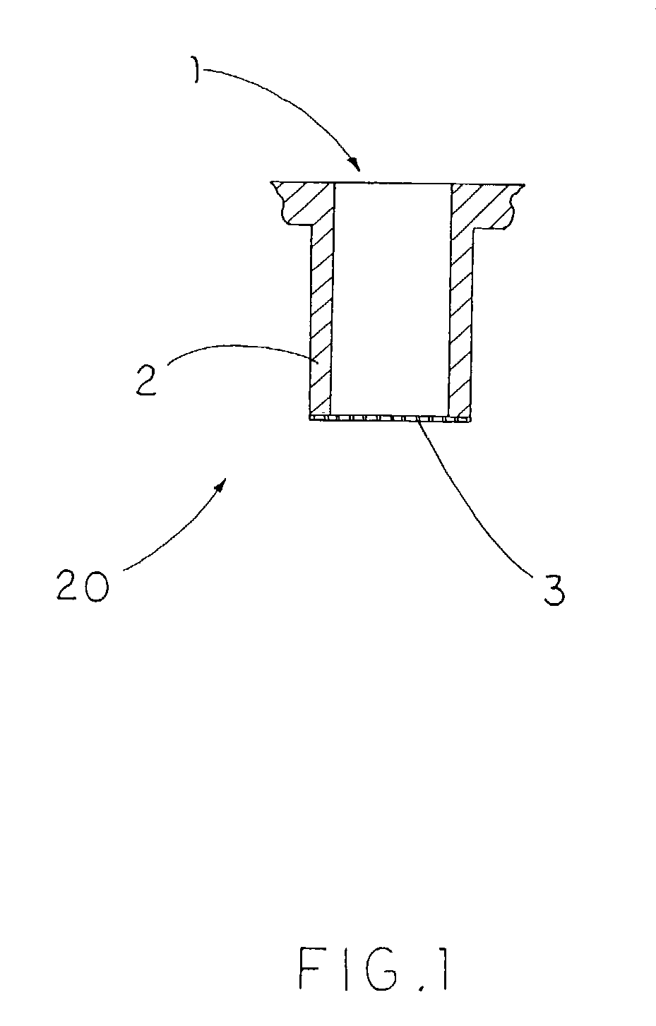
[0036] A wide variety of materials can be employed as the porous filter membrane 3, 9, the primary considerations being that they are preferably neither soluble or swellable in water. Suitable porous membrane materials provide a surface that exhibits an optimal combination of such properties as rigidity, surface area, ease of preparation and use, and cost.
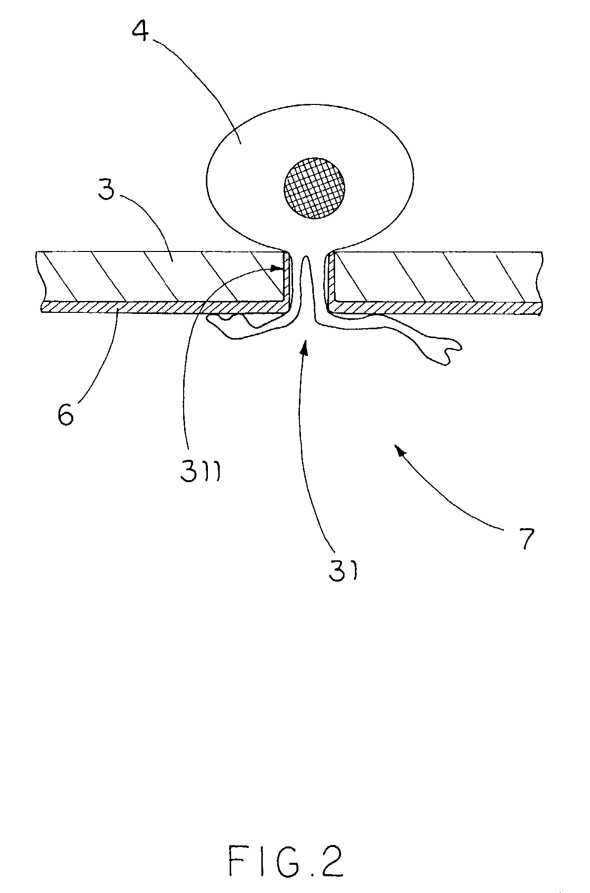
[0043] As described in more detail below, the cell adhesion factors for attracting neurite growth cones and stimulating and attaching the bodies of outgrowing neurites is preferably one or more proteins of the extracellular matrix (ECM) protein, or a preparation of ECM. The neurite culture device comprising the present invention improves the attachment and growth of the "anchorage dependent" neurites through favorable modification of the membrane surface, i.e. neurites are known to attach to substrates in a manner to some degree similar to "anchorage-dependent" cells. Such cells generally need to attach to a support surface and spread in order to grow and divide (Grinnel, F., "Cellular Adhesiveness and Extracellular Substrata", Int. Rev. Cytology 53:65-114 (1978)). Like cells, neurite attachment, growth and guidance on surfaces is mediated by polypeptides thereof. However, attachment of cells and extension and attachment of neurites to porous and non-porous surfaces and membranes is known in the art to also occur by means of static or induced charges. By example, but not by limitation, these could be poled polytetrafluoroethylene (PFE) (Vallenti, R. F. et al, Biomaterials 1989;13, 183-190), poled polyvinylidene fluoride (PVDF) (Valenti, R. F. et al, Brain Research 1989:480, 300-304) or fluoroethylenepolypropylene (FEP) (Makohliso, S. et al, J. Biomed Materials Res., 1993;27(8), 1075-1085. The electrostatic charges may be an intrinsic property of the membrane composition (as recited herein above), or may be induced asymmetrically to one side of the membrane, for instance by plasma or corona discharge (Makohliso, ibid). It is also clear to one skilled in the art that such implantation could be modified by masking of a part of the membrane surface, or otherwise directing patterns of deposition of positively charged molecules conducive to neurite outgrowth and attachment. The charge properties conducive to neurite extension and attachment may be further adjusted and modulated by additional components, such as dopants ((Preznayna, L., et al, 1991;24,5283-5287) or applied coatings of positively charged molecules.
[0045] Alternatively, positively-charged molecules employed on the bottom surfaces of the present invention can promote cell attachment to the surface by promoting ionic binding between the positively-charged molecules and cell constituents, such as negatively-charged glycoproteins and phospholipids carried on cellular surfaces.
[0054] Cell adhesion factors promote cell attachment by binding to specific receptors on the cell surface, and some cell types have receptors for more than one type of cell adhesion factor; Buck, C. A. and A. F. Horwitz, "Cell Surface Receptors for Extracellular Matrix Molecules," Ann. Rev. Cell Biol. 3:179-205 (1987). Immobilizing different types of cell adhesion factors upon the same bottom neurite surface may allow the binding of more receptors on each neurite surface than would occur with a simple type of coated cell adhesion factor, thereby possibly resulting in faster and stronger neurite attachment to the bottom surface.

Problems solved by technology

The study of neurites and neurite growth, however, is severely hampered by the difficulty of isolating and purifying these minute organelles of the neuronal cells.
Yet the lack of means to isolate and purify sufficient neurite material, and the lack of uniform and highly reproducible methods for neurite characterization, have severely impeded an understanding of the role of these neurite organelles in development, injury and disease states.
However, the means to accomplish the correct separation of the neurites from neuronal cell body is by no means obvious.
Absent this isolation or purification, assessing the properties and responses of the pseudopodia would be either more difficult or not possible.
Neurites are also very delicate, unlike pseudopodia, so for both signaling and recovery purposes they must attach securely to the underside of the membrane.
However, the filter's coating is non-directional and used for cell attachments on both sides of the filter.

Method used


Image

  • Apparatus and method for purification and assay of neurites
  • Apparatus and method for purification and assay of neurites
  • Apparatus and method for purification and assay of neurites

Examples


example 2

Preparation of Cells and Induction of Neurite Extension

[0069] Thirty minutes before completion of ECM coating of the neurite culture wells, the neuronal cells are removed from the culture dishes with dilute detachment buffer. The neuronal cells are resuspended at 3.times.10.sup.6 cells / ml in warm migration / adhesion buffer containing 0.2% RIA grade BSA.

[0070] Exactly 2.5 ml of warm migration / adhesion buffer is added to each well of a 6-well dish. The Corning Plate with coated porous filter membrane is removed from the Laminin solution and excess buffer is shaken off (the membrane need not be rinsed). Exactly 1.5 ml of the cell suspension is added to the upper chamber / membrane and immediately placed into a well of a 6-well dish containing 2.5 ml of migration / adhesion buffer. The neuronal cells are allowed to extend neurites to the lower chamber for 4-72 hours at 37.degree. C. 16 hours is found to be the best and most convenient time point for staining. The porous filter membranes are ...

example 3

Preparation of Purified Neurites with 1% SDS Lysis Buffer

[0071] To isolate neurite proteins from the bottom of the porous filter membrane, the porous filter membrane is removed from the methanol fixitive, gently rinsed in excess PBS at room temperature and gently shaken to remove excess PBS. A cotton swab is used to remove cell bodies from the top of the porous filter membrane. To obtain pure neurites, it is important to remove all of the cell bodies and debris from the top, especially to remove them at around the edges of the porous filter membrane where it attaches to the plastic chamber. Gently rinse the filter in excess PBS to remove all cell debris and repeat the once. Dry the outer edges of the chamber with a kimwipe with extra care to avoid touching the bottom of the porous filter membrane containing the neurites. Place a drop of 100-200 .mu.l of the 1% SDS Lysis Buffer (pH 7) containing the 1 mM vanadate and protease inhibitors on 3.times.3 piece of flat parafilm and place b...

example 4

Preparation of the Back (Top) of Polarized Cell

[0072] To isolate proteins from the top of the porous filter membrane (i.e. neuronal cell bodies), remove the porous filter membrane from fixitive and gently rinse in excess PBS at room temperature. Shake off excess PBS and use a cotton swab to remove neurites from the bottom of the porous filter membrane. As discussed previously, it is important to remove all debris from the bottom of the porous filter membrane prior to lysis. Rinse in excess PBS to remove all debris and repeat the step using a new cotton swab. Dry the inner and outer edges of the chamber with a kimwipe with extra care not to touch the top of the porous filter membrane containing the cell bodies. Cover the top of the porous filter membrane with 175 .mu.l of Lysis Buffer and scrape the cell bodies from the porous filter membrane with a cell scraper. Remove Lysis Buffer from the top of the porous filter membrane with a pipet with the tip being cut off and place into a 1....


PUM

PropertyMeasurementUnit
sizeaaaaaaaaaa
sizeaaaaaaaaaa
diameteraaaaaaaaaa

Abstract

An apparatus and method for purification and assay of neurites is useful for separation and analyses of extension organelles and / or protrusion of cells for purification, production, observation, and quantification of neurites in the neurobiology field. The present invention provides a pore-sized controlled porous filter membrane which outspace side surface is coated with a cell adhesion layer to form an adhesion surface, and combines the neuronal cells with the porous filter membrane in an aqueous environment under conditions in which outgrown neurites from cell bodies of the neuronal cells are attracted to and grow on the adhesion surface, wherein the outgrown neurites of the neuronal cells pass through pores provided in the porous filter membrane to the adhesion surface while each of the pores has a size smaller than the cell bodies of the neuronal cells so as to prevent the cell bodies of the neuronal cells passing through the pores and remaining on an opposing side surface of the porous filter membrane.

Description

CROSS REFERENCE OF RELATED APPLICATION[0001] This is a regular application of a provisional application, application No. 60 / 416,090, filed on Oct. 5, 2002.BACKGROUND OF THE PRESENT INVENTION[0002] 1. Field of Invention[0003] The present invention relates to an apparatus and method in the field of cell biology that is useful for separation and analyses of extension organelles and / or protrusions of cells, and more particularly to the purification, production, observation, and quantification of neurites in the neurobiology field.[0004] 2. Description of Related Arts[0005] Neurites are cellular organelles generated by stimulated neuronal cell bodies. Neurites lengthen in response to specific contact stimuli and can ultimately mature into fully functional axons and dendrites. Compounds and biomolecules have been implicated in the ability to support the sprouting of neurites from a neuronal cell, a process also referred to as "neurite outgrowth", which is essential in neural development a...

Claims


Application Information

Patent Timeline
Patent Type & Authority Applications(United States)
IPC IPC(8): B01L3/00C12M1/12C12M3/06C12N5/00C12N5/0793
CPCB01L3/50255B01L2300/0829C12N2533/52C12N5/0068C12N5/0619C12M25/04C12M23/20
Inventor LENG, JAYCURRY, RUSSELL
Owner INNOVATIV CELL SYST
Learn More