
Novel methods for the in-vitro identification, isolation and differentiation of vasculogenic progenitor cells

a vasculogenic progenitor cell and in-vitro identification technology, applied in the field of new vasculogenic progenitor cell identification, isolation and differentiation methods, can solve the problems of unsuitable for many clinical applications, difficult to maintain embryonic stem cells in culture, and high cos

Inactive Publication Date: 2003-10-16
TECHNION RES & DEV FOUND LTD
View PDF0 Cites 33 Cited by
  • Summary
  • Abstract
  • Description
  • Claims
  • Application Information

AI Technical Summary

Benefits of technology

[0032] According to a further aspect of the present invention there is provided a method of relieving or preventing a vascular disease or condition in a mammalian subject, the method effected by obtaining a population of vasculogenic progenitor cells; and administering the vasculogenic progenitor cells into the subject under conditions suitable for stimulating differentiation of the vasculogenic progenitor cells into endothelial and smooth muscle cells.

Problems solved by technology

Embryonic stem cells are difficult to maintain in culture, tending to spontaneously differentiate.
However, attempts to directly extrapolate from mouse to human EC systems have met with disappointing results, indicating that many developmental processes and requirements are species specific (see, for example, Reubinoff B E et al, Nat. Biotechnolog. 2000;18:399-404).
However, all of the abovementioned methods for differentiation of human ES require either coculturing with non-human cells or embryoid body formation prior to appearance of endothelial phenotypes, and immunofluorescent cell sorting for selection according to endothelial cell markers, rendering them both costly and unsuitable for many clinical applications.
However, the transgenic ES cell experiments described were restricted to mouse embryoid body cells only, and neither human nor any other primate embryo cells were used.
Furthermore, selection, according to the disclosure, is on the basis of Vezf1 expression, thus failing to overcome the abovementioned limitations of aggregation and immunofluorescent sorting.
However, although treatment of human embryonic tissues is proposed, no examples using human adult or embryonic cells are presented, and no methods for culture or selection of non-aggregated embryonic stem cells, designed to overcome the abovementioned limitations, are disclosed.
Compared with mouse ES cells, monkey and human pluripotent cells have proven to be much more fragile, and do not respond to the same culture conditions and manipulations.
Prior art studies describe various methods suitable for isolation of progenitor cells of specific cell type lineages from ES cells, however, such methods are typically extremely complex and costly.
All of the abovementioned methods suffer from similar disadvantages: initial ES differentiation into progenitor cells involves many complex manipulations and interactions with the stromal cell layer, live host tissues or other EB cells.
Furthermore, selection according to expression or display of cell surface markers is inefficient, requiring even more extensive manipulation, incurring great expense for reagents and detection equipment, and endangering the vitality and sterility of the progenitor cells.
WO0129206 A1 to Cibelli, J et al), these methods are cumbersome and costly.
Previous methods of isolating such progenitor cells have produced progenitor populations which lack desirable proliferation capabilities, limiting their practical application (Reubinoff, B E et al Nat Biotech 2000;18:399-404, and Schuldiner, M et al PNAS USA 2000;97:11307-312).
However, these vessel-like structures typically lack the normal complex vascular / mural organization characteristic of normal blood vessels.
To date, most tissue engineering methods have relied on microporous supports and vascularization from the host to provide permanent engraftment and transfer of oxygen and nutrients, with varying and often unpredictable results, especially where thick, complex tissues (e.g. liver) are concerned.
However, none have been successful in engineering viable, implantable vascularized tissues.
However, it was noted that the epithelial and smooth muscle cells lack the proliferative capacity of the smaller, vasculogenic progenitor cells.
Current methods for the isolation of vascular cell types rely upon cell surface marker detection, immunofluorescence and flow cytometry (see, for example, Kevil E G and Bullard D C Acta Physiol Scand 2001;173:151-57), making the preparation of vascular cells for experimentation and primary culture cumbersome, expensive and inefficient.

Method used


Image

  • Novel methods for the in-vitro identification, isolation and differentiation of vasculogenic progenitor cells
  • Novel methods for the in-vitro identification, isolation and differentiation of vasculogenic progenitor cells
  • Novel methods for the in-vitro identification, isolation and differentiation of vasculogenic progenitor cells

Examples


example 1

Isolation and Enrichment of Human Vasculogenic Progenitor Cells from Human Stem Cells

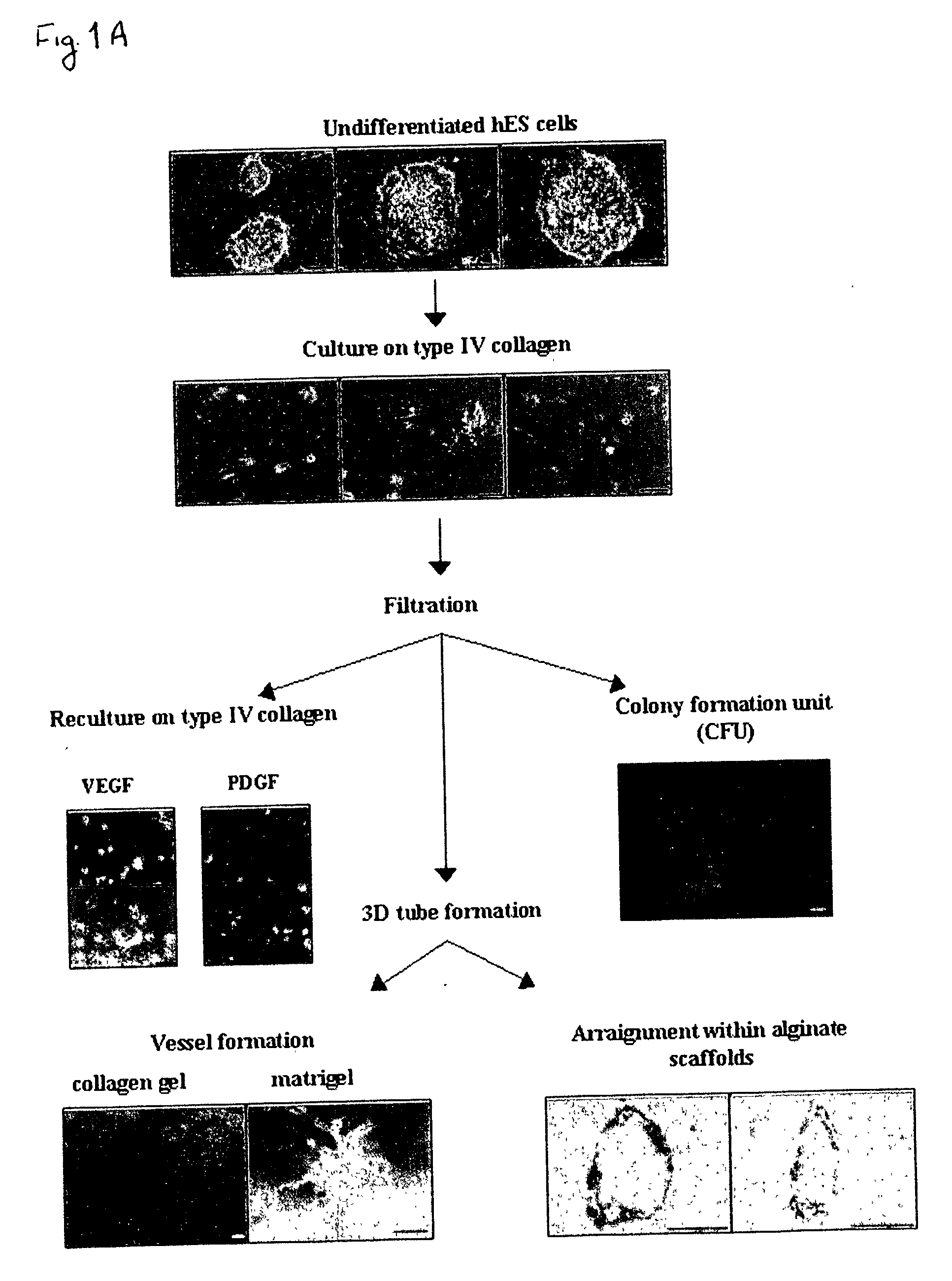
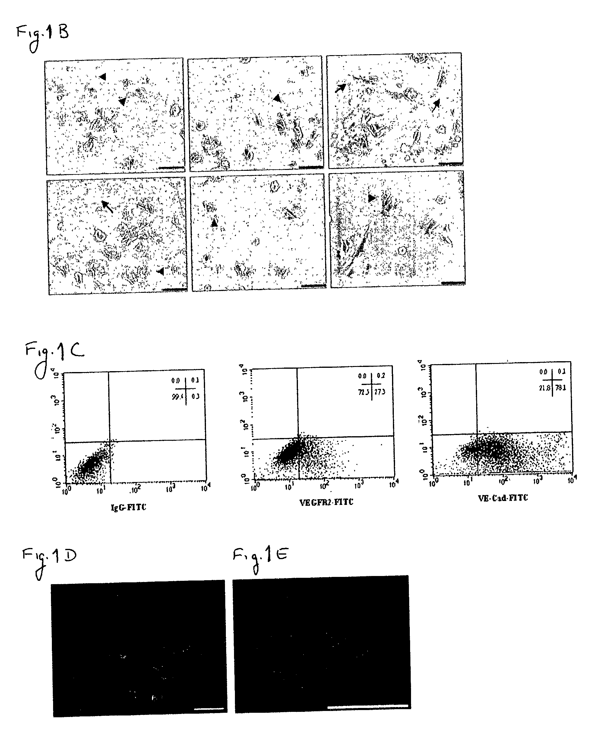
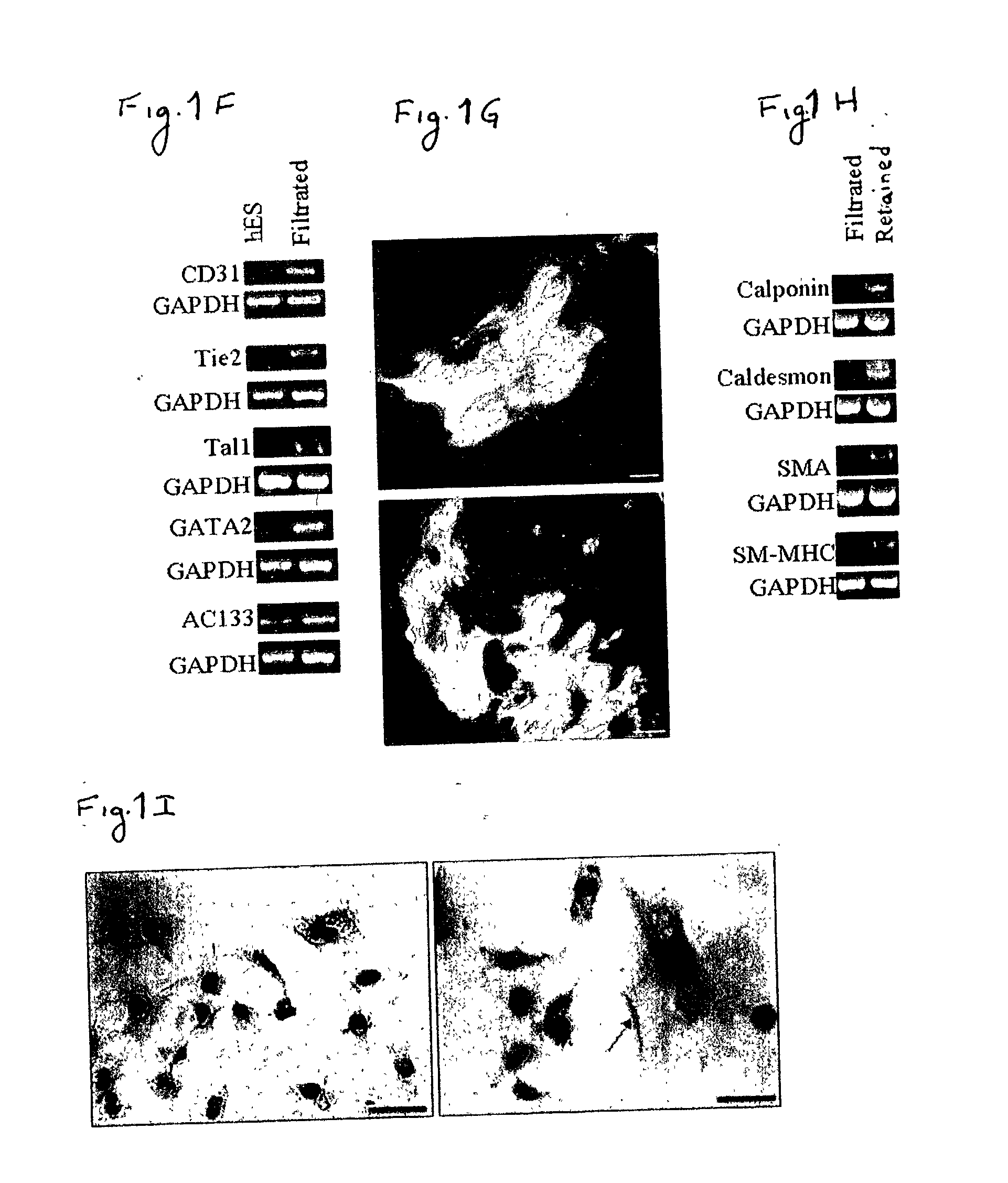
[0163] Despite the overwhelming importance of human stem cell technology to research and medicine, application of discoveries made in research with non-human species to human stem cells has been painstakingly difficult, requiring great ingenuity and much effort. While murine embryonic stem cell (mES) lines, for example, retain their pluripotency in culture, and may be predictably manipulated to differentiate in vitro into cells of mesodermal, endodermal and ectodermal lineage, in vitro differentiation in human and other primate ES cell lines has been characterized by inconsistency, disorganization, and lack of synchrony, obviating successful in vitro tissue organization (see, for example, Thompson, et al Curr Top Dev Biol 1998; 38:133-165). In pursuing the isolation of vasculogenic progenitor cell from human embryonic stem cells, initial human ES mesodermal differentiation was attempted in a novel t...

example 2

In Vitro Induction of Endothelial, Smooth Muscle and Hematopoietic Cell Differentiation of Human Vasculogenic Progenitor Cells

[0170] In order to study the differentiation potential of the vasculogenic progenitor cells, cells were recultured on type IV collagen coated dishes, at a lower cell seeding concentration (2.5.times.10.sup.4 cells / cm.sup.2). Smooth muscle cell differentiation was induced by adding platelet-derived growth factor BB (hPDGF-BB), which has been found to induce SMC differentiation in murine (mES), but not human stem cells (Gittenberger-de Groot A. C et al PNAS 2000;97:11307). After 10-12 days of culture both spindle-like shaped and epithelioid phenotype cells were detected in the culture, along with a concomitant induction of expression smooth muscle cell markers. RT-PCR analysis detected upregulation of specific smooth muscle markers such as smooth muscle .alpha.-actin (SMA), smooth muscle myosin heavy chain (SM-MHC), calponin, SM22, and caldesmon (FIG. 2A, v-SMC...

example 3

In-vitro Vasculogenesis and Blood Cell Formation by ESH Cells

[0172] Crucial events characteristic of vasculogenesis have been induced in vitro using murine embryonic stem cell-derived embryoid bodies (see, for example, Feraud O et al Lab Investig 2001;81: 1661-89), however efforts to emulate vasculogenic processes in vitro using human pluripotent stem cells have been largely unsuccessful. To study the in-vitro vascularization potential of human vasculogenic progenitor (ESH) cells we used two different 3-dimensional models: type I collagen gel and Matrigel, which have been used to promote 3D vessel-like formation from endothelial cells (Mardi JA and Pratt B M, B.M.J Cell Biol.1988; 106:1375; Kubota Y et al J Cell Biol 1988;107:1589).

[0173] Aggregation of the ESH cells, in the presence of hVEGF and hPDGF-BB supplemented differentiation medium, prior to seeding into type I collagen (FIG. 3A) or on Matrigel (FIG. 3B) clearly induces sprouting and tube-like structures associated with ear...


PUM

PropertyMeasurementUnit
Timeaaaaaaaaaa
Pore sizeaaaaaaaaaa
Vascularizationaaaaaaaaaa

Abstract

There is provided a simplified and inexpensive method for the in-vitro identification, isolation and culture of human vasculogenic progenitor cells. The method and the progenitor cells isolated thereby can be used for in-vitro vascular engineering, treatment of congenital and acquired vascular and hematological abnormalities, for evaluation and development of drugs affecting vasculo- and angiogenic processes, and for further investigation into tissue differentiation and development.

Description

[0001] This application claims the benefit of priority from U.S. Provisional Patent Application No. 60 / 372,429, filed Apr. 16, 2002.FIELD AND BACKGROUND OF THE INVENTION[0002] The present invention relates to novel methods for the isolation and culture of vasculogenic progenitor cells from stem cells and, more particularly, to methods for use of vasculogenic progenitor cells in tissue engineering, research and diagnostics.[0003] Recently, techniques have been developed which allow human embryonic stem cells to proliferate indefinitely in culture, enabling experimentation with induction of differentiation in a directed, tissue-specific manner (Itskovitz-Eldor, J et al Mol Med 2000;6:88-95, Reubinoff B E at al Nat Biotech 2000;18:399-404, Schuldiner M et al PNAS USA 2000;97:11307-12). Human embryonic stem cell growth and development is being carefully studied, and the rapidly accumulating knowledge is being employed in a variety of innovative therapeutic applications including in-vitr...

Claims


Application Information

Patent Timeline
IPC IPC(8): A61K35/12A61K45/00A61L27/00A61P7/02A61P9/00A61P9/10A61P17/02A61P35/00A61P37/06C12NC12N5/074C12N5/08
CPCA61K35/12C12N2506/02C12N5/0692A61K45/00C12N5/06C12Q1/00A61P7/02A61P9/00A61P9/10A61P17/02A61P35/00A61P37/06A61P43/00C12N5/0691
Inventor ITSKOVITZ-ELDOR, JOSEPHGERECHT-NIR, SHARON
Owner TECHNION RES & DEV FOUND LTD
Learn More