
Device and method for performing a biological modification of a fluid

a technology of biological modification and fluid, applied in the direction of biochemistry apparatus, medical devices, biochemistry apparatus and processes, etc., can solve the problems of high mortality risk of patients with liver or kidney failure, high risk of liver or kidney failure replacement, and extremely complex liver function,

Inactive Publication Date: 2003-07-10
MITRANI EDUARDO
View PDF2 Cites 52 Cited by
  • Summary
  • Abstract
  • Description
  • Claims
  • Application Information

AI Technical Summary

Benefits of technology

[0025] Kidney transplantation is considered the preferred mode of treatment for patients lacking kidney function, although it has its own set of inherent drawbacks. While routinely performed in many medical centers, it still carries with it all the risks associated with intra-abdominal surgery. In addition, transplant recipients are often treated with immuno-suppressant drugs for a prolonged period after surgery to prevent organ rejection, a practice which places them at increased risk of infection. Most problematic is the chronic shortage of available kidneys for transplantation, and the difficulty in matching donated organs to suitable recipients. Even in the case of a successful transplant, the recipient has only one kidney. Supplementation of the function of a transplanted kidney with a device such as that described in the present invention could increase this post transplantation survival time by reducing the load on the transplanted kidney.
[0027] There is thus a widely recognized need for, and it would be highly advantageous to have, a device and method for supplementing, augmenting or replacing liver function, kidney function, or both liver and kidney function, devoid of the above limitations. Such a device and method would allow patients to have a better quality of life while awaiting transplantation and also serve to increase their life expectancy after receipt of a new organ and in some instances to allow for their own organ to recover while being supported by the device.
[0032] Further according to the teachings of U.S. Pat. No. 5,888,720, populations of cells are grouped in a manner that preserves the natural affinity of one cell to another so that in vivo epithelial and stromal tissue architecture is preserved. For example, in skin micro-organ cultures, keratinocytes of the epidermis remain associated with the stroma and the normal tissue architecture is preserved including the hair follicles. Such an association facilitates intercellular communication. Many types of communication takes place among animal cells. This is particularly important in differentiating cells where induction is defined as the interaction between one (inducing) and another (responding) tissue or cell, as a result of which the responding cells undergo a change in the direction of differentiation. Moreover, inductive interactions occur in embryonic and adult cells and can act to establish and maintain morphogenetic patterns as well as induce differentiation (Gurdon (1992) Cell 68: 185-199).

Problems solved by technology

Patients with liver or kidney failure are at high risk for mortality if immediate intervention is not effected.
Clearly, liver function is extremely complex and is unlikely to be replaced by a solely mechanical or a chemical device at this time.
Firstly, there are obvious safety and regulatory concerns about using immortalized cell lines in clinical practice.
Secondly, immortalized cells would not be expected to rein all the normal physiological characteristics of primary hepatocytes, particularly after industrial scale expansion (Sussman et al., Artif Organs, 18:90-6, 1994).
In previous attempts, cells from livers have usually been disassociated using enzymes is such as collagenase, which disrupts the normal micro architecture of the liver.
Some attempts have been used to use liver pieces, but the shape of these pieces have not been designed for proper surface area to volume ratios necessary for optimal tissue maintenance (Lie et al., Res Exp Med (Berl) 185:483-94, 1985).
One current limitation is the ability of current methods of culturing mammalian liver cells to provide conditions which allow cells to assemble into tissues which simulate the spatial three-dimensional form of actual tissues in the intact organism.
Conventional tissue culture processes limit, for similar reasons, the capacity for cultured tissues to express a highly functionally specialized or different statconsidered crucial for mammalian cell differentiation and secretion of specialized biologically active molecules of research and pharmaceutical interest.
In the liver, the unique juxtaposition of diverse cell populations and matrix components in harmony with the angio-architecture results in a delicate bioecological system.
It is therefore unlikely that standard cultures of hepatocytes will perform even the minimal liver functions.
None of the prior art organ assist devices, or related devices in the prior art, uses micro-organ cultures or cryo-micro-organs in order to perform a biological modification of a fluid.
Treatment is limited to supportive care, dialysis and transplantation.
Even the continuous dialysis often used in these situations is inadequate.
For most patients, timely transplantation is not an option.
Unfortunately, current dialysis methods represent a clinically poor and economically expensive solution to kidney failure.
First, it requires the use of expensive equipment and skilled paramedical personnel to operate the equipment.
Second, since treatments are generally conducted several times per week on an outpatient basis, costs are high and patients are required to remain close to their treatment facility.
In addition, patients undergoing treatment must remain on a strict diet between treatment sessions, often leading to medical crises due to poor compliance.
As a result, some long term hemodialysis patients suffer venous collapse in the most easily accessible veins of the arm.
While routinely performed in many medical centers, it still carries with it all the risks associated with intra-abdominal surgery.
In addition, transplant recipients are often treated with immuno-suppressant drugs for a prolonged period after surgery to prevent organ rejection, a practice which places them at increased risk of infection.
Most problematic is the chronic shortage of available kidneys for transplantation, and the difficulty in matching donated organs to suitable recipients.
Unfortunately, the recent discovery of porcine endogenous retroviruses (pervs) has raised serious doubts about the advisability of xenotransplantation even if it becomes technically feasible.
Second, because of the three-dimensionality of the explant, various cells can continue to actively grow and most important to fully differentiate and function, in contrast to cells in monolayer cultures, which grow to confluence, exhibit contact inhibition, and cease to grow and divide and their function is limited.
Once the organ has been frozen it is not possible to reach all cells through the circulation network since those channels are now frozen.
When removed from the body, normal adult organs lack the system support and circulation necessary to provide adequate exchange of nutrients and gases to each cell in the organ.
The membrane or the like allows passage of nutrients and small vital molecules including oxygen, glucose and hormones between the micro-organ culture and the fluid being treated, but does not allow passage of agents of the immune system such as white cells and, if required, antibodies.

Method used


Image

  • Device and method for performing a biological modification of a fluid
  • Device and method for performing a biological modification of a fluid
  • Device and method for performing a biological modification of a fluid

Examples


example 1

Preparation of Liver Micro-Organ Cultures

[0226] Mouse micro-organ cultures from liver were prepared as follows. Organs were removed and with scalpels, were cut to an appropriate width of 2 mm, length of 3 mm, and sliced using a tissue chopper or other suitable cutting means into sections of 300 micrometers thick. These micro-organs were placed in a 24-well microplate in 400 ml of Dullbeco's Minimal Essential Medium (DMEM) in the absence of fetal calf serum (FCS) under 5% CO.sub.2 at 37.degree. C., under constant shaking at 12 rpm for periods of one to eight days. Twenty micro-explants were grown per well.

example 2

Measurement of Cell Proliferation in Liver Micro-Organ Cultures

[0227] Micro-organ cultures from several mouse organs were dissected and cultured in a humidified incubator at 37.degree. C. in the absence of serum using micro-organ cell culture technique, as described in example 1. To assess cell division, incorporation of tritiated thymidine was measured using standard protocols (Kobayashi, et al. (1994, J Biomater Sci Polym Ed 6(4):325-42). These results show that DNA synthesis occurs during the culture period (FIG. 5). In addition, mouse liver micro-organ cultures were grown as described in example 1 for 14 days and pulsed for 4 hours with bromodeoxyuridine, fixed, and stained with a fluorescent antibody to bromodeoxyuridine to label mitotic nuclei (Sigma Chemical). Nuclei that are actively synthesizing DNA were observed in these cultures (data not shown). This result demonstrates prolonged viability of the MCs in vitro and their capacity to proliferate ex vivo.

example 3

Albumin is Produced by Mouse Hepatocytes in Micro-Organ Cultures

[0228] Primary mouse hepatocytes grown in micro-organ cultures as described in example 1, remain functional for at least four weeks, as assayed by secretion of albumin and production of urea (see FIG. 6). Mouse hepatocytes in micro-organ cultures produce relatively large amounts of albumin as tested both by ELISA and by calorimetric methods (kit No 631, Sigma Chem. Co. St. Louis Mo.). The histogram shown below displays the amount of albumin secreted per 10.sup.6 cells per hour. Note that even after one month in culture the rate of albumin production remains high, particularly in comparison to two other conventional culture conditions. A is data taken from Nyberg et al. (Cell Transplant, 2:441-52, 1993) and B data from Shatford et al. (J. Surg. Res., 53:549-57, 1992). This result demonstrates that liver MCs retain important physiologic functions over time.


PUM

PropertyMeasurementUnit
molecular weightaaaaaaaaaa
temperatureaaaaaaaaaa

Abstract

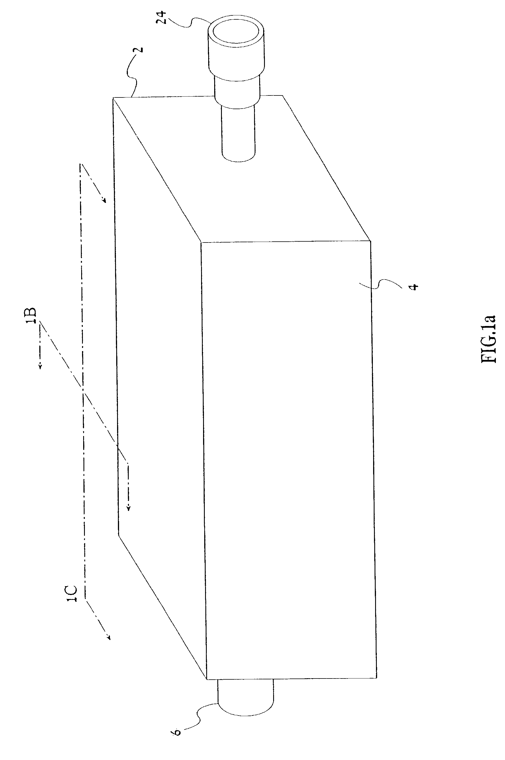
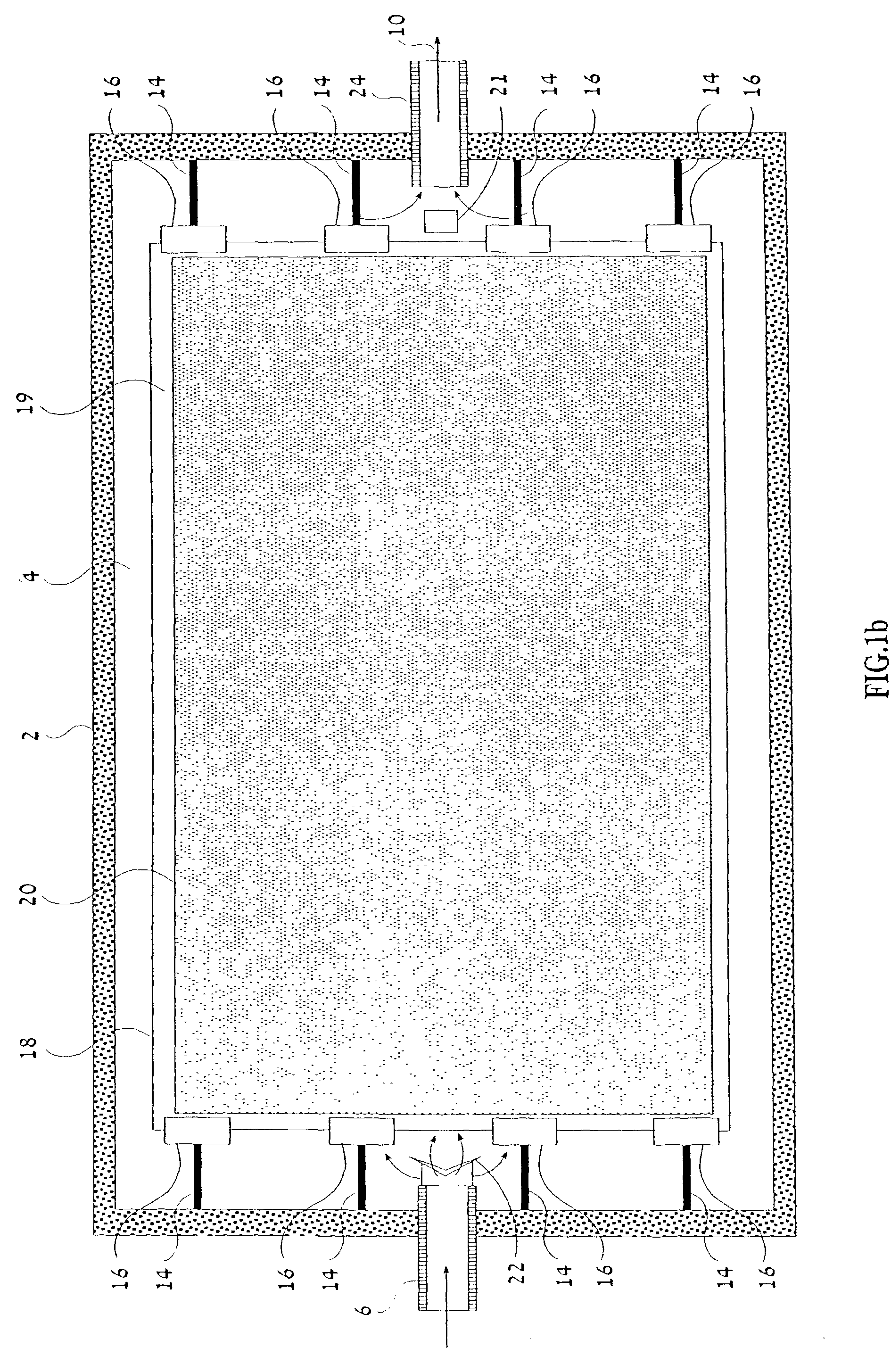
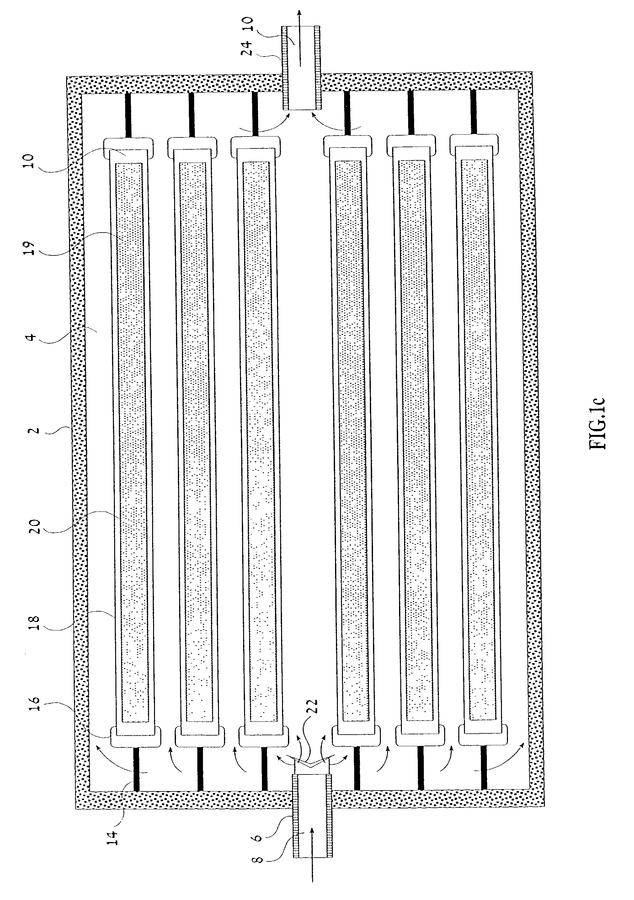
A device for performing a biological modification of a fluid, the device includes (a) a chamber having an inlet for intake of the fluid and an outlet for outflow of the fluid; and (b) a collection of micro-organ cultures of at least one organ for performing the biological modification of the fluid, each individual micro-organ culture of the collection including cells and having dimensions, such that cells positioned deepest within the individual micro-organ culture are at least about 100 micrometers and not more than about 225 micrometers away from a nearest surface of the individual micro-organ culture, thereby in vivo organ architecture (organ structure) of organ units (e.g., acinus of liver) is maintained within each individual micro-organ culture, the collection of micro-organ cultures being located within the chamber and the collection of micro-organ cultures being in contact with at least a portion of the fluid flowing through the chamber.

Description

[0001] This is a continuation-in-part of U.S. patent application Ser. No. 09 / 341,630, filed Jul. 15, 1999, which is a U.S. national phase of PCT / US98 / 00594, filed Jan. 9, 1998, which is an international phase of U.S. patent application Ser. No. 08 / 783,903, filed Jan. 16, 1997, now abandoned, which is a continuation-in-part of U.S. patent application Ser. No. 08 / 482,364, filed Jun. 7, 1995, now abandoned, which is a continuation-in-part of U.S. patent application Ser. No. 08 / 341,409, filed Jan. 31, 1995, now U.S. Pat. No. 5,888,720, issued Mar. 30, 1999. The teachings of U.S. provisional Pat. Application No. 60 / 145,095, filed Jul. 23, 1999, is incorporated herein by reference.FIELD AND BACKGROUND OF THE INVENTION[0002] The present invention relates to a device and method for performing a biological modification of a fluid and, more particularly, to an artificial installation, which supplements, augments or replaces organ function. Specifically, the artificial installation may contain...

Claims


Application Information

Patent Timeline
Patent Type & Authority Applications(United States)
IPC IPC(8): A61M1/16A61M1/34
CPCA61M1/1678A61M1/3489A61M1/34A61M2205/3331
Inventor MITRANI, EDUARDO
Owner MITRANI EDUARDO
Learn More