Single cell fluorescence in situ hybridization in microfluidic droplets

a technology microfluidic droplets, which is applied in the field of single cell fluorescence in situ hybridization in microfluidic droplets, can solve the problems of inability to retain intracellular spatial resolution of transcripts, few methods available that allow dynamic investigation of immune cell interactions, and take 2-3 days to complete, so as to reduce the overall cost of the procedure, reduce the duration of the assay, and facilitate the effect of mixing reaction kinetics

Active Publication Date: 2022-06-07
NORTHEASTERN UNIV
View PDF12 Cites 0 Cited by
  • Summary
  • Abstract
  • Description
  • Claims
  • Application Information

AI Technical Summary

Benefits of technology

[0080]Another advantage of the invention is the rapid mixing and reaction kinetics of the microfluidic droplet system, which reduce assay duration significantly ≤10 hrs while minimizing sample and reagent volume and reducing overall cost of the procedure 10 to 20 fold. In conventional FISH assays, hybridization takes a prolonged period of time (usually overnight) because the reaction relies on diffusion-limited hybridization. In some embodiments, the present method reduces by at least 40% the quantity of labeled probes necessary for completing an immunocytochemical assay or a FISH assay. In some embodiments, the present method reduces by at least 50% the time necessary for the completion of an immunocytochemical assay or a FISH assay.
[0081]The size of a cell-containing microdroplet can vary in the range from about 50 microns to about 900 microns in diameter, and is substantially determined by: the size of the polymer scaffold to which the cells are bound; device design (such as the size of the trapping sites in an array); and the flow rates of the oil and aqueous phases. Exemplary flow rates are as follows. For the flow of oil into the oil inlet: 650-1100 μl / hr, 650-1000 μl / hr, or 800-1000 μl / hr. For the flow of cell suspension into the cell inlet: 200-300 μl / hr, 250-300 μl / hr, or 300-400 μl / hr. Suitable flow rates can be readily ascertained and optimized by routine experimentation with a given device.
[0082]By coordinating individual inlet flow rates and optimizing initial cell density (such as in the range of about 1 million / mL to about 2 million / mL), large numbers of droplets can be routinely obtained which co-encapsulate the desired number of types of individual cells. Each microdroplet can include one or more cells. For example, the average number of cells in each droplet can be one or more, such as 1, 2, 3, 4, 5, 6, 7, 8, 9, 10, or more. The number of cells encapsulated in the microdroplets is a Poisson probability-regulated process. It is expected that within any population of microdroplets a number of microdroplets will have more or fewer cells than the average for that population.
[0083]In some embodiments, a single cell type is encapsulated in each droplet (though more than a single cell of that type can be encapsulated in each droplet). In some embodiments, the single cell type is a tumor cell, and the present method and device can be used for clinical or preclinical assessment of an antitumor treatment, such as an antitumor drug. In some embodiments, after droplet formation and consequent cell encapsulation, a solution containing a known or potential antitumor agent is contacted with the droplet array. The tumor cell can be evaluated in a number of parameters (e.g., viability, growth, proliferation, development, motility, and interactions with the polymer scaffold or with extracellular matrix components) for the desired period of time. In addition, gene and / or protein expression can be assessed, allowing the correlation between phenotypic markers of cell activity and gene up / down-regulation and protein expression. Genes of interest include genes encoding tumor antigens NY-ESO-1, mesothelin, PSA, MART-1, MART-2, Gp100, tyrosinase, p53, ras, MUC1, SAP-1, survivin, CEA, Ep-CAM, Her2, BRCA1 / 2 and combinations thereof. By assessing gene expression for any desired length of time before, during and after contacting a tumor cell with a treatment, it is possible to investigate how the treatment changes (or fail to change) the transcription and / or translation of any gene. This information can be used to assess the effects of effective and ineffective cancer treatments on gene expression of cancer cells, including specific subsets of cancer cells, such as cancer stem cells or the cells of a particular patient. In addition, by allowing the correlation between gene and protein expression, the present device and methods allow a deeper understanding of the spatial and temporal relations between gene expression and protein production in a cell.
[0084]In some embodiments, the cells are derived from a human or non-human subject displaying or suspected of displaying a chromosomal abnormality, such as aneuploidy, or structural alterations such as deletions, duplications, translocations, inversions, insertions, and rings. In some embodiments, the claimed method is employed to detect a disease associated with a chromosome abnormality, such as leukemias, lymphomas, Down
[0085]Syndrome, Turner Syndrome, Klinefelter Syndrome, Cri-du-Chat Syndrome, and Williams Syndrome. In some embodiments, the method is employed for diagnosis of graft-versus-host disease (GVHD). In some embodiments, the claimed method is employed for the prenatal or preimplantation diagnosis of chromosomal abnormalities in a fetus or embryo. In some embodiments, the claimed method is employed to identify gene expression patterns associated with embryo development and cell fate decisions, as well as the correlation of such patterns with protein expression and cell activity.

Problems solved by technology

However, such approaches to single cell analysis rely on genetic material extracted from cells, which do not permit retention of intracellular spatial resolution of the transcripts.
However, conventional FISH assays require highly trained personnel, take 2-3 days to complete, and are expensive (approximately $90 per slide), mainly because of the high cost of the probes.
However, there are currently few methods available that allow dynamic investigation of immune cell interactions, and other types of interactions among freely dispersed cells, without physically constraining non-adherent cells.

Method used


Image

  • Single cell fluorescence in situ hybridization in microfluidic droplets
  • Single cell fluorescence in situ hybridization in microfluidic droplets
  • Single cell fluorescence in situ hybridization in microfluidic droplets

Examples


example 1

dic Device Fabrication

[0092]The microfluidic devices were fabricated by standard soft-lithography protocols. The device design was formulated using CAD (CAD / Art Services, Bandon, Oreg.) and printed on a transparency photomask (Fine Line Imaging, Colorado Springs, Colo.). The design was transferred to clean silicon wafers via UV photolithography utilizing a negative photo resist SU-8 2100 (MicroChem, Newton, Mass.), which was spin-coated on the wafers to obtain a layer of 150 μm height. The wafers served as master templates for elastomeric device fabrication. The prepolymer poly(dimethylsiloxane) (PDMS) (Sylgard 184, Dow Corning, Midland, Mich.) was mixed with the silicone elastomer curing agent at 10:1 ratio (w / w), dispensed over the wafer, degassed and cured for 12 hours at 65° C. The PDMS layer containing the design network was then peeled from the wafer and separated into individual devices. Microscope slides were subjected to plasma oxidation for 30-60 sec and bonded with the PD...

example 2

of Discrete Microscale Agarose Matrix

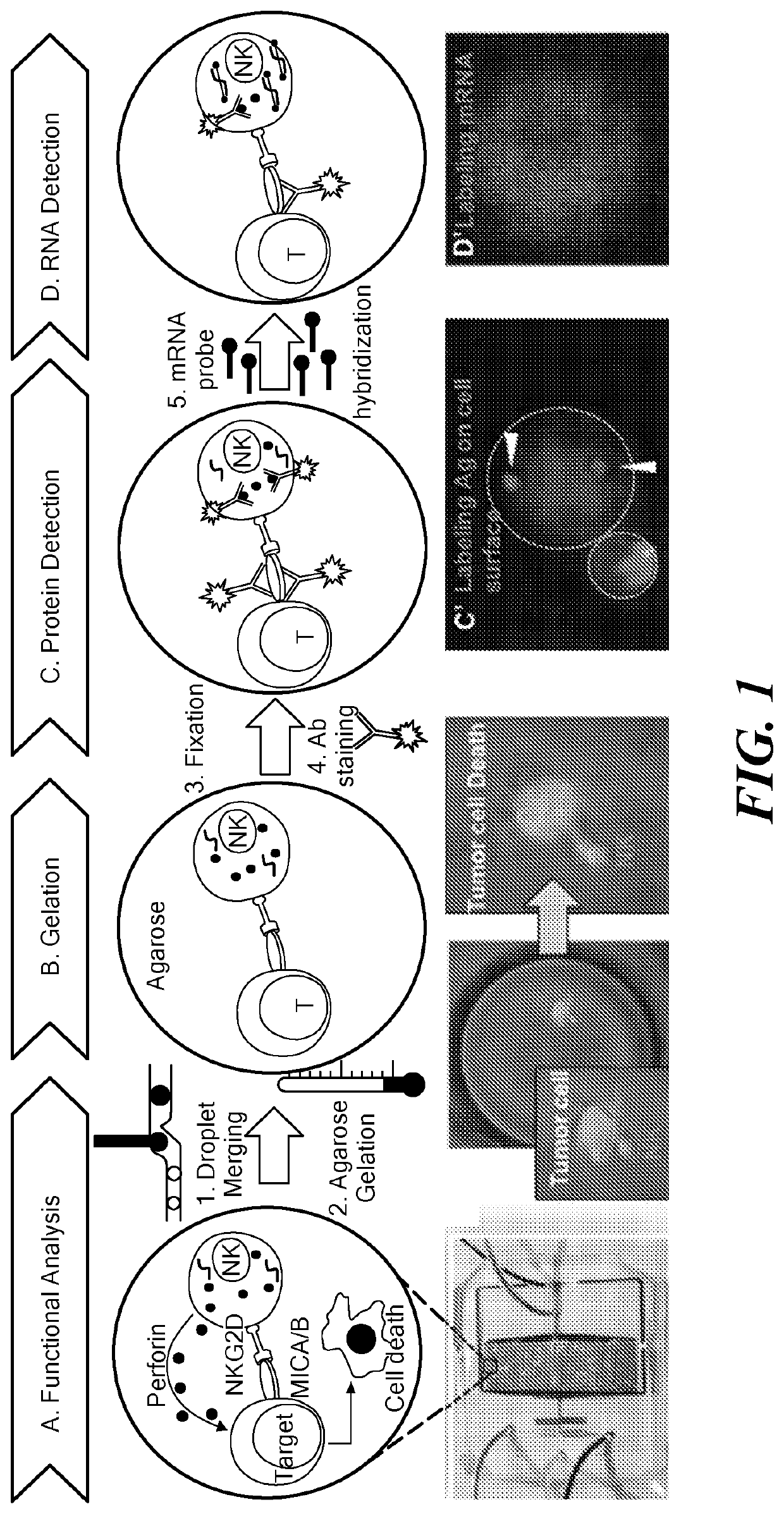
[0094]Cultured cells were purchased from commercially available sources (e.g., American Type Culture Collection (ATCC, Manassas, Va.)) and maintained in RPMI-1640 medium supplemented with 10% fetal bovine serum and 1% Antibiotic-Antimycotic solution (Corning Cellgro, Manassas, Va.). All cells were grown at 37° C. under 5% CO2 in a humidified atmosphere. Cells were routinely passaged every three days and harvested at a density of 1×10⁶ viable cells / mL. A 1.8% ultra-low temperature gelling agarose solution was merged with the formed droplets and subsequently arrested in the docking array. Cooling of the array resulted in gelation of agarose (FIG. 2C). The agarose droplets remained stable in the array for days under perfusion.

example 3

nce in Situ Hybridization in Microdroplets

[0095]Detection of ABCB1 mRNA in droplet-encapsulated live MCF-7 cells was performed using SmartFlare nanoprobes (EMD Millipore, Billerica, Mass.). Cy5-conjugated ABCB1 mRNA (#SF-2394, Millipore) stock solution was diluted as per the manufacturer's instructions. The final ratio of cells to the diluted probe was maintained at 1×10⁶ cells: 100 μL. The probes and cells were incubated through separate inlets and maintained in the dark for 24 h. The cells were periodically imaged using a Cy5 filter (FIG. 2D).


PUM

PropertyMeasurementUnit
diameteraaaaaaaaaa
temperatureaaaaaaaaaa

Abstract

The invention provides a device, method, and system for high throughput detection of nucleic acid expression in individual cells. Cells are encapsulated in aqueous microdroplets which are merged with a biocompatible matrix, allowing on-chip fluorescence in situ hybridization on both adherent and non-adherent cells. The invention also provides multiplexed detection of nucleic acids, proteins, and cellular activity. The device and methods can be used to assess cellular interactions and to test the effects of antitumor agents.

Description

CROSS REFERENCE TO RELATED APPLICATIONS[0001]This application claims the priority of U.S. Provisional Application No. 62 / 361,035 filed Jul. 12, 2016 and entitled “FISH-DROPS: FLUORESCENCE IN SITU HYBRIDIZATION IN MICROFLUIDIC DROPLETS”, the whole of which is hereby incorporated by reference.STATEMENT REGARDING FEDERALLY SPONSORED RESEARCH OR DEVELOPMENT[0002]This invention was made with government support under grant CA174401 awarded by the National Institutes of Health. The government has certain rights in the invention.BACKGROUND[0003]The goal of personalized medicine is to deliver highly specialized healthcare to every patient based on their unique biological characteristics. To this end, it is necessary to devise single cell analysis technologies that allow investigation of multiple aspect of cellular responses. Technical advances in single cell analysis have allowed researchers to explore key questions in cellular heterogeneity in areas such as immune response, intratumoral var...

Claims


Application Information

Patent Timeline
Patent Type & Authority Patents(United States)
IPC IPC(8): C12Q1/6841B01L3/00G01N21/64
CPCC12Q1/6841B01L3/502784G01N21/6458B01L2200/0668B01L2300/069B01L2300/087B01L2300/0816B01L2300/0819B01L2300/0867B01L2300/0883B01L2400/0487B01F33/3021C12Q2563/107C12Q2563/159C12Q2565/515C12Q2565/601C12Q2565/629
Inventor SARKAR, SAHELIKONRY, TANIA
Owner NORTHEASTERN UNIV
Why Eureka
Try Eureka