Microneedle patch for drug delivery or biological fluid collection and preparation method thereof

A biological fluid and microneedle sticking technology, which is applied in the direction of medical devices, microneedles, and devices introduced into the body, can solve the problems of difficult biological fluid collection, many processing steps, and high processing costs, and achieve simple process and fluid communication The effect of extended time and low processing cost

Pending Publication Date: 2021-11-02
广州市智芯禾科技有限责任公司
View PDF0 Cites 0 Cited by
  • Summary
  • Abstract
  • Description
  • Claims
  • Application Information

AI Technical Summary

Problems solved by technology

Conventional microneedle patch devices have the following disadvantages: 1. It cannot fit well with irregular skin, such as silicon-based microneedle arrays, because the substrate is hard; 2. The overall processing cost is high and the process Complicated, such as hollow microneedles, the overall processing procedures are more, and need to be carried out in a super clean environment; 3, the function is single, such as solid microneedles can only achieve drug delivery by coating the surface of the drug, and very Difficult to collect biological fluids; 4. Hollow microneedles are routinely used to collect biological fluids. The main problem is that the channels are easily blocked due to the entrapment of skin tissue

Method used


Image

  • Microneedle patch for drug delivery or biological fluid collection and preparation method thereof
  • Microneedle patch for drug delivery or biological fluid collection and preparation method thereof
  • Microneedle patch for drug delivery or biological fluid collection and preparation method thereof

Examples


Embodiment 1

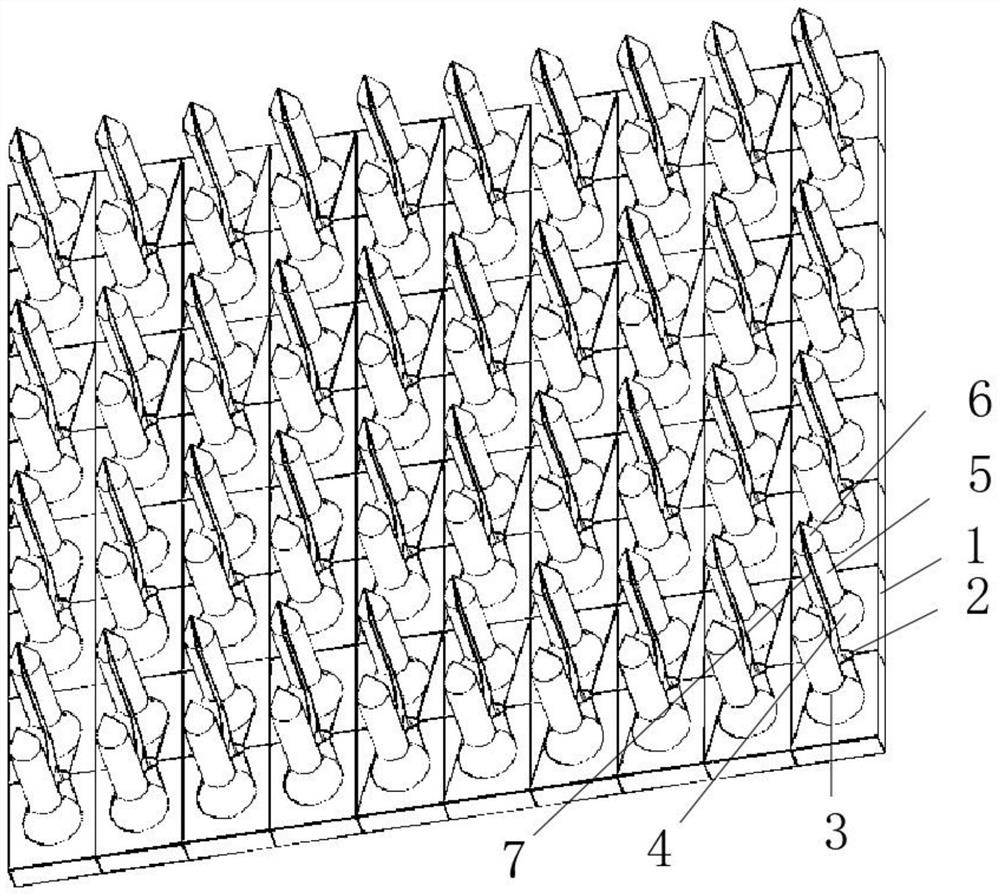
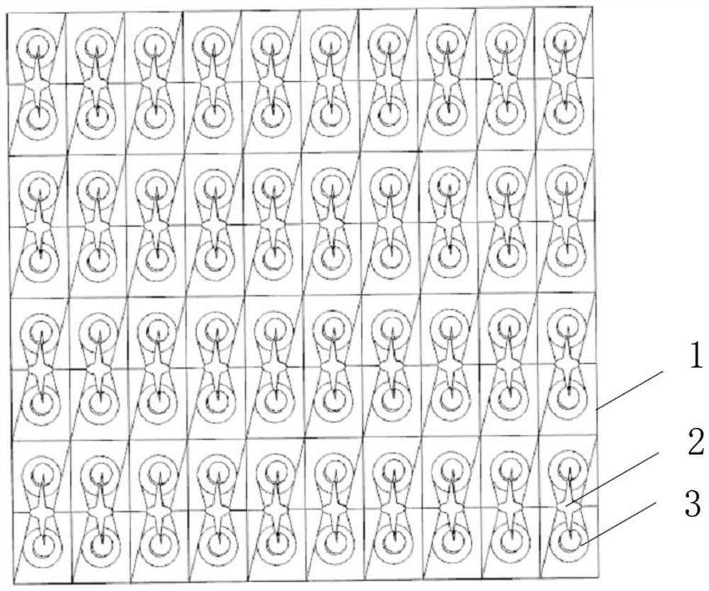
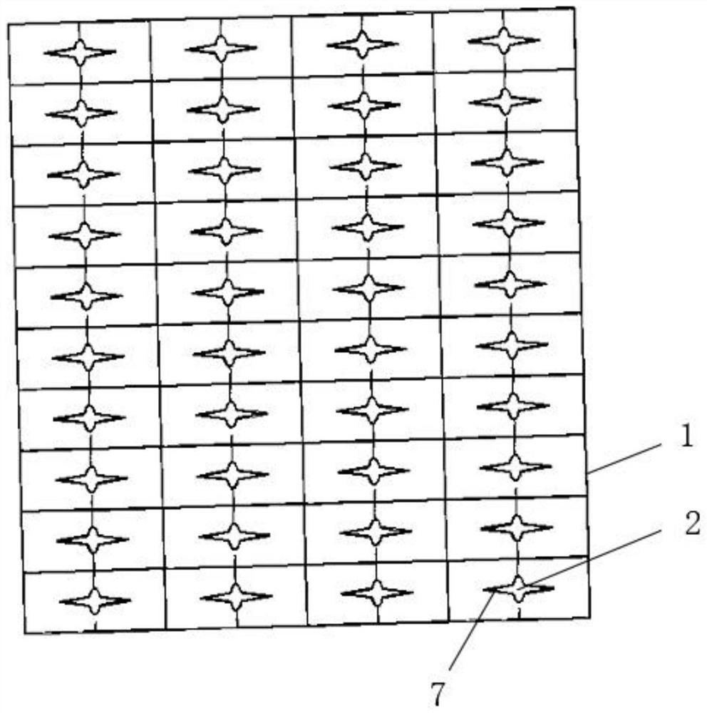
[0032] This example gives a specific implementation of a microneedle patch for drug delivery or biological fluid collection, such as Figure 1-3 As shown, including the patch base 1, a number of liquid storage pools 2 are uniformly opened on the patch base 1, and microneedles 3 are arranged on both sides of the liquid storage pools, and the side of the microneedles 3 close to the liquid storage pool 2 is provided with a diversion The channel 7 and the diversion channel 7 are connected with the reservoir 2. The microneedle 3 includes a base 4 connected to the patch substrate 1, a main body 5 arranged above the base 4, and a tip 6 arranged on the top of the main body 5.

[0033] Further, the patch substrate 1 adopts a rectangular structure.

[0034] Further, the base 4 adopts a conical structure, the main body 5 adopts a cylindrical structure, and the tip 6 adopts a conical structure.

[0035] Further, one end of the diversion channel 7 is located at the apex of the tip 6 , and...

specific Embodiment approach

[0040] This example provides a specific implementation of a method for preparing a microneedle patch for drug delivery or biological fluid collection, including the following steps:

[0041] Make templates;

[0042] Make microneedle patches.

[0043] Further, making a template includes the following steps:

[0044] Model the three-dimensional graphic structure of the microneedles through drawing software;

[0045] Import the designed graphic structure into the laser direct writing system for three-dimensional laser lithography;

[0046] Injecting photoresist into the substrate;

[0047] After exposure to the laser, unexposed areas of the negative-tone resist and exposed areas of the positive-tone resist were removed in a developer bath to obtain a template for the microneedle patch.

[0048] Further, making a microneedle patch includes the following steps:

[0049] Inject the molten No. 1 polymer material into the template to form the microneedle area, and the No. 1 polym...


PUM


Abstract

The invention relates to a microneedle patch for drug delivery or biological fluid collection, which comprises a patch substrate, a plurality of liquid storage tanks are uniformly formed in the patch substrate, microneedles are arranged on the two sides of each liquid storage tank, flow guide channels are formed in the sides, close to the liquid storage tanks, of the microneedles, and the flow guide channels communicate with the liquid storage tanks; the microneedle comprises a base connected with the patch substrate, a main body arranged above the base and a tip end arranged at the top of the main body. The invention further discloses a preparation method of the microneedle patch, the microneedle patch is suitable for the technical field of transdermal drug delivery and transdermal biological sampling, firstly, the patch substrate is made of a flexible material and can be well attached to the skin; secondly, a way of opening a channel on the side surface of the microneedle is adopted, so that flow can be well guided, and channel blockage caused by interception of skin tissues is avoided; in addition, the whole manufacturing process does not need to be carried out in a super-clean environment, the overall machining cost is low, and the technology is simple and convenient.

Description

technical field [0001] The invention belongs to the technical field of transdermal administration and transdermal biological sampling, in particular to a microneedle patch for drug delivery or biological fluid collection and a preparation method thereof. Background technique [0002] Although hypodermic needles are commonly used for blood sampling and analysis, there are limitations to the application of this technology. Hypodermic needles are painful and scary for adults and children alike, require skilled medical operators to collect blood samples, and there are significant delays between blood extraction and test results. Additionally, blood analysis requires access to pathology infrastructure, which may not be available in remote areas or in emergency situations. Difficult venous access is encountered in different patient cohorts, namely infants, children, elderly, obese individuals with poor venous permeability, and emergencies such as shock. Drugs that are not bioava...

Claims


Application Information

Patent Timeline
IPC IPC(8): A61M37/00
CPCA61M37/0015A61M2037/0023A61M2037/0046A61M2037/0053A61M2037/0061A61M2037/0038
Inventor 夏凯邓杨
Owner 广州市智芯禾科技有限责任公司
Why Eureka
Try Eureka