Biosensor capable of being modified repeatedly and used for detecting tumor cells and preparation method of biosensor
A biosensor and tumor cell technology, applied in the field of electrochemical cell sensors, electrochemical biosensors and their preparation, can solve the problems of high experimental cost and cumbersome operation, and achieve high sensitivity, good selectivity, and good linearity
- Summary
- Abstract
- Description
- Claims
- Application Information
AI Technical Summary
Problems solved by technology
Method used
Image
Examples
Embodiment 1
[0029] Example 1: Preparation of cell sensor nanoprobes
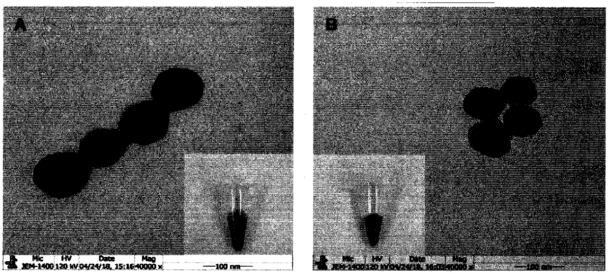
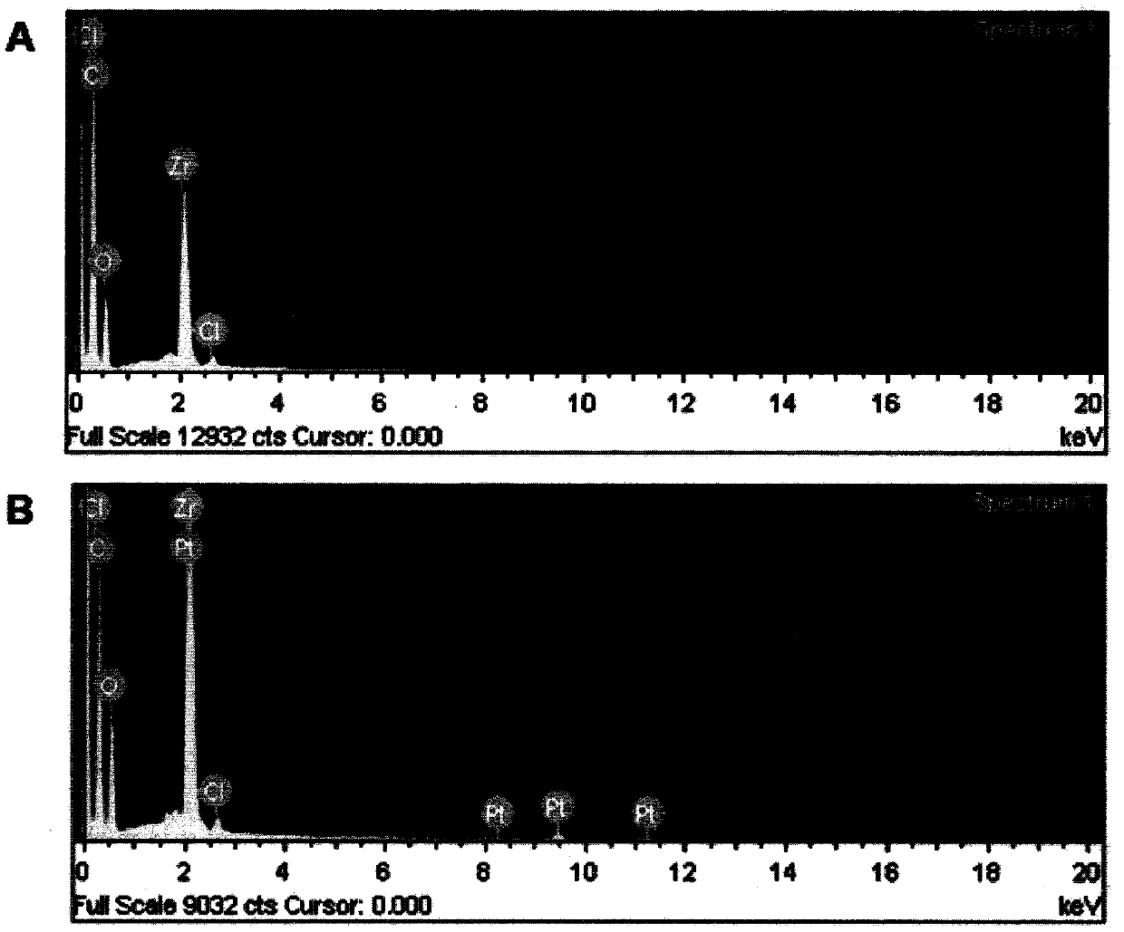
[0030] Add 0.15 g ZrOCl to the round bottom flask 2 ·8H 2O, 0.05g trichloropropyl phosphate (TCPP), 1.4g benzoic acid and 50mL N,N-dimethylformamide (DMF), stirred and reacted at 90°C for 5h. Afterwards, centrifuge at 12000 rpm for 30 min, and wash thoroughly with DMF three times. The product was stored in DMF solution, protected from light. Subsequently, 1 mL of H 2 PtCl 6 (19.31mM) was added to PCN-224 nanoparticles (2.5mg mL -1 ), stirred in 20 mL of water for 1 h. Then, add 2 mL of NaBH with vigorous stirring 4 (4mgmL -1 ), and then stirred for 3h to obtain PCN-224-Pt nanoparticles. Finally the mixture was centrifuged and washed 3 times with water. The obtained PCN-224-Pt particles were redispersed in water, some of them were used to verify the accuracy and catalytic performance of the compounds, and others were used to synthesize hybrid nanoprobes. The preparation of PCN-224-Pt nanomaterials was verified...
Embodiment 2
[0032] Example 2: Preparation of Tetrahedron-Aptamer
[0033] Take 10 μM tetrahedral single-stranded aptamer AS1411-A, tetrahedral single-stranded AS1411-B, AS1411-C and AS1411-D in four PCR tubes, add an equal volume of 1mM trichloroethyl phosphate Ester, reaction 1h. The above solutions were all mixed in the same PCR tube, reacted at 95°C for 10 minutes, and immediately cooled to 4°C for 10 minutes to obtain the three-dimensional structure of the tetrahedron-linked aptamer AS1411. The three-dimensional structure of tetrahedron-connected aptamer MUC1 can be obtained by the same method as MUC1-A, MUC1-B, MUC1-C and MUC1-D. The prepared tetrahedra were characterized using 3% agarose gel. Electrophoresis was performed at 100 V for 75 min at room temperature. After separation, the gel was imaged using a fluorescent gel imaging system and the results are shown in Figure 4 .
Embodiment 3
[0034] Embodiment three: the preparation of sensor
[0035] After soaking the gold electrode in piranha washing solution for 15 minutes, rinse it with water, and then polish it with 0.05 μm alumina suspension, and then conduct ultrasound, ethanol and deionized water for 3 minutes each. Then activate with 0.5M sulfuric acid until the signal is stable, and the activation range of sulfuric acid is -0.2 to 1.5V. Then a certain concentration of specific tetrahedron-aptamer solution was added dropwise to the electrode, and left overnight at 4°C. Then take out the electrode and rinse it with ultrapure water. It should also be soaked in ultrapure water and placed at 4°C during storage.
PUM
 Abstract
Description
Claims
Application Information
 - R&D Engineer
- R&D Manager
- IP Professional
- Industry Leading Data Capabilities
- Powerful AI technology
- Patent DNA Extraction
Browse by: Latest US Patents, China's latest patents, Technical Efficacy Thesaurus, Application Domain, Technology Topic, Popular Technical Reports.
© 2024 PatSnap. All rights reserved.Legal|Privacy policy|Modern Slavery Act Transparency Statement|Sitemap|About US| Contact US: help@patsnap.com


