An adaptive multi-part scanning imaging method and system based on deep learning

A technology of deep learning and scanning imaging, applied in the field of nuclear medicine imaging, can solve problems such as complex optimization, difficult to really play a role, difficult to quantify standards, etc.

Active Publication Date: 2021-05-18
ATOMICAL MEDICAL EQUIP FO SHAN LTD
View PDF7 Cites 0 Cited by
  • Summary
  • Abstract
  • Description
  • Claims
  • Application Information

AI Technical Summary

Problems solved by technology

There are two main problems with the above-mentioned principles and research work guiding the scanning protocol: the first is that it lacks an intuitive and clear connection with the clinical application goals of imaging-diagnosis or efficacy evaluation, thus making it difficult to have a truly accurate, personalized and Quantitative standards that are generally recognized by clinics are generated; on this basis, the selection of specific scanning acquisition procedures and parameters is greatly influenced by subjective factors such as user habits and operator experience in practice, which further leads to the questioning of the clinical value of personalized acquisition procedures , so that it is difficult to really play a role
On the basis of obtaining relatively sufficient input information, how to select parameters and procedures to achieve the ideal compromise of the above-mentioned comprehensive conditions is also a relatively complicated optimization problem.

Method used


Image

  • An adaptive multi-part scanning imaging method and system based on deep learning
  • An adaptive multi-part scanning imaging method and system based on deep learning
  • An adaptive multi-part scanning imaging method and system based on deep learning

Examples


Embodiment 1

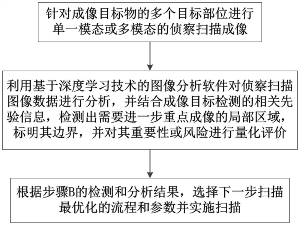
[0051] The adaptive multi-part scanning imaging method based on deep learning in this embodiment is applied to multi-modal imaging equipment mainly based on nuclear medicine, such as figure 1 shown, including the following steps:

[0052] Step A, performing single-modal or multi-modal reconnaissance scanning imaging for multiple target parts of the imaging target;

[0053] Step B, using the image analysis software based on deep learning technology to analyze the reconnaissance scanning image data in step A, and combining the relevant prior information of imaging target detection, detecting the local area that needs to be further focused on imaging, marking its boundary, and Quantitative evaluation of its importance or risk;

[0054] Step C, according to the detection and analysis results of step B, select the process and parameters to be optimized for the next scan and implement the scan.

[0055] Preferably, the nuclear medicine-based multimodal imaging equipment is a SPECT...

Embodiment 2

[0073] The adaptive multi-part scanning imaging system based on deep learning in this embodiment is applied to multi-modal imaging equipment mainly based on nuclear medicine, and the multi-modal imaging equipment based on nuclear medicine is embedded with technology based on deep learning. image analysis software, and the adaptive multi-part scanning imaging system based on deep learning includes the following modules:

[0074] The reconnaissance scanning imaging module is used to perform single-modal or multi-modal reconnaissance scanning imaging for multiple target parts of the imaging target;

[0075] The quantitative evaluation module is used to analyze the reconnaissance scan image data generated by the reconnaissance scan imaging module by using image analysis software based on deep learning technology, and combine the relevant prior information of imaging target detection to detect local areas that need further key imaging, Mark its boundaries and quantify its importanc...


PUM


Abstract

The invention discloses an adaptive multi-part scanning imaging method and system based on deep learning, which is applied to multi-modal imaging equipment mainly based on nuclear medicine, and includes the following steps: Step A, aiming at multiple imaging targets Perform single-modal or multi-modal reconnaissance scanning imaging of the target site; step B, use image analysis software based on deep learning technology to analyze the reconnaissance scanning image data in step A, and combine relevant prior information to detect further Focus on the local area of ​​imaging and quantify its importance or risk; step C, based on the results of step B, select the optimized process and parameters to implement the next scan. The self-adaptive multi-part scanning imaging method proposed by the present invention maximizes the image diagnostic value contributed by the unit scanning time and / or radiation dose, realizes individual optimized precise image inspection for multi-part scanning and imaging applications such as tumors, and improves diagnostic efficiency. Has important practical value.

Description

technical field [0001] The invention relates to the technical field of nuclear medicine imaging, in particular to an adaptive multi-part scanning imaging method and system based on deep learning. Background technique [0002] SPECT (Single Photon Emission Computed Tomography, Single Photon Emission Computed Tomography) and PET (Positron Emission Tomography, Positron Emission Tomography) are two imaging technologies in nuclear medicine, which generate radioactive indicators through gamma photon detection and imaging technology. Static or dynamic images that track the uptake, distribution and excretion of drugs in humans or animals, so as to provide functional information of related systems, organs, and tissues in humans or animals, and in some cases, reveal the biochemical reactions of special cells at the molecular level process, so as to be used in clinical medical diagnosis and basic medical research. The main difference between SPECT and PET lies in the gamma photon coll...

Claims


Application Information

Patent Timeline
Patent Type & Authority Patents(China)
IPC IPC(8): A61B6/03A61B6/06A61B5/055
Inventor 陈思
Owner ATOMICAL MEDICAL EQUIP FO SHAN LTD
Why Eureka
Try Eureka