
Hemostasis material for clinical internal medicine to stop bleeding and preparation method thereof

A hemostatic material, a technique of internal medicine, applied in medical science, surgical adhesives, bandages, etc., can solve problems such as difficulty in ensuring compatibility, absorption, easy adhesion and remaining in tissue blood vessels, increasing blood concentration and viscosity, etc. To achieve the effect of promoting wound tissue healing and coagulation, excellent biocompatibility, and promoting dissolution

Inactive Publication Date: 2019-04-19
CHONGQING MEDICAL & PHARMA COLLEGE
View PDF11 Cites 13 Cited by
  • Summary
  • Abstract
  • Description
  • Claims
  • Application Information

AI Technical Summary

Problems solved by technology

However, the research found that the absorbable carrier has a wide range of options, and it is difficult to ensure good compatibility and absorbency after electrospinning, and the antibacterial property during storage is also poor.
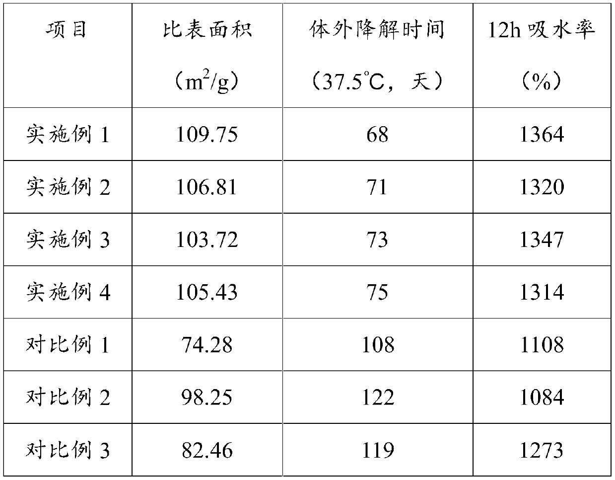
[0006] In summary, the existing hemostatic materials for clinical internal medicine have the following problems: 1. They cannot stop bleeding through physical, physiological and chemical effects, and have poor compatibility with tissues, and the effect of promoting tissue healing is not obvious; 2. Adsorption and coagulation by carriers When the molecule is used for hemostasis, the absorption and degradation performance of the carrier is poor, and it is easy to adhere and remain in the tissue blood vessels; 3. Solvents and additives will be introduced during the preparation of the material, and the inaccurate process control will not achieve a uniform and good specific surface area, which will not be optimal. Absorb water in the blood to increase blood concentration and viscosity to achieve the purpose of hemostasis

Method used


Image

  • Hemostasis material for clinical internal medicine to stop bleeding and preparation method thereof
  • Hemostasis material for clinical internal medicine to stop bleeding and preparation method thereof

Examples


Embodiment 1

[0037] The hemostatic material for clinical hemostasis in this embodiment is prepared by loading medical glue on modified chitosan fibers, and then through the process of rapid cooling at low temperature-radiation cross-linking-freeze-drying. The medical glue is prepared by stirring and mixing methyl cyanoacrylate and polylactic acid at a mass ratio of 7:1.

[0038] Wherein, the preparation method of modified chitosan fiber comprises the following steps:

[0039] 1) Softening: put the natural chitosan crude product in a three-necked flask, connect a mechanical stirrer and a thermometer, add 8-10 times the amount of deionized water, add sodium hydroxide powder under stirring until the pH is 7.2-7.5, add Acylated amino acid hydrolase with 0.1%-0.3% natural chitosan quality, heat up to 62-68°C at a rate of 3-5°C / min, keep warm and stir for 40-50min, let stand for 30-50min, together with softener Feed into a plate and frame filter press for pressure filtration, and the filtrate i...

Embodiment 2

[0049] The hemostatic material for clinical hemostasis in this embodiment is prepared by loading medical glue on modified chitosan fibers, and then through the process of rapid cooling at low temperature-radiation cross-linking-freeze-drying. The medical glue is prepared by stirring and mixing ethyl cyanoacrylate and polylactic acid at a mass ratio of 8:1.

[0050] Wherein, the preparation method of modified chitosan fiber is identical with embodiment 1. The preparation method of the softening agent is as follows: in parts by weight, 52 parts of vinyl acetate-ethylene copolymer emulsion, 15 parts of triethanolamine, 6 parts of pure chitosan, and 62 parts of deionized water are mixed and stirred evenly, and left to stand to form gel powder , placed in a reaction kettle at 55°C, stirred at a speed of 260r / min for 35min, passed through high-temperature airflow and dried into softener powder. The natural antioxidant is vitamin E, and the antibacterial agent is chitin.

[0051] W...

Embodiment 3

[0057] The hemostatic material for clinical hemostasis in this embodiment is prepared by loading medical glue on modified chitosan fibers, and then through the process of rapid cooling at low temperature-radiation cross-linking-freeze-drying. The medical glue is prepared by stirring and mixing ethyl cyanoacrylate and polylactic acid according to the mass ratio of 7.2:1.

[0058] Wherein, the preparation method of modified chitosan fiber is identical with embodiment 1. The preparation method of the softening agent is as follows: in parts by weight, 47 parts of vinyl acetate-ethylene copolymer emulsion, 13 parts of triethanolamine, 7 parts of pure chitosan, and 63 parts of deionized water are mixed and stirred evenly, and left to stand to form gel powder , placed in a reaction kettle at 58°C, stirred at a speed of 260r / min for 40min, passed through high-temperature airflow and dried into softener powder. The natural antioxidant is catechol, and the antibacterial agent is hinoki...


PUM

PropertyMeasurementUnit
Specific surface areaaaaaaaaaaa

Abstract

The invention discloses a hemostasis material for the clinical internal medicine to stop bleeding and a preparation method thereof. The hemostasis material is prepared by using a modified chitosan fiber loaded medical glue to be subjected to a low temperature rapid cooling-radiation ray irradiation cross-linking-freezing drying process. The modified chitosan fiber is obtained by softening, grafting, antibacterial process and electrospinning of natural chitosan, and has a good specific surface area, adsorption and an antibacterial property; the medical glue is prepared by mixing a cyanoacrylatecompound and polylactic acid. The hemostasis material has excellent biocompatibility, being non-toxic and non-irritating, is suitable for various complicated bloody wounds in the clinical internal medicine treatment process, is convenient to use, has good biodegradability and absorbability, can significantly promote wound tissue healing and coagulation, and is suitable for large-scale popularization and application in surgical or internal medicine surgery hemostasis process.

Description

technical field [0001] The invention relates to the technical field of medical supplies materials, in particular to a hemostatic material for clinical hemostasis and a preparation method thereof. Background technique [0002] During the operation, how to quickly and thoroughly stop the bleeding, reduce the amount of bleeding, and keep the operative field clear is the basic operation core in the process of clinical surgery or clinical medical surgery. In recent years, medical absorbable hemostatic materials have attracted more and more attention from the pharmaceutical industry, and related pharmaceutical companies have also continued to increase research and development costs. At present, the commonly used absorbable hemostatic materials include fibrin glue, gelatin sponge, oxidized cellulose, microfibrous collagen, chitosan and calcium alginate fiber, etc. They differ in their mechanism of action and application methods, and their hemostatic effects and application fields. ...

Claims


Application Information

Patent Timeline
IPC IPC(8): A61L24/08A61L24/04A61L24/00A61L26/00
Inventor 苏湲淇龙波徐露刘小东
Owner CHONGQING MEDICAL & PHARMA COLLEGE
Learn More