A micro-nano composite structure surface matching the size of circulating tumor cells, its preparation method and its application
A micro-nano composite structure, tumor cell technology, applied in micro-structure technology, micro-structure devices, manufacturing micro-structure devices and other directions, can solve the problems of limiting the capture efficiency of the substrate, neglecting the size of target cells, complex preparation, etc. Simple preparation and high capture efficiency
- Summary
- Abstract
- Description
- Claims
- Application Information
AI Technical Summary
Problems solved by technology
Method used
Image
Examples
Embodiment 1
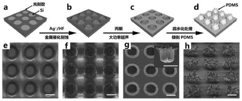
[0034] In this example, MCF-7 cells and Hela cells are used as cells to be captured and reference cells (MCF-7 cells have the membrane antigen EpCAM on the surface, but Hela cells do not). Since the sizes of the two are mainly distributed in 10-20 μm, so, In this embodiment, a circular hole array is selected as the mask plate. The array is arranged in a square, with a period of 20 μm and a pore size of 10 μm, to prepare a columnar array arranged in a square, and capture the above two types of cells on the surface of the micro-nano composite structure. The invented capture system is further elaborated and verified. The specific steps for efficiently capturing circulating tumor cells on the surface of the micro-nano composite structure are as follows:
[0035] (1) Construction of micro-nano composite structure surface
[0036] Sonicate silicon wafers with acetone, chloroform, ethanol, and water at a power of 70W for 5 minutes in sequence. After blowing dry with nitrogen, spin t...
Embodiment 2
[0049] The micro-nano composite structure capturing MCF-7 cells described in Example 1 was soaked in a phosphate buffer solution containing 2.5% glutaraldehyde at 4° C. overnight to fix the captured cells. Subsequently, the cells were dehydrated with a series of ethanol aqueous solutions with different volume fractions (0, 30%, 50%, 70%, 90%, 100%) with a certain gradient, and dried at room temperature. The 10nm gold film was sputtered, and the fixed MCF-7 cells were characterized by a Hitachi (SU8020 FE-SEM) scanning electron microscope, and the results were as follows Figure 4shown. The cell body is trapped between the four pillars and interacts with the micron-scale array, and a large number of pseudopodia protrude from the position where the cell contacts each pillar, indicating that the micro-nanostructure on the cell surface can interact with the micro-nanostructure on the pillar surface . Therefore, the micro-nano composite structure matching the size of circulating ...
Embodiment 3
[0051] The micro-nano composite structure matching the size of circulating tumor cells described in Example 1 was named PDMS-1. And using the same method as described in Example 1, replace the photolithography template, and obtain two kinds of and cycle with a period of 10 μm, a column width of 5 μm, a column height of 10 μm and a period of 60 μm, a column width of 30 μm, and a column height of 10 μm. The tumor cell size-mismatched structures were named PDMS-2 and PDMS-3, respectively. Use the same method in Example 1 to carry out metal-catalyzed etching on blank silicon wafers to obtain silicon nanowire arrays, ultrasonically pulverize silicon nanowires, and pour PDMS prepolymers, solidify, cool and peel off, and obtain rough PDMS surface structures, named as RPDMS. Scanning electron microscope (Hitachi, SU8020FE-SEM) photos of the four PDMS structures are shown in Figure 5 shown. Figure 5 (a)-(d) correspond to the structures of RPDMS, PDMS-1, PDMS-2, and PDMS-3, respect...
PUM
 Abstract
Description
Claims
Application Information
 - Generate Ideas
- Intellectual Property
- Life Sciences
- Materials
- Tech Scout
- Unparalleled Data Quality
- Higher Quality Content
- 60% Fewer Hallucinations
Browse by: Latest US Patents, China's latest patents, Technical Efficacy Thesaurus, Application Domain, Technology Topic, Popular Technical Reports.
© 2025 PatSnap. All rights reserved.Legal|Privacy policy|Modern Slavery Act Transparency Statement|Sitemap|About US| Contact US: help@patsnap.com


