Cell line for scanning peptide and non-peptide GLP-1 (Glucagon-likepeptide-1) analogues as well as preparation method and application thereof
A technology of GLP-1R and GLP-1R-, which is used in the field of screening peptide and non-peptide GLP-1 analogs of cell lines and their preparation and application, which can solve the problem of immune response, increase the economic burden of patients, and reduce the price Expensive and other issues, to achieve the effect of reducing false positives, reducing detection costs, and low cost
- Summary
- Abstract
- Description
- Claims
- Application Information
AI Technical Summary
Problems solved by technology
Method used
Image
Examples
Embodiment 1
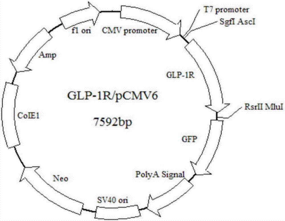
[0032] Example 1 Construction of GFP fluorescence-labeled human GLP-1R eukaryotic expression vector GLP-1R / pCMV6, establishment of GLP-1R / U2OS cell line, and determination of the average intensity change of fluorescent spots in GLP-1R / U2OS cells GLP-1 is similar biological activity.
[0033] 1. Construction of GFP fluorescently labeled human GLP-1R eukaryotic expression vector GLP-1R / pCMV6:
[0034] Human GLP-1R cDNA amplification: using human GLP-1R cDNA as a template, the full-length human GLP-1R cDNA coding region was amplified by PCR, with a total length of 1392bp.
[0035] Its upstream primer: 5'-gaggcgatcgccATGGCCGGCGCCCCCGGC-3';
[0036] Downstream primer: 5'-gcgacgcgtGCTGCAGGAGGCCTGG CAAG-3'.
[0037] 2. Recover and purify the PCR product, connect it to pMD18-T Vector to obtain the recombinant plasmid GLP-1R / pMD18-T, and use restriction enzymes Sgf I and Mlu I to digest the plasmid.
[0038] 3. Digest plasmid G-D (pCMV6-AC-GFP-DR5) with restriction enzymes Sgf I a...
Embodiment 2
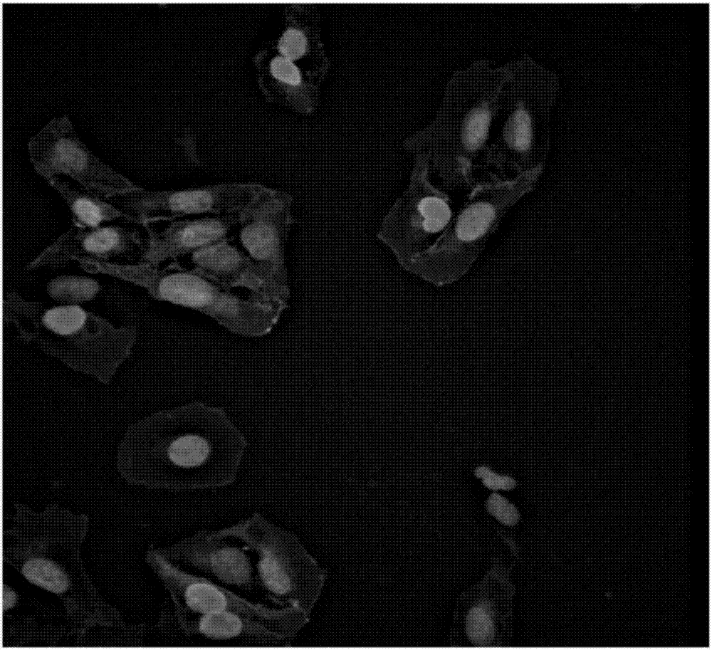
[0050] Example 2 Expression of GLP-1R on GLP-1R / U2OS cell membrane
[0051] GLP-1R / U2OS cells were cultured in 96-well culture plates at 100 μL / well (1000 cells / well) in DMEM medium containing 10% (volume fraction) calf serum at 37°C and 5% CO 2 Conditioned for 24 hours. The next day, the complete medium was removed, washed with PBS buffer, fixed with 4% paraformaldehyde at room temperature for 30 minutes, washed 3 times with PBS buffer, stained with DAPI dye in the dark for 15 minutes, washed 5 times with PBS buffer, Leave 100 μL PBS buffer in the well, and observe the fluorescence with the high content system.
Embodiment 3
[0052] Example 3 GLP-1R / U2OS cell expression GLP-1 receptor activity analysis
[0053] GLP-1R / U2OS cells were cultured in 96-well culture plates at 100 μL / well (1000 cells / well) in DMEM medium containing 10% (volume fraction) calf serum at 37°C and 5% CO 2 Conditioned for 24 hours. The next day, remove the complete medium, wash with PBS buffer, add Exendin-4 to the sample to be tested, let it stand at room temperature for 20 minutes, remove the solution in the well, fix with 4% paraformaldehyde at room temperature for 30 minutes, wash with PBS buffer for 3 Add DAPI dye for 15 minutes in the dark, wash 5 times with PBS buffer, leave 100 μL PBS buffer in the well, and observe the fluorescence change with the high-content system. The results showed that fluorescent spots were produced in the cells, while the U2OS cells not transfected with GLP-1R gene did not have any changes. The above experimental results confirm that the GLP-1R protein expressed by the recombinant GLP-1R / U2O...
PUM
 Abstract
Description
Claims
Application Information
 - R&D Engineer
- R&D Manager
- IP Professional
- Industry Leading Data Capabilities
- Powerful AI technology
- Patent DNA Extraction
Browse by: Latest US Patents, China's latest patents, Technical Efficacy Thesaurus, Application Domain, Technology Topic, Popular Technical Reports.
© 2024 PatSnap. All rights reserved.Legal|Privacy policy|Modern Slavery Act Transparency Statement|Sitemap|About US| Contact US: help@patsnap.com


