
Tissue engineering cartilage composite stent and preparation method

A composite scaffold and tissue engineering technology, applied in tissue regeneration, additive processing, medical science, etc., can solve the problems of insufficient mechanical strength of hydrogel scaffolds, achieve good biodegradability, biocompatibility, and good mechanical strength Effect

Active Publication Date: 2017-05-31
PEKING UNIV THIRD HOSPITAL +2
View PDF8 Cites 13 Cited by
  • Summary
  • Abstract
  • Description
  • Claims
  • Application Information

AI Technical Summary

Problems solved by technology

[0006] The technical problem to be solved by the present invention is to provide a tissue engineered cartilage composite scaffold and preparation method for compounding hydrogel on a three-dimensional scaffold for the defect of insufficient mechanical strength of the existing hydrogel scaffold

Method used


Image

  • Tissue engineering cartilage composite stent and preparation method
  • Tissue engineering cartilage composite stent and preparation method
  • Tissue engineering cartilage composite stent and preparation method

Examples


preparation example Construction

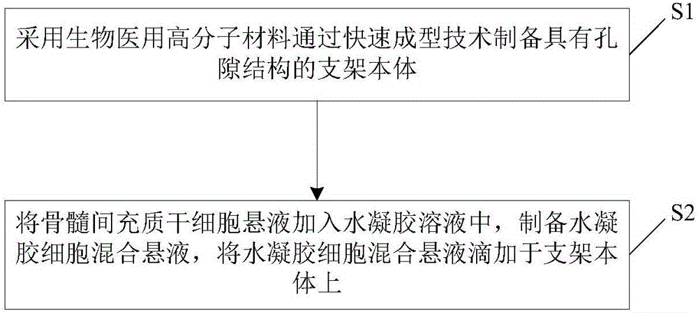
[0054] see figure 2 , is a flow chart of the preparation method of the tissue engineered cartilage composite scaffold according to a preferred embodiment of the present invention. The tissue engineered cartilage composite scaffold as mentioned above can be prepared by this method. Such as figure 2 Shown, the preparation method of this tissue engineered cartilage composite scaffold comprises the following steps:

[0055] S1. A stent body 1 with a pore structure is prepared by using a medical polymer material through rapid prototyping technology. The medical polymer materials include but not limited to polycaprolactone (PCL), polylactic acid (PLA), polyglycolic acid (PGA), polylactic-co-glycolic acid (PLGA), polyethylene glycol (PEG) and poly Ether ether ketone (PEEK) and other materials. The scaffold prepared by rapid prototyping technology, especially fused deposition modeling, has a porous structure connected in three dimensions. The porosity and pore structure of the i...

Embodiment 1

[0070] 1. Set the weight average molecular weight (Mw) to 60000gmol -1 The PCL is placed in the nozzle of the fused deposition modeling 3D printer and heated to 130°C, and is ready to print at an air pressure of 800kPa.

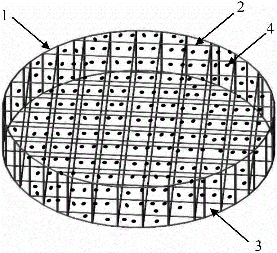
[0071] 2. Set the fiber diameter to 350 μm, the fiber spacing to 350 μm, and print out the cylindrical stent body at a speed of 0.88 mm / s. The cylinder has a diameter of 9 mm and a thickness of 2 mm.

[0072] 3. Dissolve PLGA-PEG-PLGA triblock copolymer and phosphate buffered saline solution at 4°C to form a triblock copolymer solution, wherein the mass fraction of PLGA-PEG-PLGA triblock copolymer is 14% . The PLGA-PEG-PLGA triblock copolymer in this experiment was synthesized and provided by Changchun Institute of Applied Chemistry, Chinese Academy of Sciences.

[0073] 4. Mix the triblock copolymer solution and the bone marrow mesenchymal stem cell suspension at 20°C to form a mixed hydrogel cell suspension that wraps cells; wherein, the bone marrow mese...

Embodiment 2 to 14

[0076] Examples 2 to 14 were carried out in substantially the same manner as Example 1 except for the contents of Table 1 below.


PUM

PropertyMeasurementUnit
Fiber diameteraaaaaaaaaa
Cell concentrationaaaaaaaaaa

Abstract

The invention relates to a tissue engineering cartilage composite stent and a preparation method. The composite stent comprises a stent body of a pore structure and hydrogel; the stent body is prepared from a medical macromolecule material by rapid prototyping; the hydrogel is compounded in the pore structure of the stent body and the surface of the stent body, and the hydrogel is mixed with bone marrow mesenchymal stem cells. By compounding the stent body prepared from the hydrogel and the medical macromolecule material, the prepared tissue engineering cartilage composite stent not only has good mechanical strength of the medical macromolecule material, but also provides a good microenvironment for material interchange of the bone marrow mesenchymal stem cells in the stent and on the surface of the stent and differentiation to chondrocytes by keeping a high water content through the hydrogel.

Description

technical field [0001] The invention relates to the technical field of biomaterials and tissue engineering, in particular to a composite scaffold for tissue engineering cartilage and a preparation method thereof. Background technique [0002] Articular cartilage is a tissue without blood vessels, nerves, and lymph. This structural feature limits the self-repair ability of damaged cartilage tissue. Articular cartilage has little or no effective self-repair ability after injury, and the continued development of the injury will lead to progressive articular cartilage destruction and osteoarthritis. [0003] Currently, the commonly used cartilage repair techniques include bone marrow stimulation, autologous osteochondral transplantation, allogeneic osteochondral transplantation, and autologous chondrocyte transplantation. Currently, cartilage tissue engineering technology is one of the best ways to regenerate articular cartilage. [0004] Appropriate scaffold materials, seed c...

Claims


Application Information

Patent Timeline
IPC IPC(8): A61L27/48A61L27/18A61L27/52A61L27/54A61L27/56A61L27/58B33Y10/00B33Y40/00
CPCA61L27/18A61L27/48A61L27/52A61L27/54A61L27/56A61L27/58B33Y10/00B33Y40/00A61L2300/64A61L2430/06A61L2300/412C08L67/04C08L71/02
Inventor 余家阔王少杰丁建勋张磊
Owner PEKING UNIV THIRD HOSPITAL
Learn More