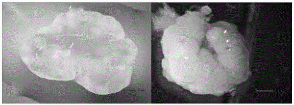
Preparation method of mouse ECMs, obtained mouse ECMs from different sources and mouse ovarium in-vivo regeneration method
A technology of in vivo regeneration and mice, applied in tissue regeneration, prosthesis, medical science, etc., can solve problems such as ovarian damage, and achieve the effect of low cost, low requirements for experimental conditions, and simple methods
- Summary
- Abstract
- Description
- Claims
- Application Information
AI Technical Summary
Problems solved by technology
Method used
Image
Examples
preparation example Construction
[0022] The invention provides a method for preparing mouse ECM, comprising the steps of:
[0023] Step 1: Collect mouse ovaries or other tissues and organs, and use different detergents and different concentrations to treat them for different times to prepare mouse ECM.
[0024] Step 2: Use sterile water and 1xPBS to wash the remaining detergent, wash each of the two cleaning solutions eight times, change the solution every two hours, and finally add double antibodies to the PBS and store at 4°C for later use.
[0025] Step 3: performing histological analysis and biochemical analysis on the prepared ECM to determine the optimal processing conditions.
[0026] The detergent is one or a combination of SDS and Triton.
[0027] The different concentrations of detergents and different treatment times are to determine the best treatment conditions according to different tissues, for example, the detergents used by the ECM of ovarian tissue are (mass volume percentage, gram per mill...
Embodiment 1
[0045] 1. Preparation and Analysis of Mouse Ovary Scaffolds
[0046] Step 1, preparation of mouse ovary scaffold
[0047]After the cervical vertebrae of 4-week-old female mice were killed, the abdomen of the mice was wiped with 75% ethanol under a stereoscopic anatomical microscope, and then the abdomen was cut open to find the ovaries along the bicornuate uterus and fallopian tubes. Gently tear off the ovarian capsule, gently remove the ovary from the root where the fat connects with the ovary with tweezers, place it in sterile PBS for cleaning, and then pre-configure it in a 50ml sterile centrifuge tube (mass volume percentage, grams per milliliter) of 1%, 0.5%, 0.2% SDS, and the obtained ovaries were placed in centrifuge tubes. The mouse ovaries were cleaned by rotating and rotating in a shaker, and the cleaning time was 6h, 12h, and 20h, respectively. After the SDS is cleaned, pour out the SDS in the centrifuge tube in the ultra-clean workbench, pour sterile water into i...
Embodiment 2
[0089] 1. Preparation and Analysis of Mouse Thymic Scaffolds
[0090] Step 1. Preparation of Mouse Thymus Scaffold
[0091] After the cervical spine of the 4-week-old female mouse was sacrificed, the mouse chest was wiped with 75% ethanol under a stereoscopic dissecting microscope, and then the chest and chest cavity were cut open to find the milky white two-lobed thymus. Gently remove the thymus from the root of the thymus with tweezers, place it in sterile PBS and wash it, then pre-configure (volume percentage) 1%, 0.5%, 0.2% TritonX-100 in a 50ml sterile centrifuge tube, and place The obtained thymus was placed in a centrifuge tube. Rotate and rotate the mouse thymus in a shaker for 10 hours, 15 hours and 20 hours respectively. After cleaning, pour out the TritonX-100 in the centrifuge tube in the ultra-clean workbench, pour sterile water into it, and clean the TritonX-100 remaining in the thymus stent. The sterile water was changed every hour, and the sterile water was ...
PUM
 Abstract
Description
Claims
Application Information
 - R&D
- Intellectual Property
- Life Sciences
- Materials
- Tech Scout
- Unparalleled Data Quality
- Higher Quality Content
- 60% Fewer Hallucinations
Browse by: Latest US Patents, China's latest patents, Technical Efficacy Thesaurus, Application Domain, Technology Topic, Popular Technical Reports.
© 2025 PatSnap. All rights reserved.Legal|Privacy policy|Modern Slavery Act Transparency Statement|Sitemap|About US| Contact US: help@patsnap.com


