Echinococcus granulosus imago diagnosis protein gene and medical uses thereof
A technology of Echinococcus granulosus and gene, applied in the field of Echinococcus granulosus adult diagnostic protein gene and its medical use
- Summary
- Abstract
- Description
- Claims
- Application Information
AI Technical Summary
Problems solved by technology
Method used
Image
Examples
Embodiment 1
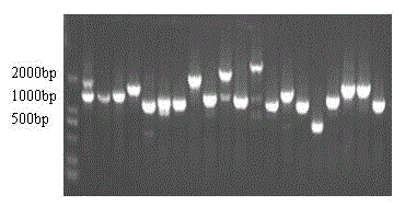
[0037] Screening, identification and prokaryotic expression of immune-related antigen genes
[0038] 1. Lambda phage plate culture
[0039] Take 30 μL of XL1-Blue strains and inoculate them into a test tube containing tetracycline LB liquid, and shake overnight at 37°C; take 4 mL of the bacterial solution and centrifuge at 5,000 g for 5 minutes, discard the supernatant, and add 200 μL of autoclaved 10 mM magnesium sulfate to resuspend the bacteria; add the resuspended Mix 200 μL of bacteria solution, 100 μL of 1×λBuffer and 100 μL of Echinococcus granulosus cDNA library, put in water bath at 37°C for 20 minutes; add LB / MgSO at 48°C 4 The top layer of agar was 3mL, mixed evenly and poured on the preheated LB plate and paved, after solidification, it was placed upside down in a 37°C incubator until needle-like phage plaques grew on the plate.
[0040] 2. Remove cross-antibody reactivity
[0041] Inoculate XL1-Blue in LB liquid medium containing tetracycline resistance and ...
Embodiment 2
[0066] Preparation of EdiagA864 Monoclonal Antibody and Polyclonal Antibody
[0067] 1. animal immunity
[0068] Take the EdiagA864 protein, add the same amount of adjuvant, fully emulsify it, inject it subcutaneously in multiple points on the back, and immunize female Balb / c mice aged 6-8 weeks. After three immunizations, the antibody titer of mouse serum was detected by indirect ELISA method, and the titer reached 200,000, and cell fusion was carried out 3 days later.
[0069] 2. cell fusion
[0070] The peritoneal cells of healthy mice were taken as feeder cells and spread on a 96-well cell culture plate, and the mouse myeloma cells and splenocytes of immunized mice were taken for cell fusion under the action of PEG.
[0071] 3. Screening and cloning of positive hybridoma cell lines
[0072] Cell culture supernatants on days 5 and 10 after fusion were collected for antibody detection. Hybridoma cell lines with positive test results were selected for cloning. Final...
Embodiment 3
[0082] Establishment of double-antibody sandwich ELISA method
[0083] 1. Horseradish peroxidase-labeled rabbit anti-EdiagA864 polyclonal antibody
[0084] The horseradish peroxidase purchased from Beijing Taitianhe Biotechnology Co., Ltd. was operated according to the instructions, and the rabbit anti-EdiagA864 polyclonal antibody was successfully labeled.
[0085] 2. Sample handling
[0086] Pretreatment of Echinococcus canis standard positive and negative samples: Weigh 1g of feces, add 5mL PBS (containing 0.3% Tween-20), mix well, ultrasonicate 3-4 times, pass through 3000g, centrifuge for 10min to take the supernatant .
[0087] 3. Detection of HRP-labeled antibody
[0088] After the standard positive and negative samples were treated, they were coated on the ELISA plate with the coating solution, blocked with 5% skimmed milk powder, and the HRP-labeled polyclonal antibody was diluted in different proportions as the secondary antibody. The reaction was terminated ...
PUM
 Abstract
Description
Claims
Application Information
 - R&D
- Intellectual Property
- Life Sciences
- Materials
- Tech Scout
- Unparalleled Data Quality
- Higher Quality Content
- 60% Fewer Hallucinations
Browse by: Latest US Patents, China's latest patents, Technical Efficacy Thesaurus, Application Domain, Technology Topic, Popular Technical Reports.
© 2025 PatSnap. All rights reserved.Legal|Privacy policy|Modern Slavery Act Transparency Statement|Sitemap|About US| Contact US: help@patsnap.com


