
A method for detecting locust microsporidiosis by pcr

A technology for locust microsporidia and locusts, applied in the field of PCR detection of locust microsporidiosis, can solve the problems of no rapid detection method of molecular biology, observation of morphological differences, time-consuming and labor-intensive sensitivity, etc., to reduce detection costs and workload, improved detection accuracy, and reduced sample volume

Inactive Publication Date: 2016-05-18
CHINA AGRI UNIV
View PDF3 Cites 0 Cited by
  • Summary
  • Abstract
  • Description
  • Claims
  • Application Information

AI Technical Summary

Problems solved by technology

However, due to the small size of Microsporidia locusts, there are no obvious morphological differences in conventional microscopic examination, and this method is time-consuming, labor-intensive and low-sensitivity, and there is no related rapid detection method for molecular biology

Method used


Image

  • A method for detecting locust microsporidiosis by pcr
  • A method for detecting locust microsporidiosis by pcr
  • A method for detecting locust microsporidiosis by pcr

Examples


Embodiment 1

[0027] The extraction of embodiment 1 locust microsporidia sample DNA

[0028] Get locust microsporidia, adopt CTAB method to extract DNA, specifically:

[0029] Take 1ml of Microsporidia locust and centrifuge to remove the supernatant. Grind the sample with electric tissue grinding rod for 20-30s; add 0.5ml CTAB buffer solution to each 1.5ml centrifuge tube, mix well, add 20μl of 20mg / ml proteinase K to each tube, and shake on the oscillator for 30s; 56℃ water bath for 6h; add an equal volume of phenol / Chloroform / isoamyl alcohol (volume ratio 25:24:1) for extraction once; 12000rpm, 10min; take the supernatant, add an equal volume of chloroform for extraction once; 12000rpm, 10min; take the supernatant, add an equal volume of isopropanol, Precipitate on ice for 20 minutes; 12000 rpm, 10 minutes, discard the supernatant; wash once with 500 μl of 75% ethanol, centrifuge slightly, discard the supernatant; wash once with 500 μl absolute ethanol, centrifuge slightly, discard the ...

Embodiment 2

[0030] The design of embodiment 2 primers

[0031] According to the known sequence (gene sequence number: GQ397125), design primers:

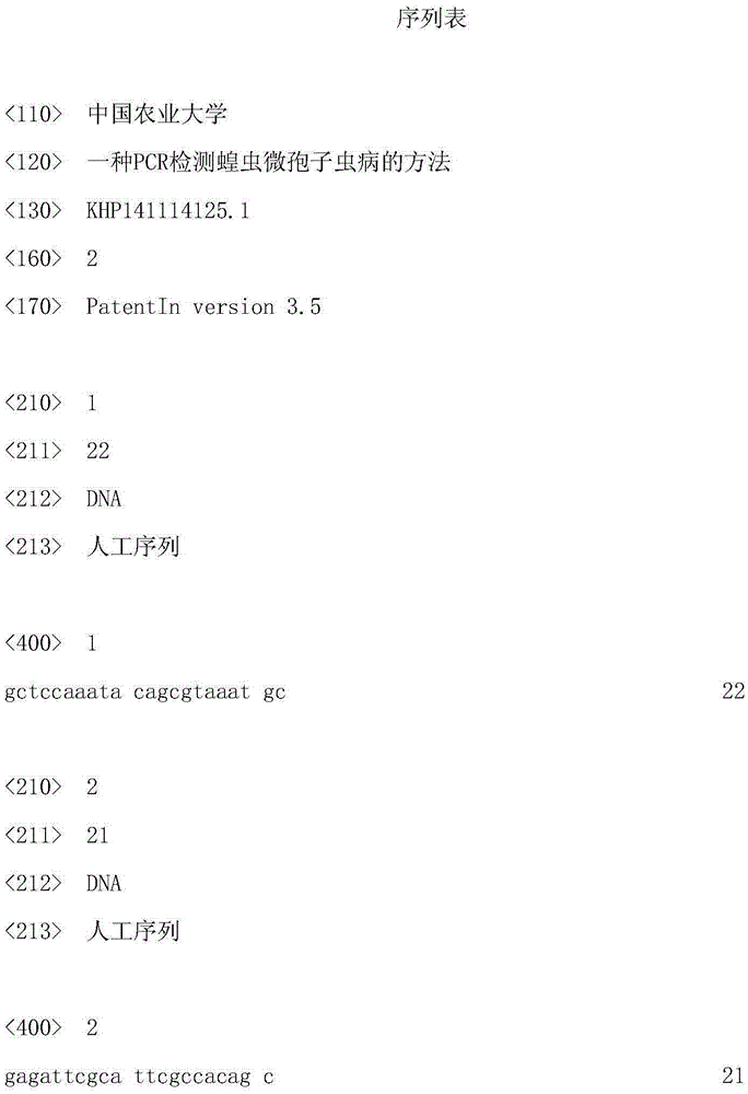
[0032] Nlos-F5: 5'-GCTCCAAATACAGCGTAAATGC-3' (SEQ ID NO: 1);

[0033] Nlos-R5: 5'-GAGATTCGCATTCGCCACAGC-3' (SEQ ID NO: 2).

Embodiment 3

[0034] The PCR amplification procedure of embodiment 3 locust microsporidia

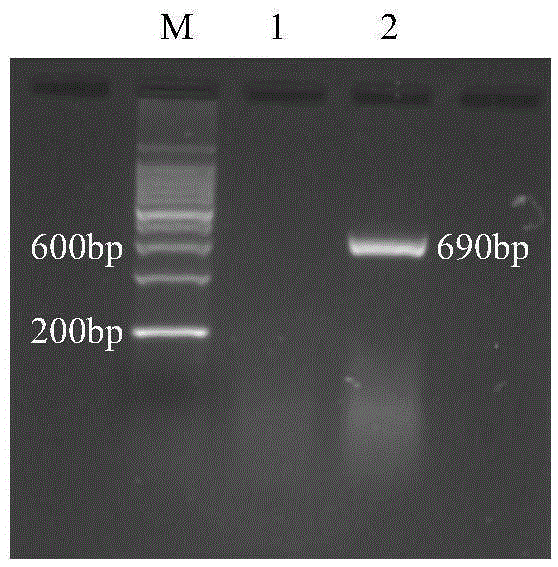
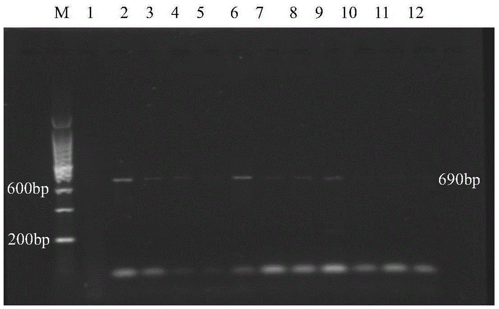
[0035] The DNA of the tested sample was used as a template, and Nlos-F5 and Nlos-R5 were used as forward and reverse primers to perform fragment amplification. The PCR reaction system is: 2×TaqMasterMix (Tiangen Biochemical Technology Co., Ltd.), 12.5 μl; 10 μM primer Nlos-F5, 1 μl; 10 μM primer Nlos-R5, 1 μl; DNA template, 1 μl; ddH 2 O, 9.5 μl; the total reaction system is 25 μl. The reaction conditions were: 95°C, 5min, 1 cycle; 94°C, 30s, 66°C, 30s, 72°C, 30s, 35 cycles; finally 72°C, 10min, 1 cycle. Take 10 μl of PCR product, electrophoresis in 1% agarose gel [containing nucleic acid dye (10000 × GeneGreen)], voltage 100V, 40min, observe the PCR diagnosis result under strong ultraviolet light (results see figure 1 ).


PUM


Abstract

The invention relates to a method for detecting microsporidiosis of locust by PCR (Polymerase Chain Reaction), and provides PCR primers and a kit for detecting microsporidiosis of locust. The method for detecting the microsporidiosis of locust by PCR is capable of performing PCR detection on locust by utilizing the primers or the kit. By means of the content disclosed by the invention, the situation of locust infected by microsporidia can be quickly determined on a large scale, and the number of samples required by detection is greatly reduced, the detection accuracy is greatly improved, the results are in the credibility, and the sensitivity can be 7*10<3> spore / ml. Compared with a traditional microscopy, the method disclosed by the invention has the advantage that the detection cost and workload can be greatly reduced.

Description

technical field [0001] The invention relates to the technical field of biological detection, in particular to a PCR detection technology for detecting locust microsporidiosis by adopting the PCR diagnosis technology. Background technique [0002] Polymerase Chain Reaction (Polymerase Chain Reaction, PCR) is an in vitro nucleic acid amplification technique developed in the mid-1980s. PCR technology has many advantages such as good specificity, high sensitivity, high yield, good repeatability, rapidity, simplicity, and easy automation; The results can even be directly observed and judged by the naked eye; with the help of this technology, technicians can amplify a sufficient amount of DNA from a hair, a drop of blood, or even a cell for analysis and research and detection and identification. What was done in days or even weeks in the past can be done in a few hours using PCR analysis technology. PCR technology is a revolutionary innovation and a milestone detection technolo...

Claims


Application Information

Patent Timeline
IPC IPC(8): C12Q1/68C12Q1/04C12N15/11C12R1/645
CPCC12Q1/686C12Q1/6888
Inventor 班丽萍张靓曹江波宋丽梅
Owner CHINA AGRI UNIV
Learn More