
Medical equipment and application thereof

A duodenum and inner covering technology, applied in obesity treatment, medical science, non-surgical orthopaedic surgery, etc., can solve the problem of damage to intestinal tissue, obstruction of duodenal bulb activity, damage, etc.

Active Publication Date: 2013-09-25
万平
View PDF11 Cites 17 Cited by
  • Summary
  • Abstract
  • Description
  • Claims
  • Application Information

AI Technical Summary

Problems solved by technology

The existing technology (utility model patent "duodenum-jejunum built-in sleeve") only considers "smooth surface, soft and dense" in terms of hose material, and does not involve the adaptability and elasticity of the ring bracket to the movement of the duodenum bulb stretching, and does not involve the use of elastic materials to prepare hoses; the inelastic tight mouth line "fixed" on the duodenal bulb along the "top of the ring bracket" further limits the effect of the ring bracket on the movement of the duodenal bulb adapt
This prior art (utility model patent "duodenum-jejunum built-in cannula") is compared with the prior art of the aforementioned invention patent "duodenal cannula and its conveyor", because of its ring-shaped bracket and its pointed The stabbing claw is only located at the duodenal bulb, although it does not block the duodenal papilla, which is the common opening of the bile duct and pancreatic duct at the lower end of the descending duodenum, but if placed on the upper edge of the duodenal bulb, when When the expansion or retraction of the gastric pylorus affects the movement of the duodenal bulb immediately below it (Zhou Lu, Ke Meiyun: "Gastrointestinal Dynamics: Basic and Clinical" pp. 381, 520, 522), the ring stent and its spiked fixed claws are bound to hinder the activity of the duodenal bulb, especially when the expansion of the gastric pylorus affects the activity of the duodenal bulb, the ring bracket and its spiked fixed claws cannot expand along the way, but penetrate into the duodenum The spiked fixed claws in the mucosa of the intestinal bulb pull the mucosal tissue of the duodenal bulb inward and towards the center, which is contrary to the activities of other tissues such as the submucosal muscle tissue of the duodenal bulb that expands centrifugally outward. , this ring-shaped bracket that cannot change or changes little along the trend and its spiked fixed claws, when the expansion or retraction of the gastric pylorus affects the movement of the duodenal bulb immediately below it, it will affect the duodenal bulb The tissue is damaged; if it is placed on the lower edge of the duodenal bulb, the expansion or retraction of the gastric pylorus will affect the lower edge of the duodenal bulb less than the upper edge of the duodenal bulb, but Even if the damage to the duodenal bulb mucosa by the ring stent and its spiked claws is ignored, the original covering effect on the duodenal bulb disappears.
[0008] Similarly, the prior art invention patent "a duodenal sleeve and its preparation method" (application date May 10, 2012, application publication date 2012 October 3, 2010), the nitinol anchoring part must also be inserted into the intestinal mucosa to directly damage the intestinal tissue

Method used


Image

  • Medical equipment and application thereof

Examples


preparation example Construction

[0054] In the preparation process of the biomimetic microarray adhesive sheet 2, the atomic force microscope etching method can also be used: flat paraffin, use the conical tip of the atomic force microscope probe to carve micropores on the surface, pour the liquid raw materials into the holes, place After cooling, the paraffin wax is removed, and the surface of the polymer after demoulding has micro-protrusions similar in structure and size to the fine forks on the gecko setae.

[0055] In the preparation process of the biomimetic microarray adhesive sheet 2, aluminum oxide template holes can also be used for injection molding: aluminum foil, placed in an acidic electrolyte, anodized, and formed into a porous aluminum oxide plate, the pore size and hole spacing can be adjusted by oxidation voltage and acid solution . Other mold injection molding methods can also be used.

[0056] The electrostatic induction etching method can also be used in the preparation process of the bi...

Embodiment 1

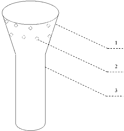
[0071] A duodenal inner membrane, which can be obtained from biocompatible biodegradable or non-biodegradable materials or / and strong hydrophobic materials, and is mainly adhered by the tubular part 2 and the horn-shaped external attachment biomimetic microarray The slice 2 consists of the ampulla 1 .

[0072] The diameter, length and thickness of the tubular part 3 match the duodenum and jejunum in different populations, the optimized diameter is 25mm, the length matches the duodenum and can extend to a section of jejunum connected to the duodenum, the length 500 mm, and the thickness of the inner coating of the tubular portion 3 is 0.1 mm. The ampulla 1 is the part connected to the tubular part 3 in a trumpet shape. The thickness of the inner coating of the ampulla 1 is optimized to be 0.1 mm. The trumpet-shaped connecting tubular part 3 is a gradual open acute angle, and the optimized angle is 45°C. The upper edge of the optimized ampulla 1 may be a wave-shaped elastic mem...

Embodiment 2

[0076] As an optimization, the ampulla 1 and the tubular part 3 of the duodenal inner membrane can be folded together or folded into a cylindrical shape in vitro. Turn inward.

[0077] As an optimization, the duodenal inner covering can be sent into the duodenum through the upper gastrointestinal tract under endoscopy and X-ray fluoroscopy, and multi-claw instruments can be used for the device (the number of claws is the same as the arrangement of the bionic microarray adhesive sheet). Matching) Centrifugally stretch the varus ampulla 1 through the center of the endoscopic forceps, and then turn the varus ampulla 1 to reset, position it on the duodenal bulb, adhere to it, and then use instruments or / and air or / and water or / and gravity or / and other methods gently push the distal end of the duodenal lining to the target position. When the intestinal contents move, the lining of the duodenum will not detach because there is no nearly vertical drag force; when the duodenal bulb ...


PUM


Abstract

The invention relates to the field of medical equipment and application thereof, and provides a duodenum internal covering film. The duodenum internal covering film is made of biocompatible biodegradable or non-biodegradable material or / and hydrophobic material and is mainly composed of an elastic ampullar portion and a tubular portion. The ampullar portion comprises a biocompatible bionic micro array adhesion piece which can achieve strong adhesion in the force application direction, does not stab intestine tissues and is capable of achieving detachment and recovery easily. The adhesion piece is good in stability, strong in adaptability to material and shapes, good in self-cleaning performance, free of damage and pollution to the intestine tissues, and capable of achieving repeated adhesion and detachment and supporting other components mutually in functions. When intestinal tract contents move, the internal covering film can not be detached due to the fact that subvertical pulling force does not exist. When the duodenum bulb is expanded and inflated, the internal covering film can not be detached due to the fact that the internal covering film does not have opposite and subvertical pulling force, and in the process of recovery, the internal covering film can be detached and recovered easily through force applied in a subvertical mode. The duodenum internal covering film can be manufactured into the medical equipment which does not damage the intestine tissues and can prevent and cure fat and diabetes.

Description

[0001] Cross references to related applications. [0002] This application can be May 5, 2012 (application number 201210136379.X), August 21, 2012 (application number 201210298363.9), January 28, 2013 (application number 201310029525.3) and March 31, 2013 (application Partial continuation of the application series filed under No. 201310107770.1). technical field [0003] The invention relates to a medical device built in the digestive tract, in particular to a duodenal inner covering film for preventing and treating obesity and diabetes without damaging intestinal tissue. Background technique [0004] Gastric bypass surgery treats obesity. Recently, it has been found that obese patients not only lose weight significantly after undergoing this operation, but also relieve their concurrent type 2 diabetes (Chinese Journal of Diabetes, 2011, 3(3):205-208): there is no need for insulin injections after surgery, and there is no need for Taking a variety of drugs can solve blood ...

Claims


Application Information

Patent Timeline
Patent Type & Authority Applications(China)
IPC IPC(8): A61F5/00
CPCA61F5/0076
Inventor 万平
Owner 万平
Learn More