
Preparation method of bone repair bracket with osteoid poriform regeneration channel structure

A channel structure, osteoid technology, applied in medical science, prosthesis, etc., can solve the problems of new tissue necrosis, poor permeability, cell apoptosis, etc., to promote bone tissue regeneration and repair, improve mechanical properties, and meet popularization and application. Effect

Inactive Publication Date: 2013-08-28
SICHUAN UNIV
View PDF11 Cites 2 Cited by
  • Summary
  • Abstract
  • Description
  • Claims
  • Application Information

AI Technical Summary

Problems solved by technology

Traditional bone tissue engineering scaffolds have an irregular isotropic pore structure with no regularity and direction orientation. The large pores are often connected only through small pores of a few microns to more than ten microns, and the pore penetration is relatively low. Low, the common problem is that new bone tissue is often formed on the outer edge of the scaffold, making it difficult for cells to penetrate into the interior of the scaffold, hindering the exchange of body fluids inside the scaffold, and eventually leading to apoptosis of cells inside the scaffold and necrosis of new tissue etc., affecting the reconstruction of tissue in large defect areas
Although these anisotropic structural scaffolds have been shown to have good characteristics and prospects in the field of tissue engineering, the current common problem of scaffolds used for bone repair is that their mechanical properties are poor, and they are not as good as mechanical properties and vascularization. Scaffold materials with bone conduction or induction can meet the needs of bone repair in clinical load-bearing parts
The scaffold pores prepared by these methods have a disordered structure, a high rate of closed pores, poor connectivity between pores, and it is difficult for the new bone tissue of the nucleus to grow into the interior of the scaffold, which affects its application effect.
In addition, such methods may leave chemical foaming agents that are not completely decomposed or removed, which will have adverse effects on the matrix or local tissues
Wang HN, et al. reported the preparation of nanoapatite-polyamide composite scaffolds with oriented pore structure by thermally induced phase transfer (J Polym Sci Part A: Polym Chem, 2008, 47: 658-699), but The stent prepared by this method has an orientation hole structure only within a few millimeters (about 3mm), and the pore size of the orientation hole is difficult to control, so it cannot be used in practice.

Method used


Image

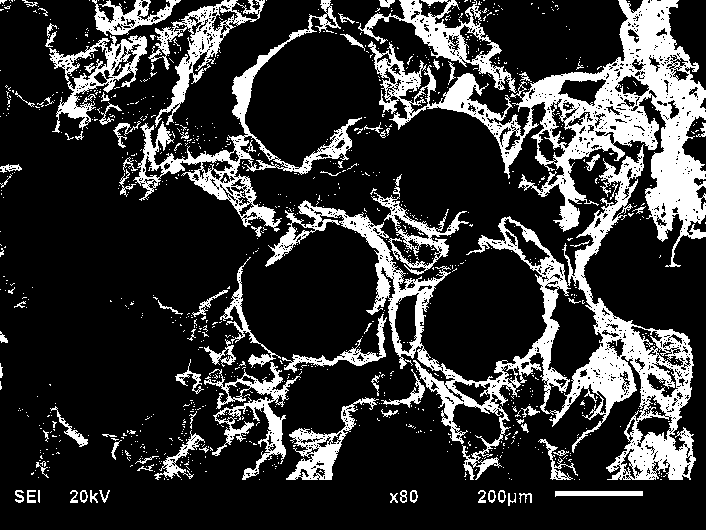
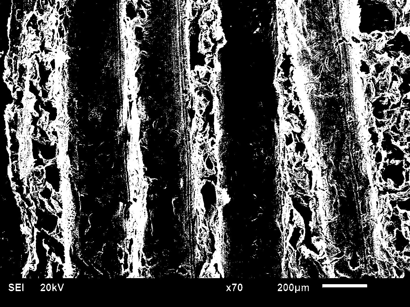
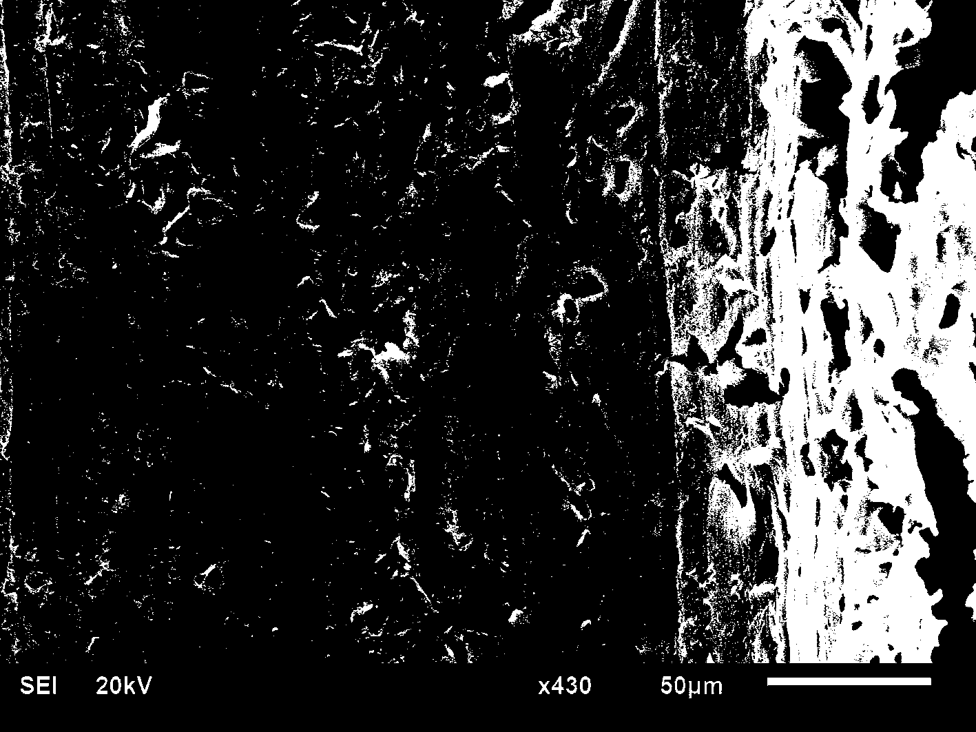
  • Preparation method of bone repair bracket with osteoid poriform regeneration channel structure
  • Preparation method of bone repair bracket with osteoid poriform regeneration channel structure
  • Preparation method of bone repair bracket with osteoid poriform regeneration channel structure

Examples


Embodiment 1

[0026] According to literature (Li JD, Zuo Y, Cheng XM, Yang WH, Wang HN, Li YB, Preparation and characterization of nano-hydroxyapatite / polyamide 66 composite GBR membrane with asymmetric porous structure, Journal of Material Science-materials in medicine, 2009, Vol.30, No.5: 1031-1038) reported the preparation of nano-apatite (n-HA)-polyamide 66 (PA66) composite slurry. Evaporate the ethanol solvent by heating, so that the n-HA-PA66 composite slurry can reach a suitable viscosity. The composite slurry is evenly coated on 304 stainless steel wires with a diameter of 300 μm, and then 40 stainless steel wires coated with slurry are bundled into a bundle and placed in a PE heat-shrinkable tube, and one end of the heat-shrinkable tube is sealed. Heat the heat shrink tubing to 90°C for 0.5 hour to allow the heat shrink tubing to start shrinking. Heating was continued at elevated temperature to 95°C and maintained for 4 hours. After the material is completely cured, remove the he...

Embodiment 2

[0028] Prepare nano-apatite-polyamide 66 (n-HA-PA66) composite material according to the method reported in CN1544099, and then use methanol as a solvent to prepare n-HA-PA66 composite slurry with suitable viscosity. The composite slurry was uniformly coated on 304 stainless steel wires with a diameter of 150 μm, and then 50 stainless steel wires coated with the slurry were bundled into a bundle and placed in a PET heat-shrinkable tube, and one end of the heat-shrinkable tube was sealed. Heat the heat shrink tubing to 80°C for 1 hour to allow the heat shrink tubing to start shrinking. Elevate the temperature and continue heating to 100°C for 6 hours. After the material is completely cured, remove the heat-shrinkable tube, soak it in deionized water for 3 hours, and pull out the stainless steel wire after the material swells. The material was then ultrasonically cleaned in deionized water for 5 times, each time for 5 minutes, and dried to obtain a bone repair scaffold with an ...

Embodiment 3

[0031] According to the method described in the literature described in Example 1, nano-apatite-polyamide 66 (n-HA-PA66) composite slurry was prepared using propanol as a solvent. Evaporate the solvent propanol by heating, so that the n-HA-PA66 composite slurry can reach a suitable viscosity. The composite slurry is uniformly coated on 304 stainless steel wires with a diameter of 500 μm, and then 40 stainless steel wires coated with slurry are bundled into a bundle and placed in a PE heat-shrinkable tube, and one end of the heat-shrinkable tube is sealed. Heat the heat shrink tubing to 110°C for 2 hours to allow the heat shrink tubing to start shrinking. Increase the temperature and continue heating to 150° C. and maintain for 3 hours. After the material is completely cured, remove the heat-shrinkable tube, soak it in deionized water for 3 hours, and pull out the stainless steel wire after the material swells. The material was then ultrasonically cleaned in deionized water f...


PUM

PropertyMeasurementUnit
Viscosityaaaaaaaaaa
Viscosityaaaaaaaaaa
Diameteraaaaaaaaaa

Abstract

The invention discloses a preparation method of a bone repair bracket with an osteoid poriform regeneration channel structure. The method comprises the steps that a filament pore forming material with the diameter corresponding to the required pore diameter is embedded into nanometer apatite and polyamide composite pulp with the viscosity of 50-500Pa.s; a solvent is heated, volatilized and solidified at 80-200 DEG C, and then the filament pore forming material in the solvent is removed; and finally a bone repair bracket body product with orientation pores / channels in the structure is obtained. The method is simple and convenient; the bone repair bracket with the orientation pores / channels can be prepared; the pore size of the prepared osteoid orientation pores / channels and the porosity of the bracket can be adjusted and controlled as required; the mechanical properties in hole directions of the orientation pores / channels of the bracket can be significantly improved; the connectivity of the pores is good; rapid vascularization, cell growth and new bone tissue growth are facilitated; and regeneration and repair of a bone tissue are promoted.

Description

technical field [0001] The invention relates to a method for preparing a bone-like apatite-polyamide composite material porous bone repair bracket with a bone-like porous regeneration channel structure. Background technique [0002] As a porous scaffold for bone repair tissue, its three-dimensional structure not only plays an important role in the selective permeability of cells, nutrients and oxygen, the removal of metabolic substances and the survival of new tissue, but also greatly affects the cell behavior and tissue reconstruction. Appropriate pore structure is the key to the optimal osteogenesis performance of porous scaffolds in bone tissue engineering. [0003] Studies have found that many tissues in the human body, including nerves, muscles, tendons, blood vessels, bones and teeth, have ordered anisotropic structures. This anisotropy is not only reflected in the structure, but also in the physiological function and mechanical properties. For example, bone tissue i...

Claims


Application Information

Patent Timeline
IPC IPC(8): A61L27/12A61L27/18
Inventor 李吉东尤福李玉宝左奕
Owner SICHUAN UNIV
Learn More