
Compositions and methods for protein display on the surface of bacteria and vesicles derived therefrom and uses thereof

A protein, cell surface technology, applied in biochemical equipment and methods, chemical instruments and methods, immunoglobulins, etc., can solve the problem of not carrying signal peptides

Active Publication Date: 2016-04-13
CORNELL RES FOUNDATION INC
View PDF6 Cites 0 Cited by
  • Summary
  • Abstract
  • Description
  • Claims
  • Application Information

AI Technical Summary

Problems solved by technology

However, the pathway by which ClyA manages to cross the bacterial IM and assemble in the OMV remains a mystery because it does not carry a conventional signal peptide (delCastillo et al., "The Escherichia coli K-12 SheA Gene Encode sa 34-kDa Secreted Haemolysin (Escherichia coli K-12 SheA gene encoding 34- kDa secreted hemolysin)", MolMicrobiol 25:107-15 (1997)), and is not processed at the N-terminus (Ludwig et al., "Analysis of the SlyA-Controlled Expression, Subcellular Localization and Pore-Forming Activity of a 34 kDa Haemolysin (ClyA) from Escherichia coli K-12 (Escherichia coli K-12 34 kDa hemolysin SlyA-controlled expression, subcellular localization and pore-forming activity analysis of ClyA)", MolMicrobiol31: 557-67 (1999))

Method used


Image

  • Compositions and methods for protein display on the surface of bacteria and vesicles derived therefrom and uses thereof
  • Compositions and methods for protein display on the surface of bacteria and vesicles derived therefrom and uses thereof
  • Compositions and methods for protein display on the surface of bacteria and vesicles derived therefrom and uses thereof

Examples


Embodiment Embodiment 1

[0093] The following examples illustrate various methods of the compositions in the methods of treatment of the invention. The examples are intended to illustrate, but in no way limit the scope of the invention. EXAMPLES Example 1 - Bacterial Strains, Plasmids and Growth Conditions

[0094]The bacterial strains and plasmids used in these examples are described in Table 1. Table 1. Bacterial Strains and Plasmids

[0095] Using DHA as a donor, the dsbA::Kan allele was introduced into JC8031 via P1vir transduction to prepare cell strain JCA. The PCR-amplified clyA gene was ligated with pBAD18-Cm between SacI and XbaI sites to construct plasmid pClyA. The gene encoding gfpmut2 was inserted between the XbaI and HindIII sites (Crameri et al., "Improved Green Fluorescent Protein by Molecular Evolution Using DNA Shuffling (by using DNA shuffling molecular evolution to improve green fluorescent protein)", NatBiotechnol14: 315-9 (1996) and DeLisa et al., "GeneticAnalysisoftheTw...

Embodiment 2

[0096] Human epithelial cervical carcinoma (HeLa) cells were obtained from the American Type Culture Collection (ATCC#CCL-2) and cultured in Dulbecco's modified Eagles minimal medium supplemented with 10% NuSerum and 1% penicillin / streptomycin (DMEM). Maintain cells at 37°C, 95% air, 5% CO 2 in a humidified atmosphere. For fluorescence microscopy experiments, cells were grown on 12-mm round glass coverslips for 2 days prior to the experiment. Example 3 - Subcellular Fractionation

Embodiment 3

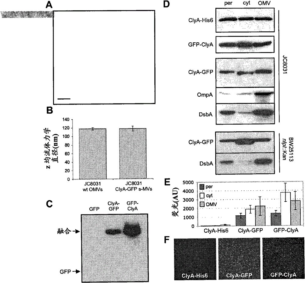
[0097] By cold osmotic shock procedure (coldosmotic shock procedure) (Kim et al., "Twin-Arginine Translocation of Active Human Tissue Plasminogen Activator in Escherichiacoli (Twin-Arginine Translocation of Active Human Tissue Plasminogen Activator in Escherichia coli)", Applied and Environmental Microbiology 71: 8451-8459 (2005 ), which is hereby incorporated by reference in its entirety), cytoplasmic and periplasmic fractions were prepared from cells expressing the fusion protein, and the residual pellet was collected as the insoluble fraction after removal of the soluble fraction. Example 4 - Isolation of Bacterial Vesicles


PUM

PropertyMeasurementUnit
diameteraaaaaaaaaa

Abstract

The present invention relates to compositions and methods for displaying proteins and polypeptides on the surface of cells and vesicles. Also disclosed herein are methods and compositions for the delivery of drugs and vaccines utilizing the cell surface display systems of the present invention.

Description

[0001] This application claims the benefit of US Provisional Patent Application Serial No. 60 / 939,506 filed May 22,2007. [0002] The subject matter of this application was made with support from the United States Government under the National Institutes of Health under grant number NIBIBR21EB005669. The U.S. government has certain rights. field of invention [0003] The present invention relates to compositions and methods for the display of proteins and polypeptides on the surface of cells and vesicles. Background of the invention [0004] Protein translocation is a highly conserved process essential to all life. Extracellular secretion of virulence factors is a strategy used by invading bacteria to establish a colony niche, communicate with host cells, and modulate host defenses and responses. Bacterial protein secretion systems are characterized by membrane translocation of single proteins or other small protein complexes, with few exceptions (Christie et al., "Bacteri...

Claims


Application Information

Patent Timeline
Patent Type & Authority Patents(China)
IPC IPC(8): C12P21/04
CPCC12N15/1037C12N15/11C07K14/005C07K16/44C07K2317/622C07K2317/92C07K2319/33C12N15/113C12N2310/14C12N2320/30
Inventor M·德利萨J·金D·A·普特曼A·M·杜迪
Owner CORNELL RES FOUNDATION INC
Learn More