Norovirus immunogens and related materials and methods
a technology of immunogens and norroviruses, applied in the field of biotechnology, can solve the problems of small animal model for infection study, no vaccine or effective treatment of norrovirus, and inability to grow human norrovirus in cell cultur
- Summary
- Abstract
- Description
- Claims
- Application Information
AI Technical Summary
Benefits of technology
Problems solved by technology
Method used
Image
Examples
example 1
Materials and Methods
[0043]Plasmid Construction.
[0044]Plasmids encoding VSV N (pN), P (pP), and L (pL) genes; and an infectious cDNA clone of the viral genome, pVSV1(+), were generous gifts from Dr. Gail Wertz. Plasmid pVSV1(+) GxxL which contains Sma I and Xho I at the G and L gene junction, was kindly provided by Dr. Sean Whelan. The capsid VP1 gene of HuNoV G.IV strain HS66 (kindly provided by Dr. Linda Saif) was amplified by high fidelity PCR with the upstream and downstream primers containing VSV gene start and gene end sequences. The resulting DNA fragment was digested with Sma I and Xho I, and cloned into pVSV(+)GxxL at the same sites. The resulting plasmid was designated as pVSV1(+)-VP1, in which HuNoV VP1 gene was inserted into G and L gene junction. The VP1 gene was also cloned into a pFastBac-Dual expression vector (Invitrogen) at Sma I and Xho I sites under the control of the p10 promoter, which resulted in construction of pFastBac-Dual-VP1. All constructs were confirmed...
example 2
Recovery of Recombinant VSV Expressing HuNoV Capsid Protein
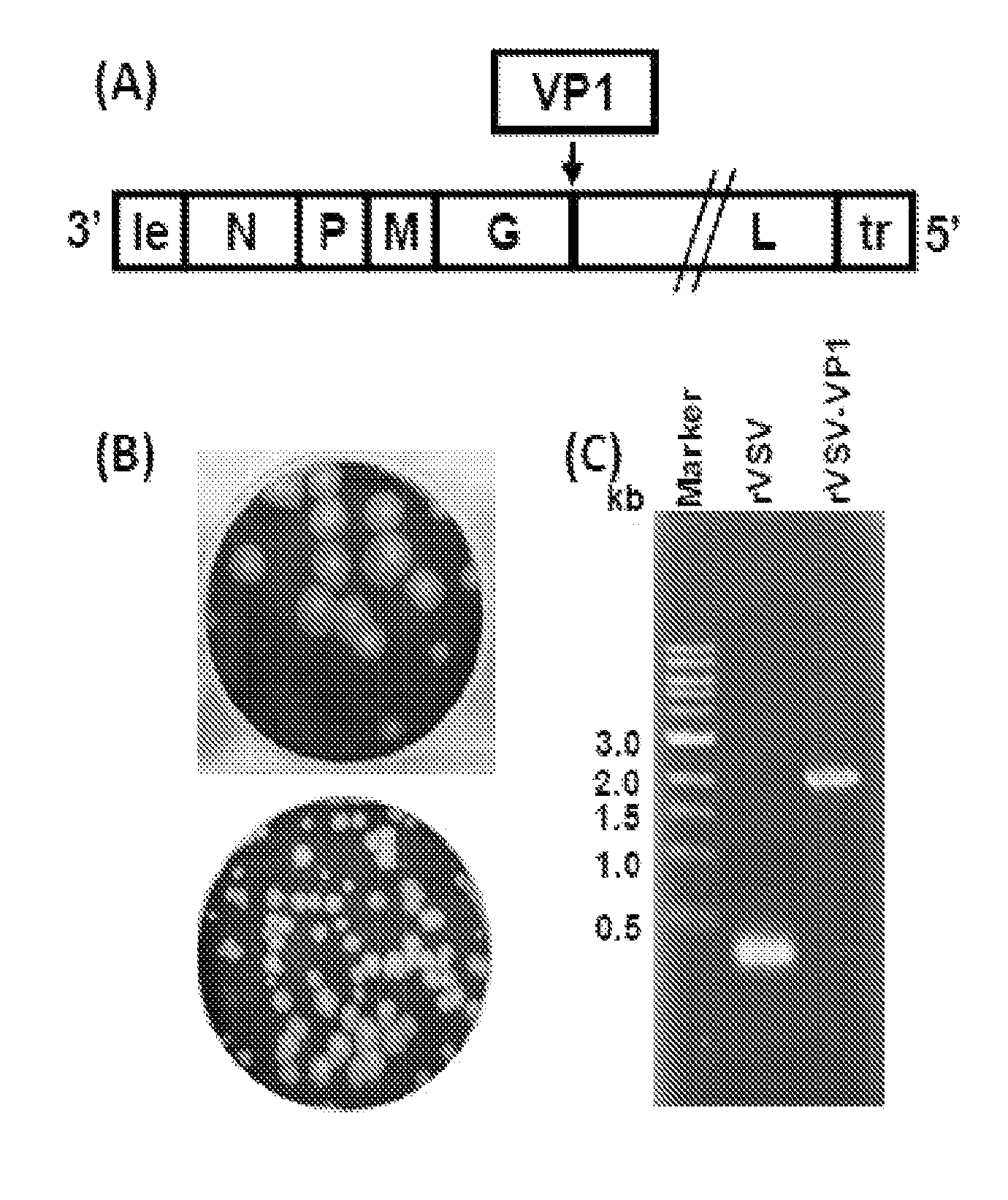
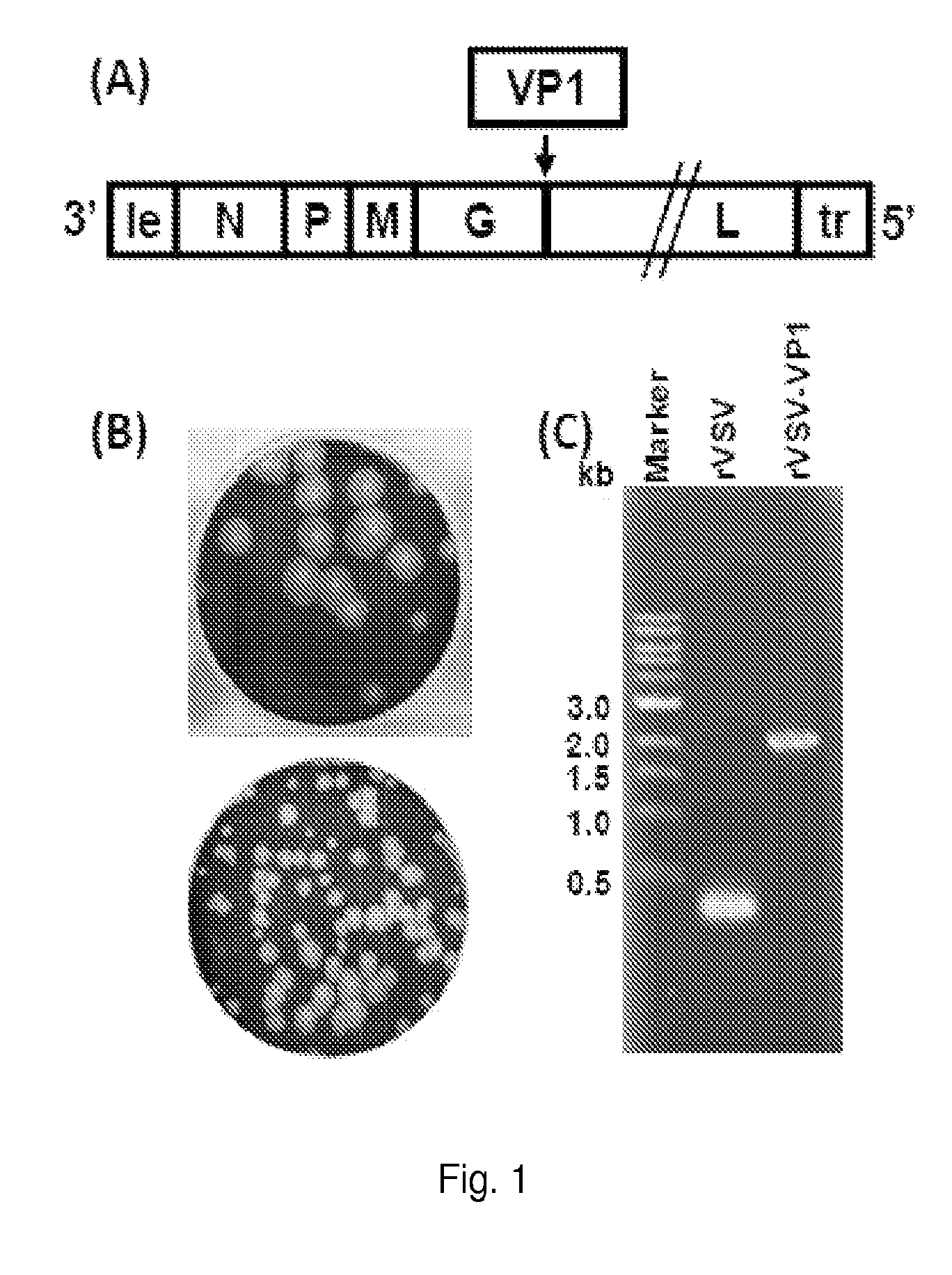
[0073]The capsid gene of human norovirus (HuNoV) strain HS66 was amplified by PCR and inserted into the gene junction of G and L in the genome of vesicular stomatitis virus (VSV) (FIG. 1A). Recombinant VSV expressing HuNoV VP1 (rVSV-VP1) was successfully recovered using a reverse genetics technique. Recombinant rVSV-VP1 formed much smaller plaques in Vero cells as compared to wild type rVSV (FIG. 1B). After 24 h of incubation, rVSV formed plaques that were 4.3±0.8 mm in diameter. However, average plaque size for rVSV-VP1 was 2.3±0.8 mm even after 48 hours incubation, suggesting that rVSVVP1 may have defect in viral growth. To confirm the recovered virus indeed containing VP1 gene, viral genomic RNA was extracted followed by RT-PCR using two primers annealing to VSV G and L genes respectively. As shown in FIG. 1C, a 2.0 kb DNA band containing VP1 gene was amplified from genomic RNA extracted from rVSV-VP1. However, a 300 by D...
example 3
Recombinant rVSV-VP1 has a Delayed Replication in Cell Culture
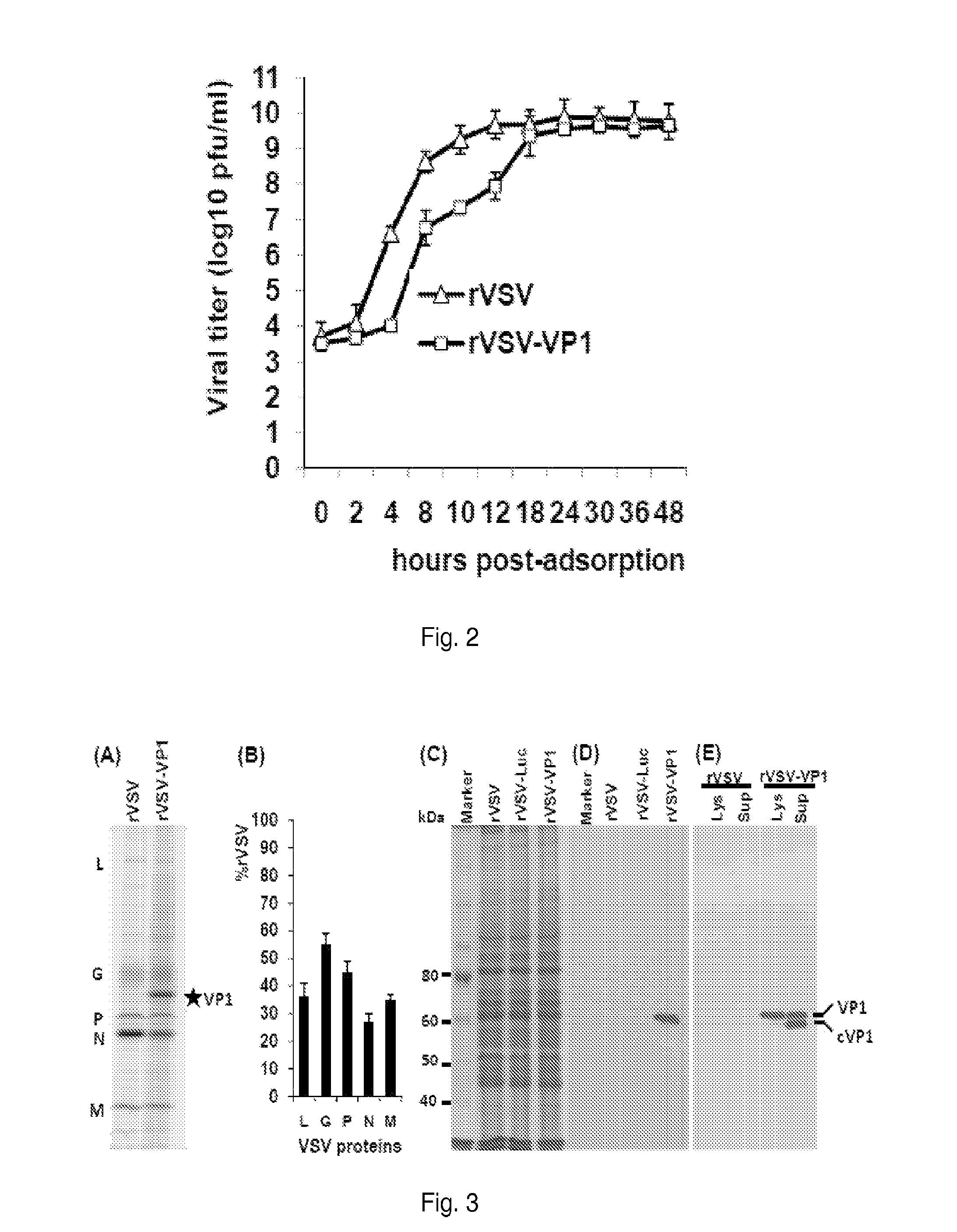
[0074]To further characterize recombinant rVSV-VP1, the inventors monitored the kinetics of release of infectious virus by a single-step growth assay in BSRT7 cells. Briefly, BSRT7 cells were infected with each of the recombinant viruses at an MOI of 10 and viral replication was determined at time points from 0-48 h postinfection. As shown in FIG. 2, rVSV-VP1 had significant delay in viral replication as compared to that of rVSV. Wild type rVSV reached peak titer (6.3×10⁹ pfu / ml) at 12 h post-infection. However, rVSV-VP1 reached peak titer of 6.3×10⁸ pfu / ml at approximately 30 h post-infection. At an MOI of 10, recombinant rVSV exhibited significant cytopathic effect (CPE) around 6 h postinfection, and cells were completely killed at 14 h post-infection. However, rVSV-VP1 showed CPE around 12 h post-infection, and cells were killed until 36 h post-infection. These results suggested that rVSV-VP1 had delayed replication an...
PUM
| Property | Measurement | Unit |
|---|---|---|
| volume | aaaaa | aaaaa |
| pH | aaaaa | aaaaa |
| OD | aaaaa | aaaaa |
Abstract
Description
Claims
Application Information
 - R&D
- Intellectual Property
- Life Sciences
- Materials
- Tech Scout
- Unparalleled Data Quality
- Higher Quality Content
- 60% Fewer Hallucinations
Browse by: Latest US Patents, China's latest patents, Technical Efficacy Thesaurus, Application Domain, Technology Topic, Popular Technical Reports.
© 2025 PatSnap. All rights reserved.Legal|Privacy policy|Modern Slavery Act Transparency Statement|Sitemap|About US| Contact US: help@patsnap.com


