
Estimating and correcting for contrast agent extravasation in tissue perfusion imaging

a tissue perfusion imaging and contrast agent technology, applied in image data processing, health-index calculation, sensors, etc., can solve the problems of confounding effect on tumor blood volume and perfusion estimation, inability to accurately estimate the extravasation of contrast agents, and inability to detect the presence of contrast agents in the intravascular spa

Active Publication Date: 2014-05-06
UNIV OSLO HF
View PDF16 Cites 33 Cited by
  • Summary
  • Abstract
  • Description
  • Claims
  • Application Information

AI Technical Summary

Benefits of technology

This approach provides more accurate estimates of extravasation rates and corrected hemodynamic parameters, reducing errors associated with tissue transit time variations and improving tumor grading and prognosis prediction.

Problems solved by technology

Many brain tumors result in disrupted blood-brain-barrier (BBB) causing the contrast agent to leave the intravascular space and leak into the interstitial space.
This extravasation has a confounding effect on the estimations of tumor blood volume and perfusion unless corrected for
Given the highly heterogeneous vasculature in tumor such as gliomas, MTT / delay values which deviate significantly from the mean MTT are very likely and may lead to incorrect estimations of contrast agent extravasation.
A disadvantage of this approach is that a very long time series is needed to correctly identify the leakage term from the apparent residue function and further that CBV leakage correction cannot be performed directly from the residue function.

Method used


Image

  • Estimating and correcting for contrast agent extravasation in tissue perfusion imaging
  • Estimating and correcting for contrast agent extravasation in tissue perfusion imaging
  • Estimating and correcting for contrast agent extravasation in tissue perfusion imaging

Examples


Embodiment Construction

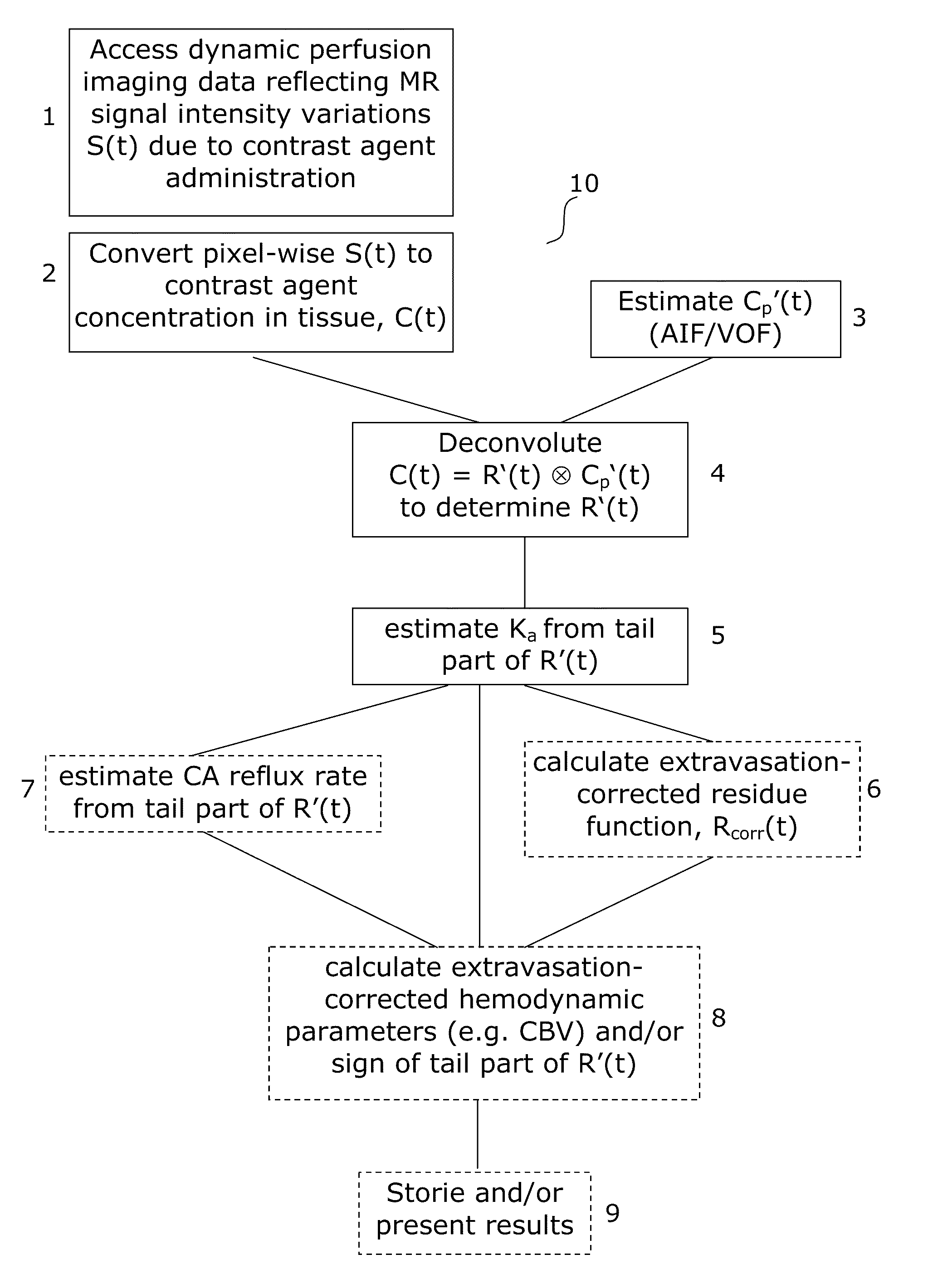
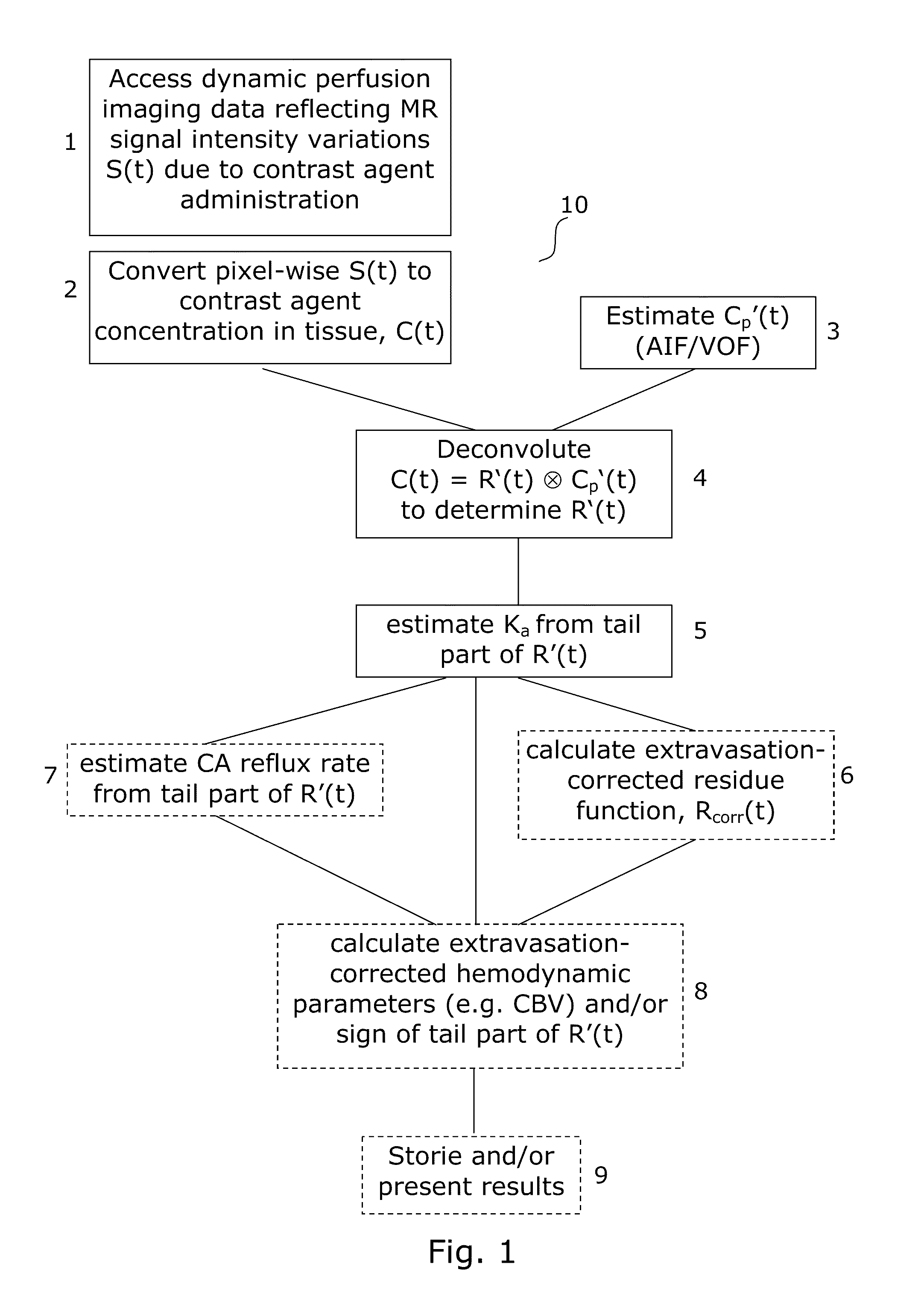
[0059]The generalized flow diagram of FIG. 1 embodies the methods for correcting perfusion image data according to the various aspects of the invention. FIG. 1 further embodies the layout of software applications according to the various aspects of the invention.

[0060]The invention can be implemented by means of hardware, software, firmware or any combination of these. The invention or some of the features thereof can also be implemented as software running on one or more data processors and / or digital signal processors.

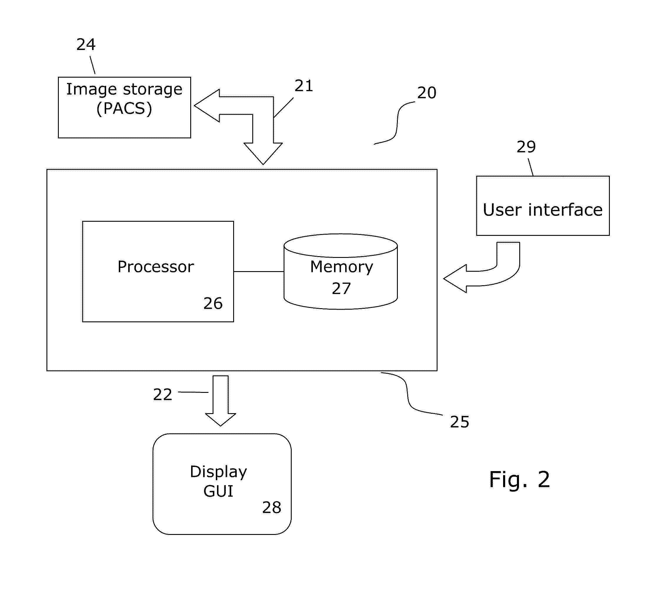
[0061]FIG. 2 illustrates a medical image analysis system 20 for implementing different embodiments for the various aspects of the invention.

[0062]The system 20 has means 21 for receiving or accessing image data to be processed or already processed (i.e. post-processed) image data from an image recording apparatus such as a CT or MR scanner and / or internal or external storage 24 holding images recorded by such apparatus such as a PACS. The means 21 may e.g. be a data ...


PUM


Abstract

The invention provides a method, an image analysis software product, and a system for medical imaging analysis for estimating contrast agent extravasation in contrast agent based perfusion imaging such as MRI dynamic contrast enhanced (DCE) imaging, and in particular correction, compensation, or visualization of extravascular leakage of contrast agent in tumors. According to the invention, the effect of extravasation is directly manifested in the tail part of an observed, apparent residue function, R′(t), obtained directly by de-convoluting the expression C(t)=R′(t)Cp′(t) with the arterial input function (AIF). A leakage rate or extravasation constant is determined directly from the tail part of the determined apparent residue function. The invention also relates to distinguishing between T1-dominant and T2*-dominant extravasation effects in perfusion imaging to from the sign of the tail part of the determined apparent residue function and to an automated method for DSC-MRI involving correction for contrast agent extravasation and partial volume effects.

Description

CROSS REFERENCE TO RELATED APPLICATIONS[0001]This application is a non-provisional of and claims the benefit of priority to U.S. provisional Patent Application Ser. No. 61 / 325,019, filed on Apr. 16, 2010, the disclosures of which are hereby expressly incorporated by reference in their entireties.FIELD OF THE INVENTION[0002]The present invention relates to perfusion analysis based on dynamic contrast enhanced (DCE) imaging, in particular correction or compensation due to extravascular leakage of the contrast agent in tumors.BACKGROUND OF THE INVENTION[0003]In DCE imaging, an MRI or CT sequence is recorded after a contrast agent (CA) has been injected intravenously. The recorded sequence consist of dynamic concentration time curves (CTC) recorded for each voxel, which shows the contrast agent induced signal (˜which can be related to relative CA concentration) in a given voxel as a function of time. Thereby, DCE imaging can be used to make a quantitative estimates of tissue perfusion a...

Claims


Application Information

Patent Timeline
Patent Type & Authority Patents(United States)
IPC IPC(8): A61B6/00
CPCG01R33/56366A61B6/486G06T7/0016A61B6/481G06T2207/10096A61B6/507A61B5/055G06T2207/30016A61B6/5217G01R33/5601G16H50/30
Inventor BJORNERUD, ATLEEMBLEM, KYRRE EEG
Owner UNIV OSLO HF
Learn More