
Hypodermic optical monitoring of bodily analyte

a technology of optical monitoring and bodily analyte, which is applied in the field of hyperodermic optical monitoring of bodily analyte, can solve the problems of increasing the burden on patients and health care resources, increasing the rate of coronary heart disease, peripheral vascular disease and stroke, and difficult to achieve near-normal glucose concentrations in many patients, so as to avoid repeated skin pricking and high cost-effective

Inactive Publication Date: 2013-12-24
ROCHE DIABETES CARE INC
View PDF35 Cites 29 Cited by
  • Summary
  • Abstract
  • Description
  • Claims
  • Application Information

AI Technical Summary

Benefits of technology

The invention is a monitoring device that can measure the concentration of glucose and dispense insulin through a single needle. Its design allows for high-rate measurements and includes all the necessary functions in a compact patch unit, eliminating the need for a separate control unit. The device is small, discreet, economical, and highly cost-effective for users. The invention can be built as a single unit or in two parts, with the disposable part containing the optical components and batteries. This simplifies the user experience and increases the manufacturer and payer's profit.

Problems solved by technology

Much of the burden of the disease to the patient and to health care resources is due to long-term tissue complications, which affect both the small blood vessels (microangiopathy, causing eye, kidney and nerve damage) and the large blood vessels (causing accelerated atherosclerosis, with increased rates of coronary heart disease, peripheral vascular disease and stroke).
In theory, returning glucose levels to normal by hormone replacement therapy using insulin injections and / or other treatments should prevent complications, but, frustratingly, near-normal glucose concentrations are very difficult to achieve and maintain in many patients, particularly those with type I diabetes.
In these patients, glucose concentration can swing between very high (hyperglycemia) and very low (hypoglycemia) levels in an unpredictable manner.
Unfortunately, the discomfort involved leads to poor patient compliance.
Testing cannot be performed while sleeping and while the subject is occupied.
In addition, the results do not give information regarding the trends in glucose levels, but rather provide only discrete readings, taken at large time intervals from one another.
NIR absorption features become significantly weaker and broader as the order increases, thereby greatly reducing the analytical utility of the short-wavelength region in terms of molecular vibrational information.
However, when the collected spectra encompass multiple spectral regions, it is not possible to match the sample thickness with each spectral region (Anal. Chem. 2005, 77, 5429-5439).
However, the signal is weak and therefore requires a highly sensitive detection system (e.g., CCD array).
In addition, the presence of other optically active molecules makes the accurate detection of glucose concentration complicated.
This method is particularly sensitive to changes in temperature.
Although optical approaches for glucose sensing are attractive, they are nevertheless often plagued by a lack of sensitivity and / or specificity since variations in optical measurements depend on variations of many factors in addition to glucose concentration.
Furthermore, non-invasive optical glucose monitors, which involve sensing of glucose levels through the skin, involve very low signal-to-noise ratio, scattering and interferences by bodily fluids and by the skin itself, causing noninvasive optical sensors to lack specificity and repeatability.
Implanted CGMs have several disadvantages, such as the need for a surgical procedure to implant the device, possibility of failure, the potential for sensor blockage, and biocompatibility problems.
Disadvantages in the performance and operation of non-invasive and fully invasive CGMs lead to the development of various minimally-invasive CGM systems.
Disadvantages of the GlucoWatch® include occasional sensor values differing markedly from blood values, skin rashes and irritation in those locations which are immediately underneath the device appearing in many users, a long warm up time of 3 hours, and skips in measurements due to sweating.
Numerous disadvantages inherent to glucose monitoring are present in CGMs which employ GOX-based reactions.
Most GOX-based devices rely on the use of oxygen as the physiological electron acceptor, and thus, are subject to errors due to fluctuations in the oxygen tension and the stoichiometric limitation of oxygen in vivo.
These and other oxidizable constituents of biological fluids can compromise the selectivity and hence the overall accuracy of the glucose concentration measurement.
Hydrogen peroxide deactivates the GOX molecules, limiting the time available for application of the sensor.
Miniaturizing the sensing technology within the cannula, which requires high levels of enzyme loading, while keeping high measurement sensitivity, remains a challenge.
Disadvantages of the microdialysis-based glucose sensors stem primarily from the constant perfusion of solution through the microdialysis probe.
This operational method requires the presence of a dedicated pump and reservoir, leading to large and bulky devices, and also necessitates high-energy consumption.
Furthermore, the relatively large size of the microdialysis catheter often causes a wound and subsequent local tissue reactions, following its insertion into the subcutaneous tissue.
Disadvantages of optics-based glucose monitoring techniques stem primarily from the indirect optical means applied for measuring analyte concentration levels.
In addition, optics-based techniques involving an electrochemical reaction possess the disadvantages inherent to glucose monitors which employ GOX-based reactions, described hereinbefore.

Method used


Image

  • Hypodermic optical monitoring of bodily analyte
  • Hypodermic optical monitoring of bodily analyte
  • Hypodermic optical monitoring of bodily analyte

Examples


Embodiment Construction

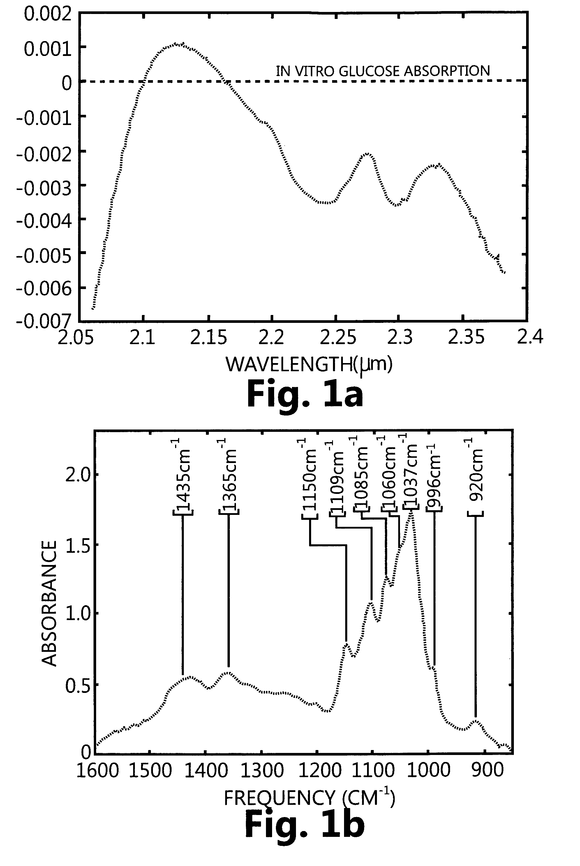
[0126]FIG. 1a shows optical absorption spectra of glucose in the NIR region for aqueous glucose after water subtraction as shown in the Journal of Biomedical Optics 5 (1), 5-16 Jan. 2000.

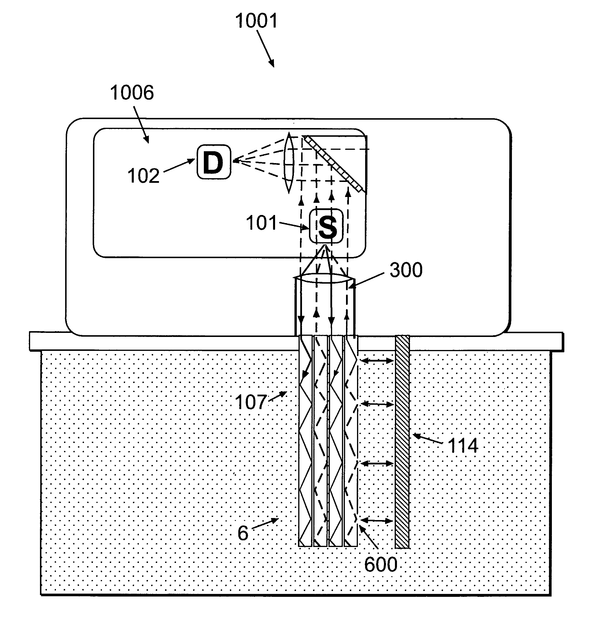
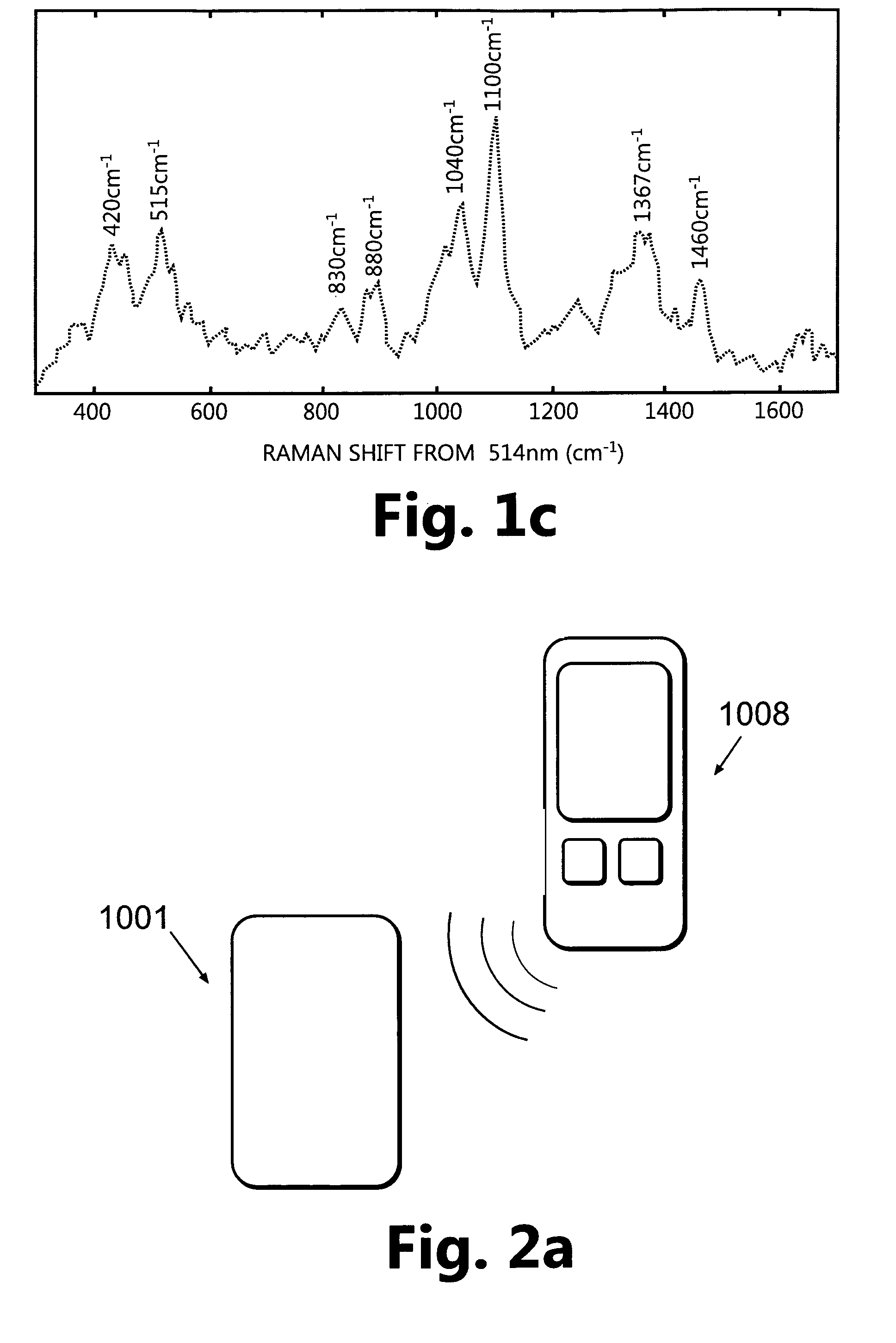
[0127]FIG. 2a shows a monitoring device composed of a patch unit (1001) and a remote control unit (1008). In accordance with one embodiment, the patch unit (1001) may be composed of a single part (FIG. 2b) or two parts (FIG. 2c): a reusable part (1) and a detachable disposable part (2), wherein each part is enclosed in its own housing. The patch unit (1001) can be programmed by the remote control unit (1008) or by the use of manual buttons (not shown) provided on the patch unit (1001).

[0128]FIGS. 3a-d show embodiments for connecting the patch unit (1001) to the user's skin (5). FIGS. 3a-b show embodiments of the patch unit (1001) that can be comprised of a single part (FIG. 3a) or two parts (FIG. 3b): a reusable (1) and a disposable part (2). The patch unit (1001) can be connected to and disconnecte...


PUM


Abstract

Disclosed is a skin adherable device for monitoring analytes in interstitial fluid. The device includes an electromagnetic radiation emitting source and a transmitter for transmitting the electromagnetic radiation between the electromagnetic radiation emitting source and the interstitial fluid. The device further includes a detector, operating electronics and a power supply. The device may include a reusable part and a disposable part.

Description

CROSS-REFERENCE TO RELATED APPLICATIONS[0001]The present application is a 35 U.S.C. §371 national stage entry of PCT / IL2008 / 001520, which has an international filing date of 20 Nov. 2008, which claims priority to U.S. Provisional Patent Application No. 61 / 004,039, entitled “Hypodermic Optical Monitoring of Bodily Analyte,” filed on Nov. 21, 2007, the disclosures of which are incorporated herein by reference in their entireties.FIELD[0002]Methods and devices for continuous monitoring of bodily analyte and continuous delivery of therapeutic fluid to the body are described herein. More particularly, optics-based devices and a methods for continuously monitor hypodermic glucose levels are described herein. Such embodiments can be coupled with an insulin delivery means and can be integrated into a closed loop or semi closed loop system.DEFINITIONS[0003]Banana-shaped, photon trajectory: a backscattering geometry which is a function of the source-detector separation and tissue's optical ch...

Claims


Application Information

Patent Timeline
Patent Type & Authority Patents(United States)
IPC IPC(8): A61B5/1455
CPCA61M5/14248A61B5/14532A61B5/1459A61B5/6833A61M2005/14268A61M2005/1726A61M2205/3561A61M2205/3592
Inventor YODFAT, OFERGESCHEIT, IDDO M.GESCHEIT, ILLAIKAIDAR, RUTHY
Owner ROCHE DIABETES CARE INC
Learn More