
Hollow nanoparticle of NBsAg large protein for drug delivery

a technology of nbsag and large protein, which is applied in the direction of inactivation/attenuation, powder delivery, viruses, etc., can solve the problems of difficult to achieve near 100% uptake, cannot be readily applied to cells or tissues of internal bodies, and cannot have a high level of specificity to cells or tissues

Inactive Publication Date: 2011-05-31
BEACLE +1
View PDF18 Cites 13 Cited by
  • Summary
  • Abstract
  • Description
  • Claims
  • Application Information

AI Technical Summary

Benefits of technology

[0014]The inventors of the present invention accomplished the present invention by successfully preparing different types of hollow protein nanoparticles displaying an antibody, and by finding that hollow nanoparticles displaying an antibody specific to the human squamous carcinoma cell was effective in the treatment of transplanted cancer when a drug encapsulating a cancer treating gene in the hollow nanoparticles was administered in laboratory animals through intravenous injection.
[0020]In this way, the hepatitis B virus surface-antigen protein modified to lose its infectivity to the hepatocytes and display an antibody is expressed in large amounts in the eukaryotic cell. With the increased amount of protein, more substance in the protein can be transported into specific cells or tissues, thereby greatly enhancing the effectiveness of the substance.
[0026]The present invention discloses a drug that can be used by a convenient method of intravenous injection to effectively treat specific diseased cells or tissues. The drug is a great leap forward from conventional disease treatment methods in that it does not require large dose or any surgical operation in disease treatment including gene therapy, and that the risk of side effect is greatly reduced. The drug is therefore usable in clinical applications in its present form.

Problems solved by technology

Despite the simplicity of these methods, they cannot be readily applied to cells or tissues of internal body, because the methods involve direct physical contact with the cells and surgically expose the site of gene transfer.
It is also difficult to achieve near 100% uptake.
A problem of the method, however, is that the liposome, which is a simple lipid, cannot have a high level of specificity to the cells or tissues, and uptake of genes in vivo is far below the required level.
However, the method suffers from a serious drawback in that the virus non-specifically infects a wide range of cells, transferring the gene to cells other than the target cell.
Further, the method has a potential risk of unexpected side effect if the viral genome is incorporated in the chromosomes.
In fact, the method is not used in initial stages of disease treatment.
In sum, none of the conventional gene transfer methods is sufficient to specifically transfer genes to a target cell and express the protein therein to produce a drug.
To this date, there has been no effective method of directly delivering a protein as a drug into a target cell or tissue.
However, these publications do not fully discuss how the method can be used to develop drugs for the treatment of diseased cells or tissues (cancer, for example).
Owning to the difficulty in specifically and safely delivering and transferring a protein (drug) into a target cell or tissue, a great burden has been put on the patients receiving treatment using such a protein drug.
Though the effectiveness of the treatment is well recognized, it has many side effects due to the non-specific action of the interferon, including high fever, loss of hair, tiredness, and immune response, which occur every time the drug is administered.
However, since systemic administration of the drug through intravenous injection may cause unexpected side effects, the hepatocyte growth factor is directly administered to the liver with a catheter.
The use of catheter requires surgery, which puts a burden on the patient if he or she must receive prolonged treatment.

Method used


Image

  • Hollow nanoparticle of NBsAg large protein for drug delivery
  • Hollow nanoparticle of NBsAg large protein for drug delivery
  • Hollow nanoparticle of NBsAg large protein for drug delivery

Examples


examples

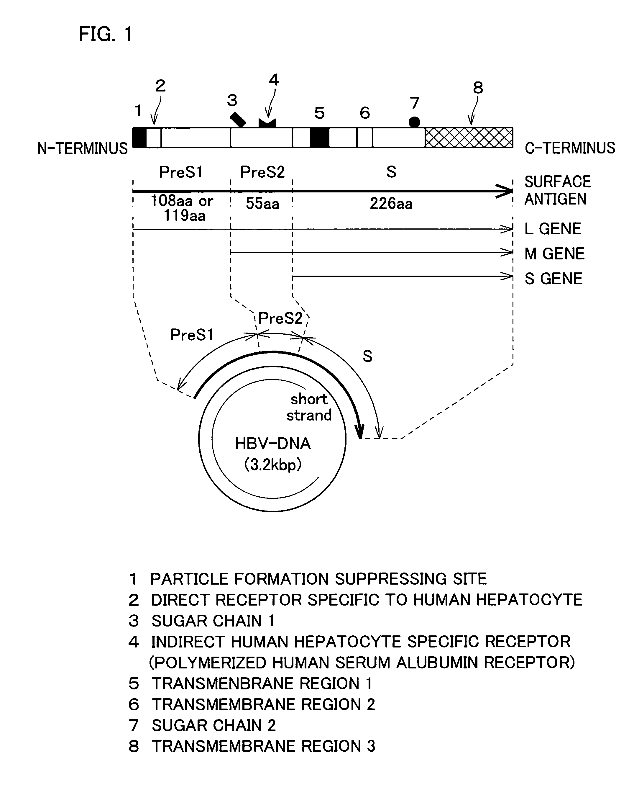
[0085]In the following, HBsAg refers to hepatitis B virus surface antigen. HBsAg is an envelope protein of HBV, and includes three kinds of proteins S, M, and L, as schematically illustrated in FIG. 1. S protein is an important envelope protein common to all three kinds of proteins. M protein includes the entire sequence of the S protein with additional 55 amino acids (pre-S2 peptide) at the N-terminus. L protein contains the entire sequence of the M protein with additional 108 amino acids (serotype y) or 119 amino acids (serotype d) at the N-terminus. In the following Examples, serotype y was used.

[0086]The pre-S regions (pre-S1, pre-S2) of HBV have important roles in the binding of HBV to the hepatocytes. The Pre-S1 region has a direct binding site for the hepatocytes, and the pre-S2 region has a polymeric albumin receptor that binds to the hepatocytes via polymeric albumin in the blood.

[0087]Expression of HBsAg in the eukaryotic cell causes the protein to accumulate as membrane p...

example a

Expression of HBsAg Particles in Recombinant Yeasts

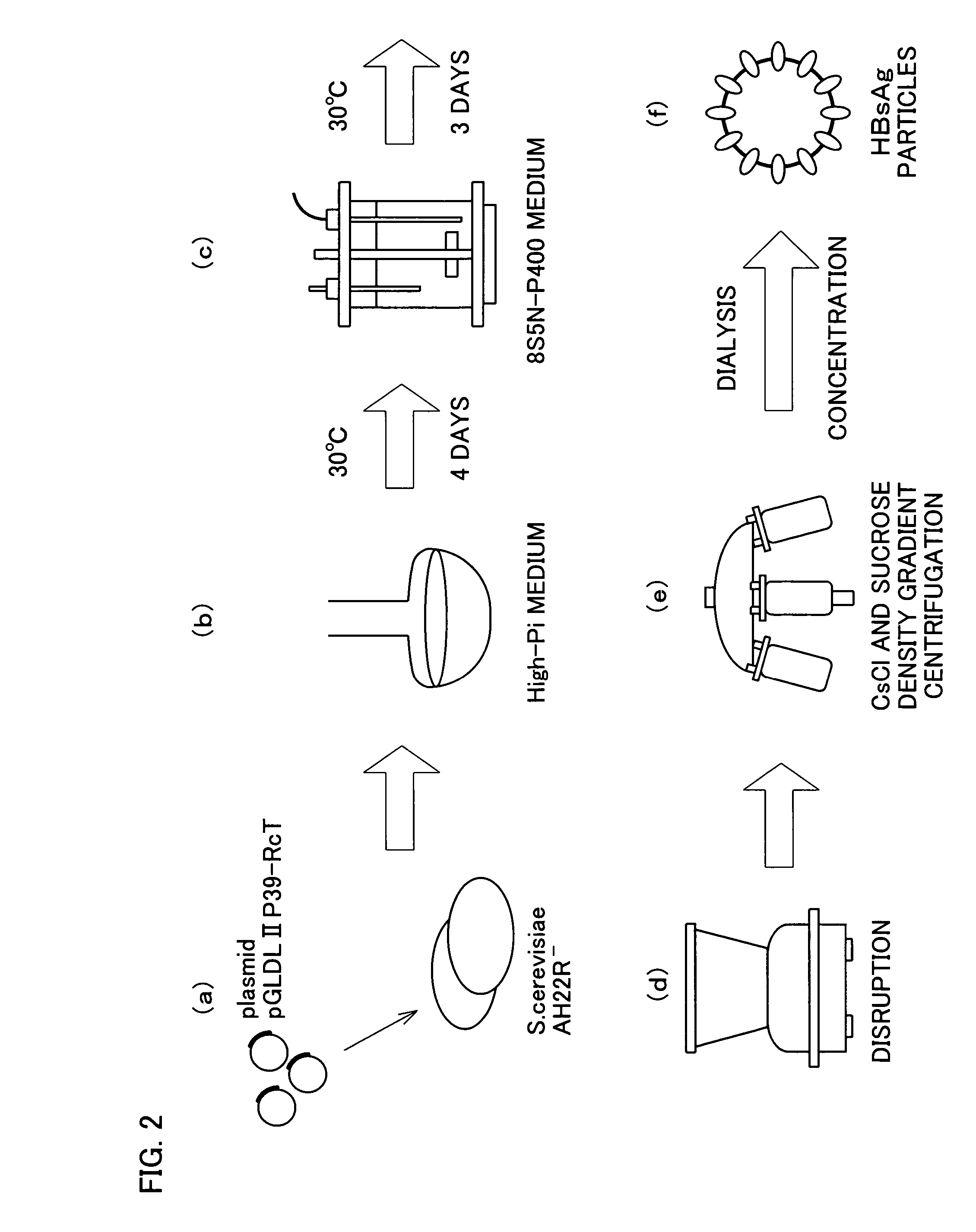
[0089]Recombinant yeasts (Saccharomyces cerevisiae AH22R− strain) carrying (pGLDLIIP39-RcT) were cultured in synthetic media High-Pi and 8S5N-P400, and HBsAg L protein particles were expressed (FIG. 2a through 2c). The whole procedure was performed according to the method described in J. Biol. Chem., Vol. 267, No. 3, 1953-1961, 1992 reported by the inventors of the present invention.

[0090]From the recombinant yeast in stationary growth phase (about 72 hours), the whole cell extract was obtained with the yeast protein extraction reagent (product of Pierce Chemical Co., Ltd.). The sample was then separated by sodium dodecyl sulfate-polyacrylamide gel electrophoresis (SDS-PAGE), and the HBsAg in the sample was identified by silver staining.

[0091]The result showed that HBsAg was a protein with a molecular weight of about 52 kDa.

example b

Purification of HBsAg Particles from the Recombinant Yeasts

[0092](1) The recombinant yeast (wet weight of 26 g) cultured in synthetic medium 8S5N-P400 was suspended in 100 ml of buffer A (7.5 M urea, 0.1 M sodium phosphate, pH 7.2, 15 mM EDTA, 2 mM PMSF, and 0.1% Tween 80), and disrupted with glass beads by using a BEAD-BEATER. The supernatant was collected by centrifugation (FIG. 2d).

[0093](2) The supernatant was mixed with a 0.75 volume of PEG 6000 solution (33%, w / w), and cooled on ice for 30 min. The pellets were collected by centrifugation at 7000 rpm for 30 min, and resuspended in buffer A without Tween 80.

[0094](3) The solution was layered onto a 10-40% CsCl gradient, and ultracentrifuged at 28000 rpm for 16 hours. The centrifuged sample was divided into 12 fractions, and each fraction was tested for the presence of HBsAg by Western blotting (the primary antibody was the anti-HBsAg monoclonal antibody). The HBsAg fractions were dialyzed against buffer A without Tween 80.

[0095...


PUM

PropertyMeasurementUnit
molecular weightaaaaaaaaaa
pHaaaaaaaaaa
pHaaaaaaaaaa

Abstract

The invention provides a therapeutic drug that uses hollow protein nanoparticles displaying an antibody against a specific cell or specific tissue. The effectiveness of the drug has been proved by animal testing. The invention also provides a therapeutic method using such a drug. In a drug according to the present invention, a substance to be transferred into a cell for treating a disease (for example, a cancer treating gene such as a thymidine kinase gene derived from simple herpes virus) is encapsulated in hollow nanoparticles of a particle-forming protein (for example, hepatitis B virus surface-antigen protein that has been modified to lack its infectivity to hepatocytes and display an antibody). The particle surface of the drug displays an antibody, such as a cancer specific antibody, that recognizes an antigen molecule displayed on the surface of a specific cancer cell.

Description

TECHNICAL FIELD[0001]The present invention relates to therapeutic drugs using antibody-displaying hollow protein nanoparticles, and to hollow protein nanoparticles. The invention particularly relates to a drug whose particle surface displays bio-recognizing molecules such as an antibody against a specific cell or tissue, and which contains particles encapsulating a substance to be transferred into a cell for treating a disease, wherein the drug allows the disease-treating substance to be specifically incorporated into a specific cell or tissue. The invention also relates to particles suitable for the drug.BACKGROUND ART[0002]In the field of medicine, there has been active research on drugs that directly and effectively act on the affected area without causing serious side effects. One area of active research is a method known as a drug delivery system (DDS), in which active ingredients of drugs or other substances are specifically delivered to a target cell or tissue, where they can...

Claims


Application Information

Patent Timeline
Patent Type & Authority Patents(United States)
IPC IPC(8): A61K39/385A61K39/29C07K14/00A61K9/50C12N15/09A61K9/51A61K39/245A61K39/395A61K47/48A61K48/00A61P35/00C07K14/02C07K16/18C07K16/28C07K16/40C12N7/04C12N15/88
CPCA61K9/5184A61K47/48869A61K48/0041B82Y5/00C07K14/005C07K16/18C07K16/2863C07K16/40C12N7/00C12N15/88A61K47/48561C12N2810/859A61K2039/55555C07K2317/622C12N2730/10122C12N2730/10123A61K47/6849A61K47/6925A61P35/00A61K39/44A61K39/395
Inventor KURODA, SHUNICHITANIZAWA, KATSUYUKIOKAJIMA, TOSHIHIDEKONDO, AKIHIKOUEDA, MASAKAZUSENO, MASAHARU
Owner BEACLE
Learn More