
Method for the in vitro diagnosis of alzheimer's disease using a monoclonal antibody

a monoclonal antibody and alzheimer's disease technology, applied in the field of neurological disorders, can solve the problems of gradual deterioration of cognitive abilities, changes in personality, behavioral problems, and eventually death, and achieve the effect of facilitating extraction of isoforms and facilitating their subsequent detection, identification and/or quantification

Inactive Publication Date: 2011-04-26
CONSEJO SUPERIOR DE INVESTIGACIONES CIENTIFICAS (CSIC)
View PDF6 Cites 21 Cited by
  • Summary
  • Abstract
  • Description
  • Claims
  • Application Information

AI Technical Summary

Benefits of technology

The invention is a monoclonal antibody that recognizes a specific sequence of β-amyloid peptide and can bind to it regardless of its form (soluble, aggregated, or denatured). The antibody can be used to detect and identify the presence of β-amyloid peptide in brain tissue or biological samples. The invention also includes fragments of the antibody that can also bind to β-amyloid peptide. The antibody can be coupled to a substance that facilitates its detection, such as a second antibody or a magnetic particle. The invention can be used for the diagnosis and monitoring of Alzheimer's disease by detecting the presence of β-amyloid peptide in biological samples.

Problems solved by technology

As the disease progresses, there is gradual deterioration of cognitive abilities, including the ability to take decisions and carry out everyday tasks, and there may be changes in personality, as well as behavioral problems.
In its advanced stages, AD leads to dementia and finally to death.
At present the disease is incurable and constitutes a major cause of mortality.
In view of the risks associated with brain biopsy in live patients, this procedure is very rarely used, and accordingly it is estimated that the error rate in in-vivo diagnosis of this disease is around 20-30%.
There are, nevertheless, some discrepancies between the studies, particularly with respect to the characteristics of the amino end of the peptides that make up the diffuse plaques, and the relative amount of amyloid deposit each of them represents.
However, it is rather unlikely that analysis of the CSF, which involves the use of an invasive technique, lumbar puncture, could be applied for the diagnosis and routine monitoring of patients with Alzheimer's disease in clinical practice.
Moreover, no experimental proof is given that demonstrates its capacity for detecting forms of the amyloid peptide present in biological fluids.

Method used


Image

  • Method for the in vitro diagnosis of alzheimer's disease using a monoclonal antibody
  • Method for the in vitro diagnosis of alzheimer's disease using a monoclonal antibody
  • Method for the in vitro diagnosis of alzheimer's disease using a monoclonal antibody

Examples


example 1

Production of Antibodies

[0058]The polyclonal antibodies EM2 and EM5 used in this study were produced in the manner reported previously [24].

[0059]The following steps were carried out for production of the EM5 monoclonal antibody:

[0060]Production of Hybridomas

[0061]BALB / c mice were injected subcutaneously with 40 μg of peptide Aβ1-4 coupled to KLH (keyhole limpet heamocyanin) and dissolved in phosphate-buffered saline (PBS) and emulsified with an equal volume of Freund complete adjuvant. The mice received three injections repeated every other week, in Freund incomplete adjuvant. Three days before fusion, the mice received an intraperitoneal injection of 25 μg of Aβ1-40-KLH in PBS. On the day of fusion, spleen cells from animals immunized with the mouse myeloma line P3 / X63-Ag.653 were fused using polyethylene glycol 1400 (Sigma), following established procedures [25]. The fused cells were distributed in sterile 96-well plates at a density of 105 cells per well and were selected in med...

example 2

Calculation of the Apparent Dissociation Constant: Affinity for Peptides Aβ1-40 and Aβ1-42

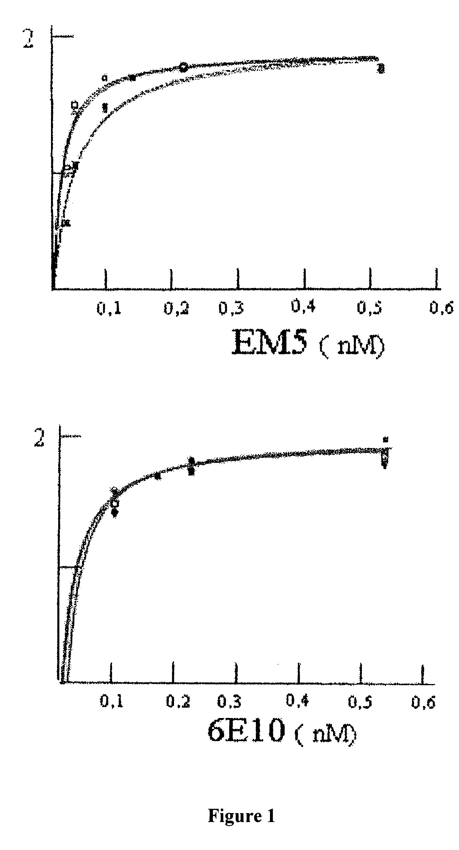
[0066]ELISA was used for investigating the affinity of EM5 and 6E10 monoclonal antibodies using recently dissolved, immobilized peptides Aβ1-40 and Aβ1-42, which had been synthesized in the WM Keck plant of Yale University using the N-t-butyloxycarbonyl methodology.

[0067]Polystyrene microtitre plates (Immulon 2, Dynex Technology Inc., Chantilly, Va.) were covered for 16 hours at 4° C. with 0.5 μg of peptide Aβ1-40 or Aβ1-42 recently dissolved or aggregated in carbonate / bicarbonate buffer pH 9.6. After blocking with Superblock (Pierce Chemical Co.), increasing concentrations of purified EM5 (0-0.5 nM in TBS-T, 100 microliters per well) were added to the wells covered with Aβ, incubating for 3 hours at 37° C. The bound EM5 was detected with the F(ab′)2 fragment of goat anti-mouse IgG conjugated with horseradish peroxidase (1:3000, Amersham). The reaction was developed for 15 minutes with 3,3′,5,...

example 3

Immunotransfer Analysis

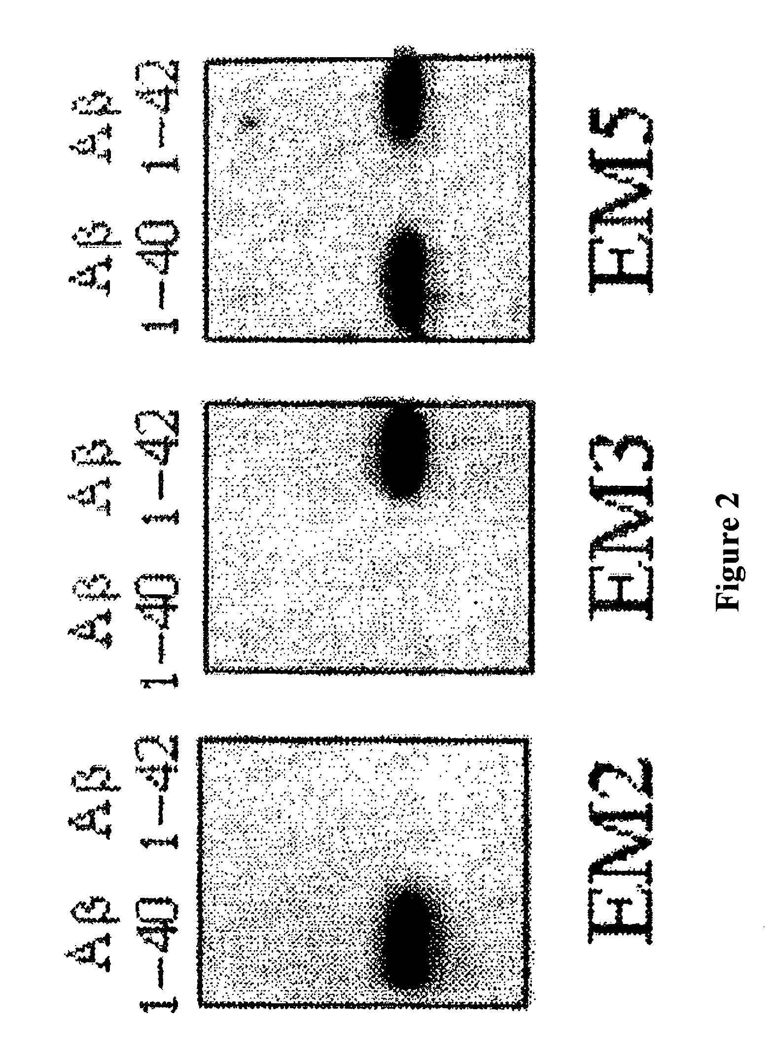
[0069]The specific recognition of Aβ1-40 and Aβ1-42 by EM5 was investigated and compared with EM2 and EM3 polygonal antibodies by means of immunotransfer analysis. For this, Aβ peptides (0.5 μg / lane) were submitted to PAGE electrophoresis in 16% acrylamide, tris-tricine-SDS. The peptides were transferred electrophoretically for 1 hour at 400 mA and 4° C. to poly(vinylidene fluoride) membranes (Immobilon-P, Millipore) using 3-cyclohexylamino-1-propanesulfonic acid, pH 11, containing 10% methanol. The membranes were blocked for 16 h at 4° C. with TBS-T containing 5% powdered skimmed milk and were then incubated for 1 hour at room temperature with 2 μg / ml of IgGs EM2, EM3 or EM5. A goat anti-rabbit IgG (EM2 and EM3) or goat anti-mouse IgG (EM5) coupled to horseradish peroxidase (Amersham) and diluted 1:2000 was used as the second antibody. Immunotransfer was visualized by means of chemoluminescence (Amersham) following the manufacturer's specifications.

[0070]The ...


PUM

PropertyMeasurementUnit
pHaaaaaaaaaa
pHaaaaaaaaaa
pHaaaaaaaaaa

Abstract

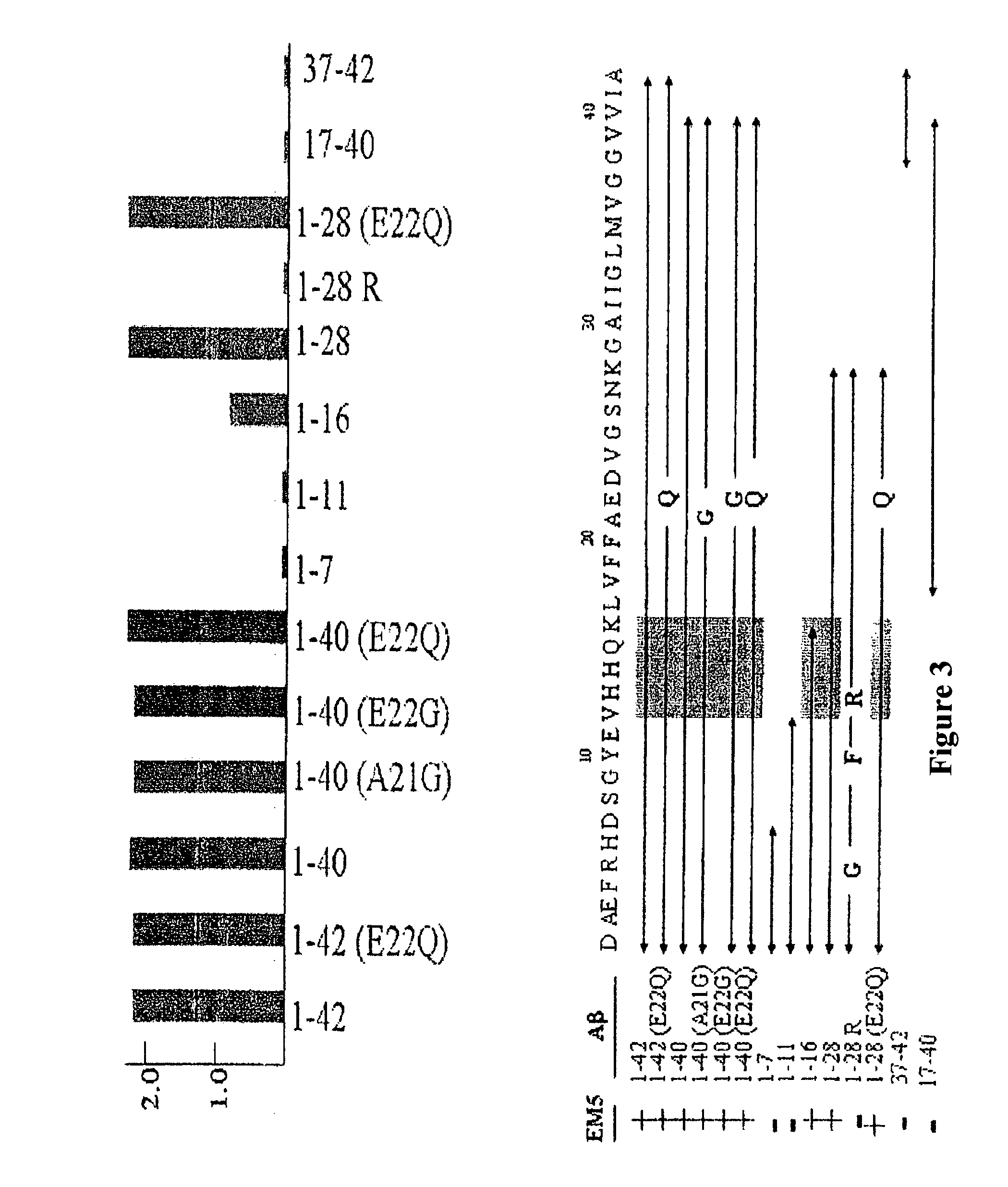
The invention relates to a method for the in vitro diagnosis of Alzheimer's disease using a monoclonal antibody. Said antibody can bind at least to amino acids 12-16 of the β-amyloid peptide, specifically detecting the neuritic plaques which are characteristic of Alzheimer's disease, without detecting diffuse plaques which are not defining characteristics of the disease. Within the neuritic plaques, the monoclonal antibody can detect a different sub-group in the composition of the different deposited isoforms of the β-amyloid peptide, which is associated with the disease progression stage. In addition, the antibody can bind to isoforms of the β-amyloid peptide in biological fluids such as urine. As a result, the inventive monoclonal antibody, the cell lines that produce said antibody and compositions containing same can be used in the in vitro diagnosis of Alzheimer's disease and in determining the disease progression stage.

Description

FIELD OF THE INVENTION[0001]The present invention relates to the diagnosis of neurological disorders, especially to the diagnosis of Alzheimer's disease by means of an antibody that is able to interact with specific peptides associated with said disease.BACKGROUND OF THE INVENTION[0002]Alzheimer's disease (AD) is a neurological disorder that causes the death of neurons in the brain. Generally it progresses slowly, commencing after age 50 years, and its first symptoms may be attributed to old age or to ordinary forgetfulness. As the disease progresses, there is gradual deterioration of cognitive abilities, including the ability to take decisions and carry out everyday tasks, and there may be changes in personality, as well as behavioral problems. In its advanced stages, AD leads to dementia and finally to death. At present the disease is incurable and constitutes a major cause of mortality.[0003]At the present time, Alzheimer's disease is normally diagnosed from the clinical picture,...

Claims


Application Information

Patent Timeline
Patent Type & Authority Patents(United States)
IPC IPC(8): G01N33/53G01N33/563C12P21/08
CPCC07K14/4711C07K16/18G01N33/6896G01N2800/2821C07K2317/92C07K2317/34A61P25/28C07K7/06G01N33/53
Inventor MENDEZ, ENRIQUE
Owner CONSEJO SUPERIOR DE INVESTIGACIONES CIENTIFICAS (CSIC)
Learn More