
Plastic cantilevers for force microscopy

a technology of force microscopy and cantilever, which is applied in the direction of measurement devices, instruments, nanotechnology, etc., can solve the problems of affecting the cost of replacement of cantilever tips can be a barrier, and the cantilever beam and the attached probe tips are subject to wear and tear during use, etc., to achieve the effect of improving the quality of sfm, improving the reliability of sfm, and being more sensitive to for

Active Publication Date: 2010-04-06
WISCONSIN ALUMNI RES FOUND
View PDF22 Cites 1 Cited by
  • Summary
  • Abstract
  • Description
  • Claims
  • Application Information

AI Technical Summary

Benefits of technology

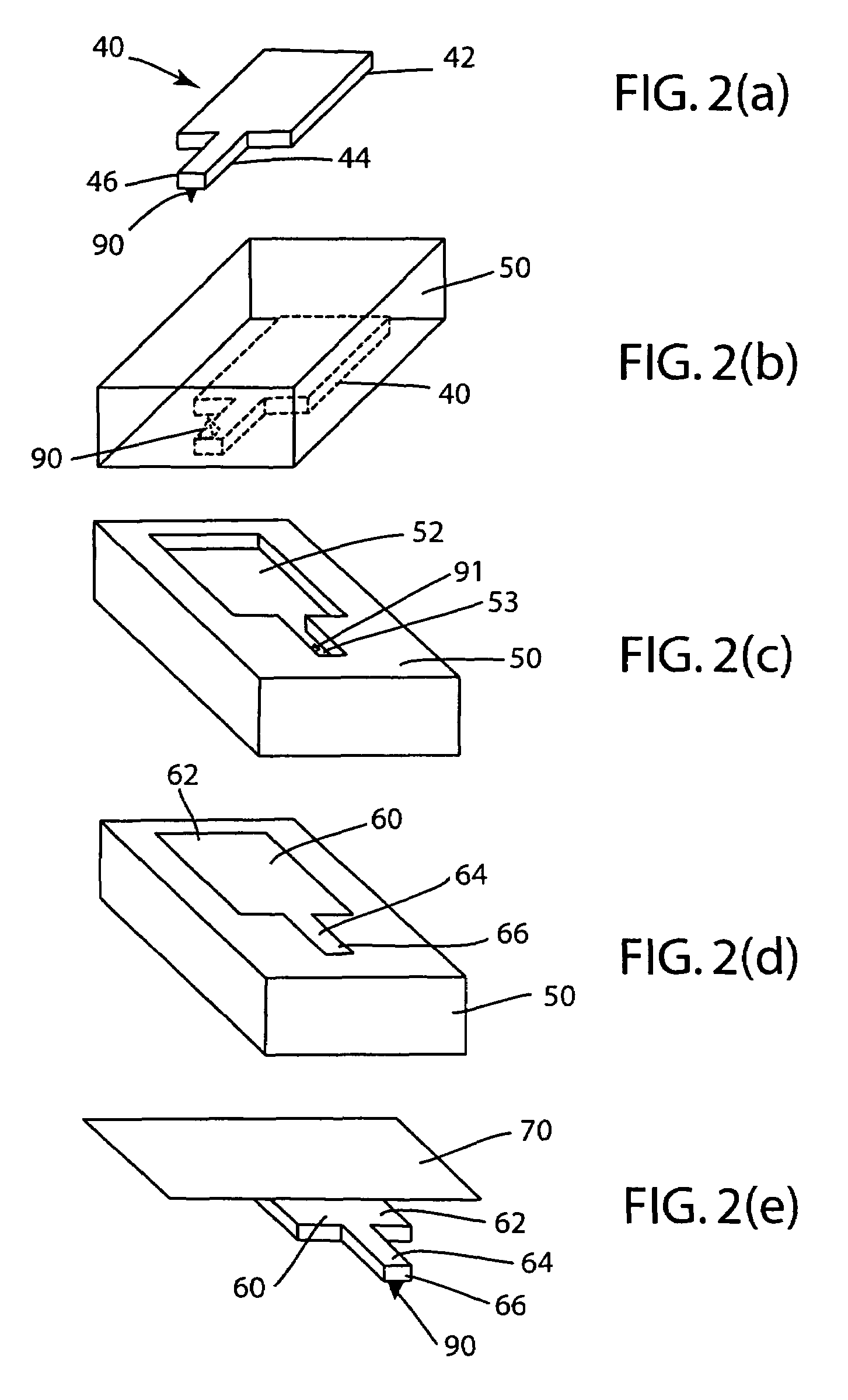
The invention is a method for making plastic cantilevers with probe tips, which can be used in scanning force microscopy or magnetic force microscopy. The method involves covering a master cantilever with a mold material, removing the master cantilever from the mold material to reveal a mold cavity, and then filling the mold cavity with plastic material to form a plastic cantilever with the probe tip. The plastic cantilever can be coated with a reflective metal, such as gold, and can be combined with magnetic or electronically conductive materials for various applications. The method is cost-effective and efficient, with cycle times on the order of 4 hours. The plastic cantilevers are compliant and can detect soft materials with low noise. The invention also provides a way to dispose of replacement cantilevers, making it easier to use for medical or biological applications.

Problems solved by technology

The cantilever beams and attached probe tips used in SFM are subject to wear and tear during use, especially in contact or intermittent contact modes where the probe tip repeatedly contacts the sample.
These types of processes are relatively expensive, so that commercially available replacement tips typically cost at least $100 for a single replacement cantilever tip.
Consequently, the cost of replacement cantilever tips can be a barrier, especially in educational or research settings.
These materials are relatively brittle and inflexible, making them relatively susceptible to damage during use compared to other materials, such as plastics, which are more pliable.
The electrical properties of these materials can also cause problems in some applications, compared to other materials, such as plastics, which are electromagnetically insulating.
Although there have been a few reports of the use of plastic probes for scanning force microscopy, replacement plastic cantilevers are not widely available commercially.

Method used


Image

  • Plastic cantilevers for force microscopy
  • Plastic cantilevers for force microscopy
  • Plastic cantilevers for force microscopy

Examples


Embodiment Construction

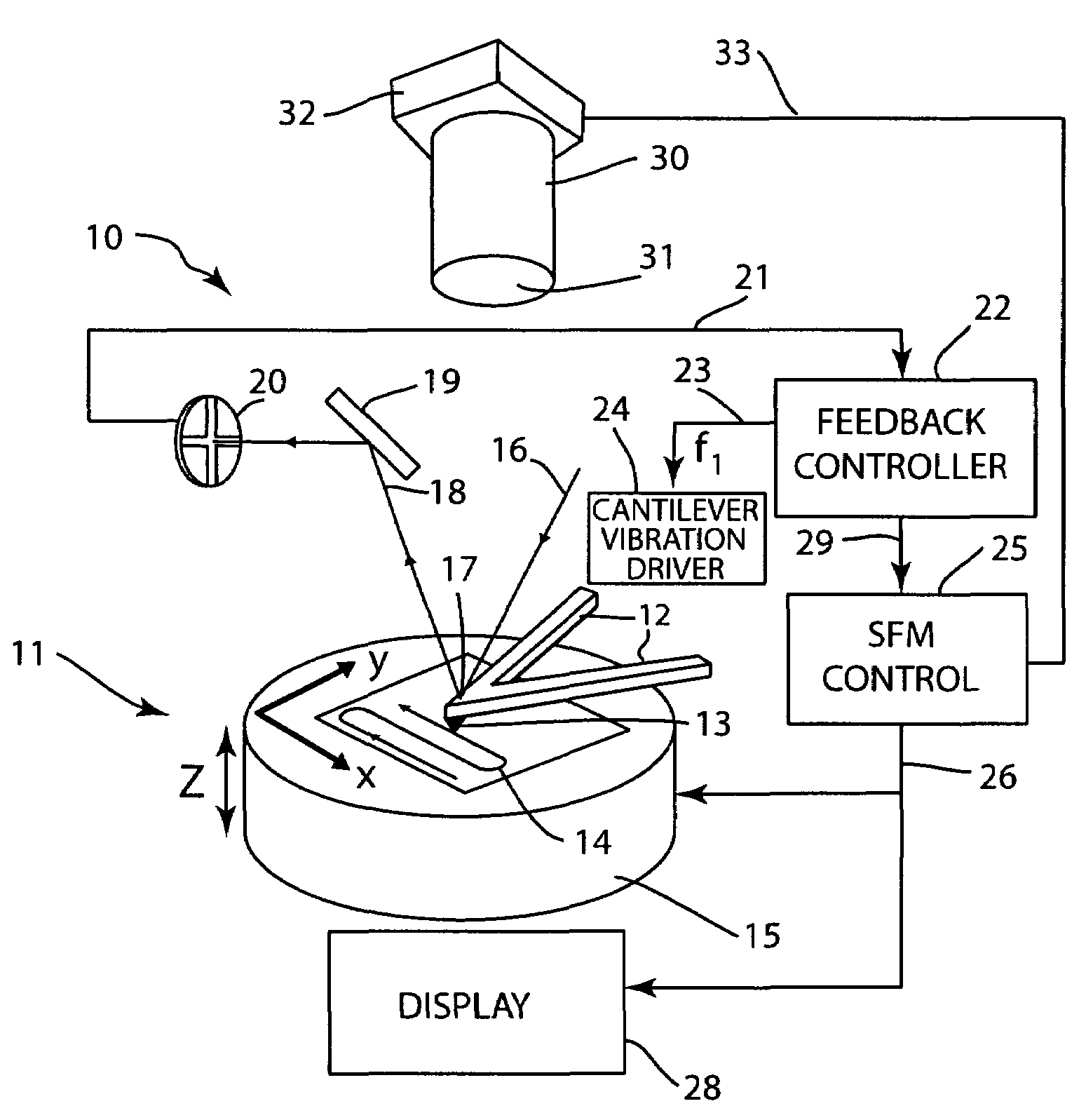
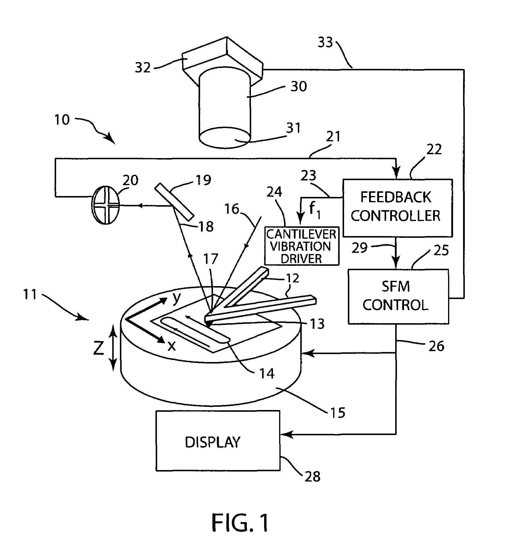
[0029]Referring to the figures, FIG. 1 is a simplified view of an exemplary scanning force microscope (“SFM”) system, shown generally at 10, which includes a scanning force microscope, shown generally at 11, having a cantilever beam 12 according to the invention that includes a scanning tip 13. The cantilever beam 12 supports the scanning tip 13 over a sample 14 supported on a scanner stage 15 that can be operated to translate the sample 14 in X, Y and Z directions, as illustrated in FIG. 1, with the Z direction being in a direction toward or away from the tip 13. Such an exemplary scanning force microscope system is described, for example, in U.S. Patent Application Publication No. 2004 / 0182140, the contents of which are incorporated by reference.

[0030]In the exemplary scanning force microscope system 10, the position and movement of the tip 13 and cantilever beam 12 can be monitored, for example, by reflecting a laser beam 16 off of the back surface 17 of the cantilever 12 and / or ...


PUM

PropertyMeasurementUnit
temperatureaaaaaaaaaa
diameteraaaaaaaaaa

Abstract

A method disclosed for producing polymer-based cantilevers for use in atomic force microscopy in a batch process. The method includes forming a mold in a mold material, for example PDMS, using a master cantilever, removing the master cantilever from the mold material to reveal a mold cavity, filling the mold cavity with plastic, for example polystyrene, to form a plastic cantilever in the mold, and removing the plastic cantilever from the mold, for example using adhesive tape or flexing the mold. At least one surface of the plastic cantilever can be coated with a reflective metal, such as gold. The plastic cantilever can be functionalized for use in magnetic force microscopy by attaching a probe tip formed of magnetic metal, for example a 10 μm nickel sphere.

Description

REFERENCE TO GOVERNMENT RIGHTS[0001]This invention was made with United States government support awarded by the following agencies: USAF / AFOSR F49620-03-1-0420. The United States has certain rights in this invention.FIELD OF THE INVENTION[0002]The present invention relates generally to a device and method for producing cantilevers for atomic force microscopy and, more particularly, to a device and method for producing polymer-based cantilevers in a batch process.BACKGROUND OF THE INVENTION[0003]The advent of scanning force microscopy (SFM), also known as atomic force microscopy (AFM), has brought an instrument capable of microscopic surface studies with atomic resolution, suited for ambient and liquid environments and a wide variety of samples. SFM is a method for observing nanoscale topography and other properties of a surface. In general, SFM scans a force sensor over a surface.[0004]SFM can be carried out in contact and non-contact modes. In a contact mode of operation, a topogr...

Claims


Application Information

Patent Timeline
Patent Type & Authority Patents(United States)
IPC IPC(8): B29C67/00
CPCB29C33/3878B29C37/0003G01Q70/14G01Q60/54B82Y35/00
Inventor VAN DER WEIDE, DANIEL W.PAULSON, CHARLES A.STEVENS, ANDREW G.
Owner WISCONSIN ALUMNI RES FOUND
Learn More