
Rapid tissue processing method and apparatus

a tissue and tissue technology, applied in the field of tissue processing, can solve the problems of insufficient impregnation, potential misdiagnosis, adverse impact, etc., and achieve the effects of rapid tissue processing, rapid tissue processing, and easy resistance to electricity flow

Inactive Publication Date: 2007-09-25
BIRKNER JEFFREY C
View PDF20 Cites 29 Cited by
  • Summary
  • Abstract
  • Description
  • Claims
  • Application Information

AI Technical Summary

Benefits of technology

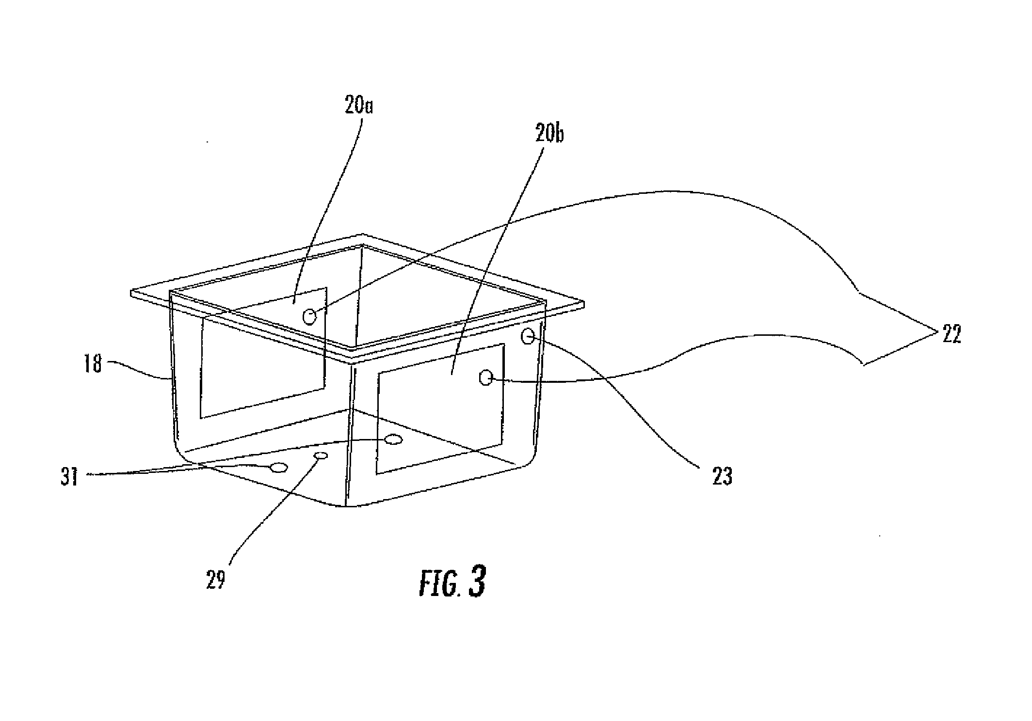
[0024]The present invention is a method for the rapid processing of a tissue sample. The process includes the four general steps of fixation, dehydration, clearing, and impregnation (infiltration). In the preferred embodiment, more than one of the steps can be performed simultaneously using a single solution. After collecting the tissue sample, the first step of the processing method is to enclose the tissue sample in a cassette which is then placed in a cassette rack, as is known in the art. The rack is placed in a processing container with conductive leads on the container walls. The container is placed in the processing unit and connected to the electrical power source. The rack is oriented in such a manner as to expose the openings in the cassettes with the flow of electrical current between the conductive leads. The container is filled with the proper processing solution or material into which is added a conductive agent to facilitate the resistive flow of electricity through the tissue specimen. It has been determined that a conductive polymer acts as the best conductive agent. During the fixation, dehydration, clearing, and impregnation steps, the present invention uses a combination of ohmic and microwave energy to heat the sample to achieve rapid tissue processing and thereby reduce the time the tissue is exposed to heat.

Problems solved by technology

A failure to preserve the micro-anatomical structure of the cellular specimen may adversely impact the analysis and lead to the potential for misdiagnosis.
Failure to dehydrate the specimen can lead to inadequate impregnation, poor ribbon formation during sectioning, clefts in tissue sections, dissociation of structures, water crystals in tissue sections, and poor staining.
If left in place, the fatty molecules would impair clearing and impregnation.
Specifically, inadequate fat removal can result in spreading artifacts of tissue sections, wrinkling of tissue sections, and poor staining.
Many of these prior art methods are undesirable for a number of reasons.
Such degradation makes analysis and diagnosis difficult or even impossible, and increases the risks of a misdiagnosis.
Conventional methods may or may not include the addition of heat energy; however, any heat used is not produced by microwave energy.
The problem with conventional (non-microwave) methods is three fold.
First, conventional methods have traditionally used hazardous chemicals that are known carcinogens to effectuate the chemical reactions during processing.
Second, all tissue types are lumped together during tissue processing so that both “bound water” and “free water” molecules are removed from the tissue sample during processing, which damages the tissue sample.
Removal of bound water causes excessive hardening and shrinkage of the tissue sample.
Fibrous tissue becomes tough and specimens comprised mostly of cells become crumbly.
In conventional tissue processing, in the removal of free water from the larger, dense segments of the tissue specimen, bound water is also inadvertently removed from smaller, less dense segments, thus degrading those segments of the specimen.
Third, for conventional processing methods processing times are usually in excess of 10 hours which means patients often have to wait until the following day for the tissue processing, analysis, and diagnosis.
This relatively long time period can be detrimental and even life threatening for patients in situations where tissue analysis is urgently required.
Because of this extended length of time, conventional tissue processing techniques may not be used for surgical analysis.
One problem with prior art microwave methods is that the heating of the tissue sample lacks uniformity.
The temperature within the hot spots can cause tissue destruction.
Within the cold spots, the temperature may be inadequate to trigger or to complete the required chemical reactions.
The result of the hot and cold spots is a lack of consistent processing of all of the cellular tissue comprising the specimen.
Such a lack of consistency may result in poor or improper staining or analysis.
The resulting potential for misdiagnosis may be dangerous and even life threatening for patients.
Another problem with microwave methods is that microwave energy may affect the bound water that physically separates macromolecules.
Although microwaves cannot ionize molecules and are too small to break molecular bonds, use of excessive microwave energy can redistribute the hydrogen bonds (chemical energy).
Excessive microwave exposure also causes thinning of the bound water layers.
This allows bound water layers to unwind and form new crosslinks, resulting in the formation of different configurations with a resultant potential for misdiagnosis.
Another problem with microwave energy in tissue processing is that microwave energy is only capable of penetrating the tissue to only a limited depth.
Thin samples are more difficult to handle and they can also be more difficult to obtain.
Attempts to overcome this limitation by increasing the amount of microwave energy to penetrate more deeply into the sample can damage the cell structure, DNA, and RNA, as discussed above.
Yet another problem with microwave energy in tissue processing is that microwave energy only affects molecules with an electric dipole.
The different concentrations of free and bound water within the specimen means that some water molecules in the tissue will be heated more than other water molecules in the tissue, thus contributing to the inconsistent heating.
Over heating of this bound water can lead to tissue distortion causing a “cooked” tissue sample.
This potential for tissue distortion after dehydration is exacerbated by the fact that most traditional processing chemicals are nonpolar.
Thus, excessive microwave energy is required to build up enough heat to bring about the desired chemical reactions.
These solvents are harsh in that they can damage the tissue sample.
This extended processing requirement has a tendency to decrease porosity, inhibiting the removal of lipids.
These traditional solvents also present a risk to the laboratory personnel.

Method used


Image

  • Rapid tissue processing method and apparatus
  • Rapid tissue processing method and apparatus
  • Rapid tissue processing method and apparatus

Examples


Embodiment Construction

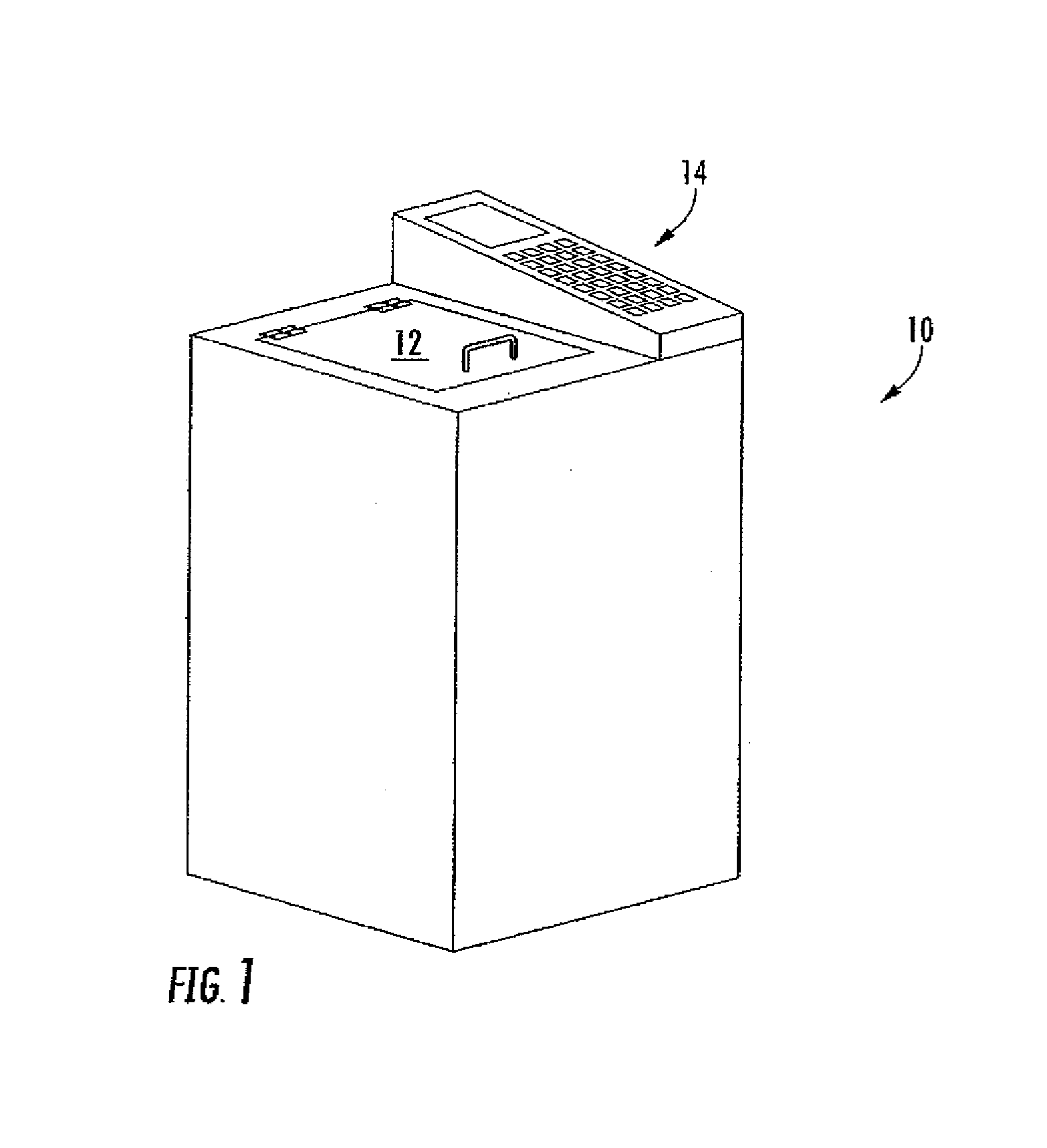
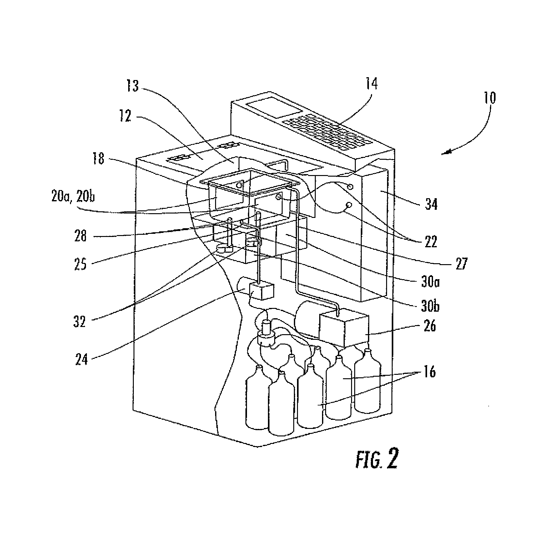
[0036]The present invention is a method for the rapid processing of a tissue sample. The steps of fixation, dehydration, clearing, and impregnation (sometimes herein referred to as infiltration) can be rapidly performed in a period of time ranging from about ten minutes to about two hours, based on the tissue type and size, and, further, depending on the selected processing protocol. FIG. 1 shows the tissue processing unit 10 having a control panel 14 and a loading door 12. The processing unit 10 can be of any size and shape so long as it contains all relevant components to process the tissue sample. The loading door 12 can be of any type so long as it provides access to the processing chamber 13. The control panel 14 comprises various controls for adjusting the temperature, run time, run cycle, and the like. Preferably, the control panel 14 also comprises a screen for displaying a variety of information, such as the temperature of processing solution inside the chamber 13 and the t...


PUM


Abstract

A rapid tissue processing method and apparatus including the steps of fixation, dehydration, clearing, and impregnation. The preferred method is performed in a processing unit that employs alternating microwave and ohmic energy to heat the process solution and tissue sample. In one embodiment, the method employs environmentally friendly green chemicals to effectuate the processing.

Description

BACKGROUND OF THE INVENTION[0001]The present invention relates to tissue processing, and more particularly it relates to a rapid tissue processing method and apparatus that uses ohmic energy and microwave energy to heat the process solution and tissue sample during processing. The present invention also relates to a tissue processing method that employs the use of environmentally friendly green chemicals.[0002]A tissue sample must be processed before it can be analyzed for diagnostic or testing purposes. This processing acts to halt the degradation of cellular structure, and to stabilize the cellular characteristics, and to sufficiently harden the tissue so that extremely fine segments may be cut therefrom for purposes of analysis. Tissue processing is well known in the art of histology.[0003]There are four general steps performed when processing a tissue sample: fixation, dehydration, clearing, and impregnation (sometimes herein also referred to as infiltration). These steps are ge...

Claims


Application Information

Patent Timeline
IPC IPC(8): G01N1/30G01N33/48
CPCG01N1/30G01N1/44G01N2001/315
Inventor BIRKNER, JEFFREY C.WOLTMAN, MICHAEL L.
Owner BIRKNER JEFFREY C
Learn More