
Therapeutic delivery systems

a technology of therapeutic compound and delivery system, which is applied in the field of therapeutic compound delivery system, can solve the problems of x-rays, which are known to be somewhat dangerous, prohibit portable examination, and large shear size, and achieve the effect of enhancing cellular uptake of therapeutic compound and increasing membrane fluidity

Inactive Publication Date: 2006-08-01
BRISTOL MYERS SQUIBB MEDICAL IMAGING
View PDF439 Cites 110 Cited by
  • Summary
  • Abstract
  • Description
  • Claims
  • Application Information

AI Technical Summary

Benefits of technology

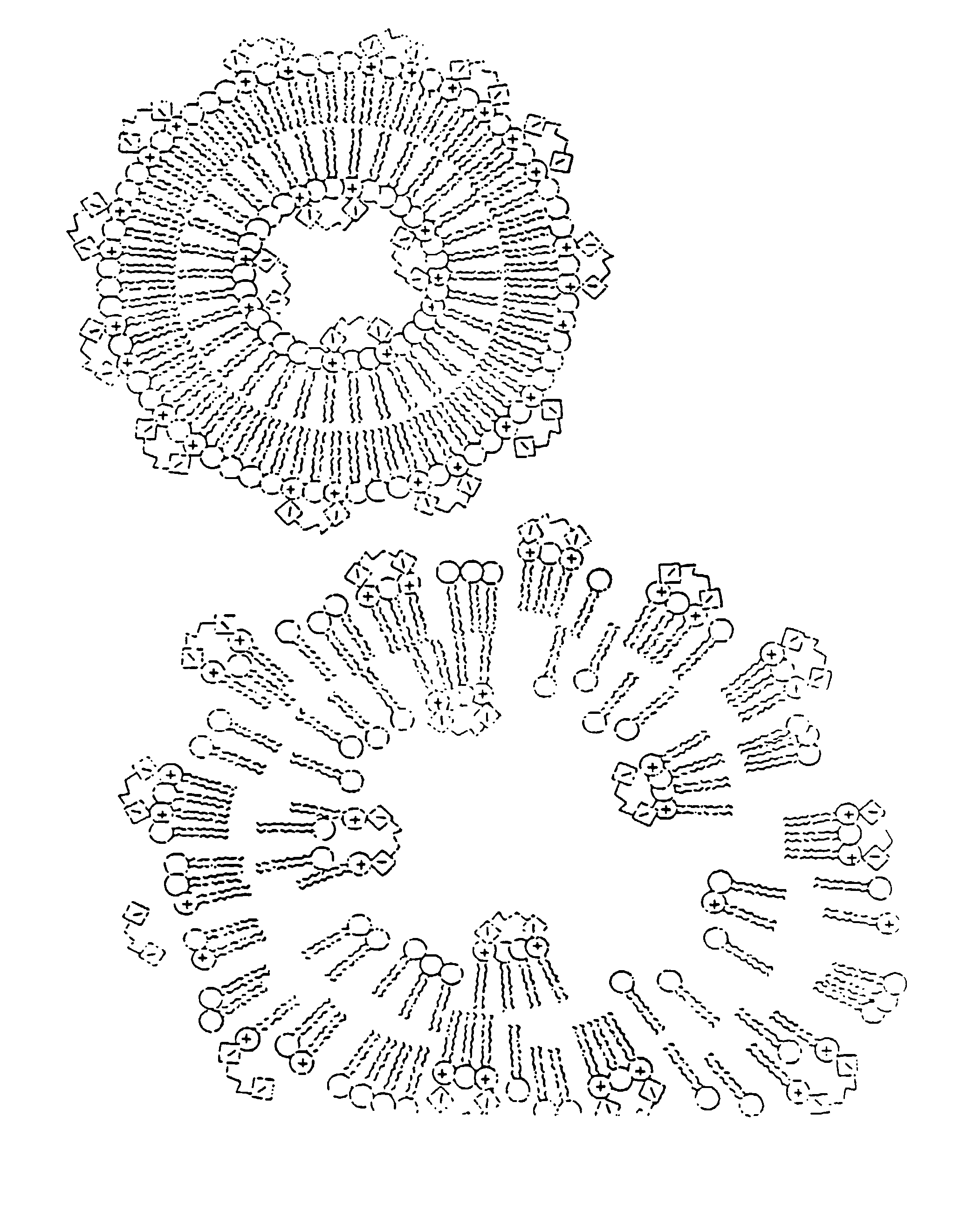
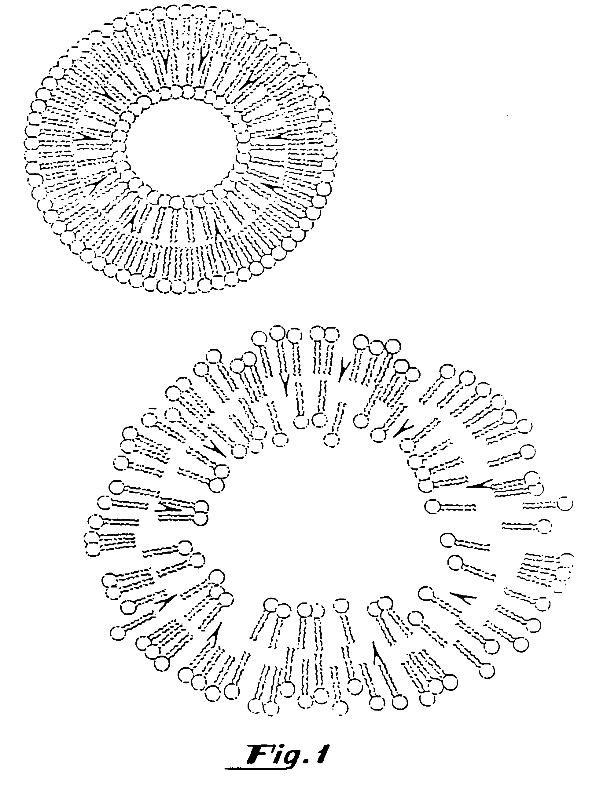
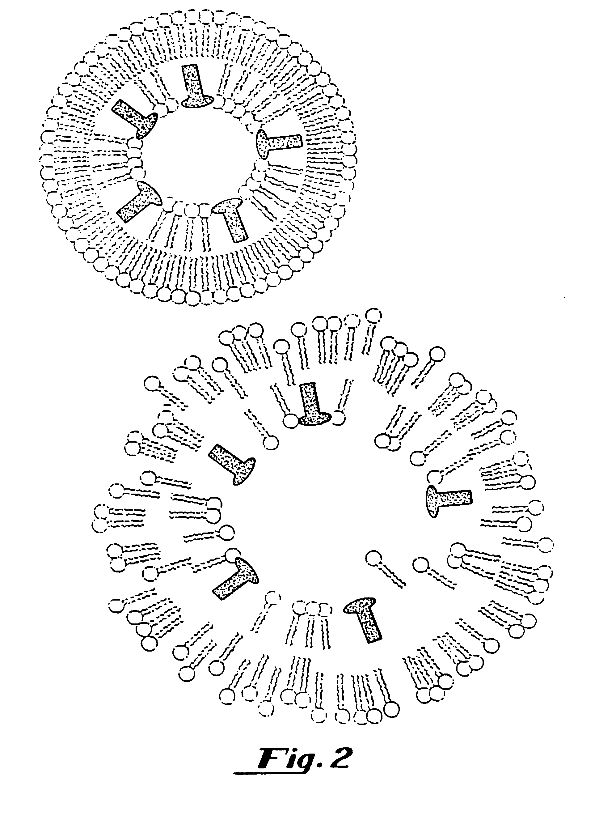
"The present invention provides therapeutic delivery systems using gas-filled microspheres that can be targeted to specific tissues in the body. The microspheres contain a temperature activated gaseous precursor which becomes a gas upon activation at a selected temperature. The microspheres can be ruptured and release the therapeutic compound using sonic energy, microwave energy, magnetic energy, or hyperthermia. The invention also contemplates methods for controlled delivery of therapeutic compounds to a region of a patient using temperature activated gaseous precursor-filled microspheres. The invention provides a simple and cost-effective method for preparing temperature activated gaseous precursor-filled microspheres. The temperature activated gaseous precursor-filled liposomes made according to the present invention can encapsulate lipophilic drugs and can be easily incorporated into the lipid layer. The invention also includes the advantage of enhancing cellular uptake of the therapeutic compound through the capture of ultrasonic energy by the gaseous precursor in the microspheres."

Problems solved by technology

X-rays, however, are known to be somewhat dangerous, since the radiation employed in X-rays is ionizing, and the various deleterious effects of ionizing radiation are cumulative.
This technique, however, has various drawbacks, such as expense and shear size of an MRI scanner rendering it stationary which prohibits portable examination.
In addition, MRI is not available at many medical centers.
Nuclear medicine techniques, however, suffer from poor spatial resolution and expose the animal or patient to the deleterious effects of radiation.
Furthermore, the handling and disposal of radionuclides is problematic.
However, despite these various technological improvements, ultrasound is still an imperfect tool in a number of respects, particularly with regard to the imaging and detection of disease in the liver and spleen, kidneys, heart and vasculature, including measuring blood flow.
The methods and materials in the prior art for introduction of genetic materials to, for example, living cells is limited and ineffective.
These methods have all been relatively ineffective in vivo and only of limited use for cell culture transfection.
None of these methods potentiate local release, delivery and integration of genetic material to the target cell.
Great strides have been made in characterizing genetic diseases and in understanding protein transcription but relatively little progress has been made in delivering genetic material to cells for treatment of human and animal disease.
A principal difficulty has been to deliver the genetic material from the extracellular space to the intracellular space or even to effectively localize genetic material at the surface of selected cell membranes.
Whole virus has been used but the amount of genetic material that can be placed inside of the viral capsule is limited and there is concern about possible dangerous interactions that might be caused by live virus.
Despite extensive work on viral vectors, it has been difficult to develop a successfully targeted viral mediated vector for delivery of genetic material in vivo.
Conventional, liquid-containing liposomes have been used to deliver genetic material to cells in cell culture but have generally been ineffective in vivo for cellular delivery of genetic material.
For example, cationic liposome transfection techniques have not worked effectively in vivo.
Despite the advances that have been made, the prior art has still not solved many of the problems inherent in the development of ultrasound contrast agents.
Gases may diffuse out of stabilizing emulsions or particle coatings and the efficacy of the product may be lost.
The large size of these particles makes it very difficult to exclude potential contaminants from the injection such as bacteria passing into the patient during the injection.
Gas-containing microspheres currently under development are generally unstable in vivo and do not persist long enough to provide ideal contrast enhancement.

Method used


Image

  • Therapeutic delivery systems
  • Therapeutic delivery systems
  • Therapeutic delivery systems

Examples


example 1

[0291]The methods described below demonstrate that a therapeutic such as DNA can be entrapped in gas-filled microspheres and that ultrasound can be used to release a therapeutic from a gas-filled microsphere. As shown below, liposomes entrapping water and DNA failed to release the genetic material after exposure to the same amount of ultrasonic energy. The presence of the gas within the microspheres results in much more efficient capture of the ultrasonic energy so it can be utilized for delivery of a therapeutic such as genetic material.

[0292]Gas-filled liposomes were synthesized as follows: Pure dipalmitoylphosphatidylcholine (DPPC), Avanti Polar Lipids, Alabaster, Ala., was suspended in normal saline and then Extruded five times through 2 micron polycarbonate filters (Nuclepore, Costar, Pleasanton, Calif.) using an Extruder Device (Lipex Biomembranes, Vancouver, Canada) at 800 p.s.i. The resulting liposomes were then dried under reduced pressure as described in U.S. Ser. No. 716,...

example 2

[0312]Binding of DNA by liposomes containing phosphatidic acid and gaseous precursor and gas containing liposomes. A 7 mM solution of distearoyl-sn-glycerophospate (DSPA) (Avanti Polar Lipids, Alabaster, Ala.) was suspended in normal saline and vortexed at 50° C. The material was allowed to cool to room temperature. 40 micrograms of pBR322 plasmid DNA (International Biotechnologies Inc., New Haven, Conn.) was added to the lipid solution and shaken gently. The solution was centrifuged for 10 minutes in a Beckman TJ-6 Centrifuge (Beckman, Fullerton, Calif.). The supernatant and the precipitate were assayed for DNA content using a Hoefer TKO-100 DNA Fluorometer (Hoefer, San Francisco, Calif.). This method only detects double stranded DNA as it uses an intercalating dye, Hoechst 33258 which is DNA specific. It was found that the negatively charged liposomes prepared with phosphatidic acid surprisingly bound the DNA. The above was repeated using neutral liposomes composed of DPPC as a co...

example 3

[0313]A cationic lipid, such as DOTMA is mixed as a 1:3 molar ratio with DPPC. The mixed material is dissolved in chloroform and the chloroform is removed by rotary evaporation. Water is added to the dried material and this mixture is then extruded through a 2 μm filter using an Extruder Device (Lipex Biomembranes, Inc., Vancouver, BC). Then positively charged gaseous precursor-filled liposomes are prepared according to the procedure provided in U.S. Ser. No. 717,084, filed Jun. 18, 1991, which is hereby incorporated by reference in its entirety.

[0314]The resulting dried, positively charged gaseous precursor-filled liposomes are rehydrated by adding PBS, saline or other appropriate buffered solution (such as HEPES buffer); a vortexer may be used to insure homogenous mixing. DNA is added and the mixture is again shaken. Since the DNA is attached to the surface of the cationic gaseous precursor-filled liposomes, unattached DNA may be removed with filtering or selective chromatography....


PUM


Abstract

Targeted therapeutic delivery systems comprising gas- or gaseous precursor-filled lipid microspheres comprising a therapeutic are described. Methods for employing such microspheres in therapeutic delivery applications are also provided.Targeted therapeutic delivery systems comprising gas- or gaseous precursor-filled liposomes having a drug encapsulated therein are preferred. Methods of and apparatus for preparing such liposomes and methods for employing such liposomes in therapeutic delivery applications are also disclosed.

Description

CROSS REFERENCE TO RELATED APPLICATIONS[0001]This application is a divisional of U.S. application Ser. No. 08 / 485,998, filed Jun. 7, 1995, now U.S. Pat. No. 6,443,898, which in turn is a divisional of U.S. application Ser. No. 08 / 160,232, filed Nov. 30, 1993, now U.S. Pat. No. 5,542,935. This application is also a continuation-in-part of U.S. application Ser. No. 08 / 159,687, filed Nov. 30, 1993, now U.S. Pat. No. 5,585,112, and U.S. application Ser. No. 08 / 159,674, filed Nov. 30, 1993, now abandoned. The disclosures of these related applications are hereby incorporated herein by reference in their entirety.BACKGROUND OF THE INVENTION[0002]1. Field of the Invention[0003]This invention relates to therapeutic delivery systems, and more specifically, to gaseous precursor-containing microspheres comprising a therapeutic compound. The invention further relates to methods for employing such microspheres as therapeutic delivery systems.[0004]2. Background of the Invention[0005]Targeted ther...

Claims


Application Information

Patent Timeline
Patent Type & Authority Patents(United States)
IPC IPC(8): A61B8/00A61K9/127A61K41/00A61K47/48A61K49/18A61K49/22A61M5/31
CPCA61K9/127A61K9/1277A61K9/1278A61K41/0028A61K47/48869A61K49/223A61K49/227A61K41/0052A61M5/3145A61K47/6925A61P43/00
Inventor UNGER, EVAN C.FRITZ, THOMAS A.MATSUNAGA, TERRYRAMASWAMI, VARADARAJANYELLOWHAIR, DAVIDWU, GUANLI
Owner BRISTOL MYERS SQUIBB MEDICAL IMAGING
Learn More