Eureka AIR delivers breakthrough ideas for toughest innovation challenges, trusted by R&D personnel around the world.

Implantable stimulator methods for treatment of incontinence and pain

Inactive Publication Date: 2005-09-06
BOSTON SCI NEUROMODULATION CORP
View PDF57 Cites 414 Cited by
  • Summary
  • Abstract
  • Description
  • Claims
  • Application Information

AI Technical Summary

Benefits of technology

[0048]It is an object of this invention to reduce or eliminate the incidence of unintentional episodes of bladder emptying (i.e. incontinence) as well as other dysfunctions of perineal structures, such as urgency and frequency, by stimulating nerve pathways that diminish involuntary bladder contractions, improve closure of the bladder outlet, and / or improve the long-term health of the urinary system by increasing bladder capacity and thus, the time period between emptying. As one example of another dysfunction of perineal structures, it is also an object of this invention to similarly reduce or eliminate the incidence of fecal incontinence.
[0049]Another object of this invention is to reduce or eliminate pelvic pain by chronically stimulating nerve pathways, such as those that derive from the sacral roots, using an implantable neurostimulator that is implanted with a minimal surgical procedure.
[0050]It is a further object of this invention to teach a method whereby a patient can receive one or more patterns of neural stimulation that can be prescribed by a physician and administered without continuous oversight by a clinical practitioner.

Problems solved by technology

Women who have their first child when over age 30 or who use the drug oxytocin for inducing labor appear to be at increased risk for urinary incontinence later on.
(Such medically-induced labor tends to subject pelvic muscles and nerves to greater forces than does natural labor.)
In addition, women who perform high-impact exercise, such as gymnasts, and softball, volleyball, and basketball players, are susceptible to urinary leakage, particularly those with a low foot arch, which, on impact, increases the shock to the pelvic area.
Patients with stress incontinence experience minor leakage from activities that apply pressure to a full bladder, such as coughing, sneezing, laughing, running, lifting, or even standing.
When the bladder reaches capacity, the nerves appropriately signal the brain that the bladder is full, but the urge to void cannot be voluntarily suppressed—even temporarily.
It cannot empty properly and so becomes distended.
Eventually this distention stretches the internal sphincter until it opens partially and leakage occurs.
Stimulation of the pudendal nerve results in increased tone of the external urethral sphincter, contributing to continence.
Urination may be initiated by spinal reflexes, but an absence of sensation and central nervous system control, and the failure of the sphincters to relax, leads to interrupted, involuntary, and / or incomplete voiding.
Detrusor instability may also occur secondary to prolonged overdistention of the bladder.
The patient voluntarily initiates urination, but the urine stream is abruptly stopped because there is synchronization between bladder contraction and urethral relaxation, leading to incomplete voiding.
Urethral obstruction can result in a similar pattern of micturition.
Cerebral lesions may also result in the loss of voluntary control of micturition.
The most common cause of urge incontinence is detrusor instability, where patients have involuntary detrusor contractions that are non-neuropathic or of unknown origin.
Surgical procedures, such as prostatectomy and transurethral resection of the prostate (TURP), can cause detrusor instability.
These conditions can result in detrusor hyperactivity by interfering with the normal flow of nerve messages between the urinary system and the CNS.
Urge incontinence results from bladder contractions that overwhelm the ability of the cerebral centers to inhibit them.
Anxiety and normal aging can also cause the bladder to become overactive.
Disorders of the cortex may result in an overactive bladder.
Potential complications of all of these surgical procedures include obstruction of the outlet from the bladder, which causes difficulty in urination and irritation.
Bladder hyperreflexia and detrusor instability have proven more difficult to treat.
These methods are limited in their portability and are often poorly accepted by patients because they are inconvenient and often associated with unpleasant skin sensations.
Further, the methods are inadequate for the treatment of urge incontinence in which continual electrical stimulation is commonly needed to diminish or inhibit the heightened reflexes of bladder muscles.
The implantable devices are relatively large, expensive and challenging to implant surgically.
Thus, their use has generally been confined to patients with severe symptoms and the capacity to finance the surgery.
The resting pressure increased in four patients, but there was no consistent measured physiologic change that could account for the symptomatic improvement.
However, this method requires, first, the arduous task of, for example, “identifying the anatomical location and functional characteristics of those nerve fibers controlling the separate functions of said bladder and external sphincter.” This identification is accomplished via a sacral route.
Such procedures may reasonably be expected to be more time-consuming than the identification and stimulation of a single neural structure.
The '639 patent does not teach a method in which identification of only one neural structure is necessary, e.g., identification of a single nerve such as the pudendal nerve, which directly controls the function of the external sphincter and not the bladder.
Thus, it is not surprising that the surgical procedures of the '639 patent are complicated by the identifying, separating, and sectioning of multiple nerves.
In addition, it is difficult or impossible to reach portions of some nerves via a sacral approach, such as the portion of the pudendal nerve that passes through Alcock's canal.
These devices may only be used acutely, and may cause significant discomfort.

Method used


Image

  • Implantable stimulator methods for treatment of incontinence and pain
  • Implantable stimulator methods for treatment of incontinence and pain
  • Implantable stimulator methods for treatment of incontinence and pain

Examples


Embodiment Construction

[0063]The following description is of the best mode presently contemplated for carrying out the invention. This description is not to be taken in a limiting sense, but is made merely for the purpose of describing the general principles of the invention. The scope of the invention should be determined with reference to the claims.

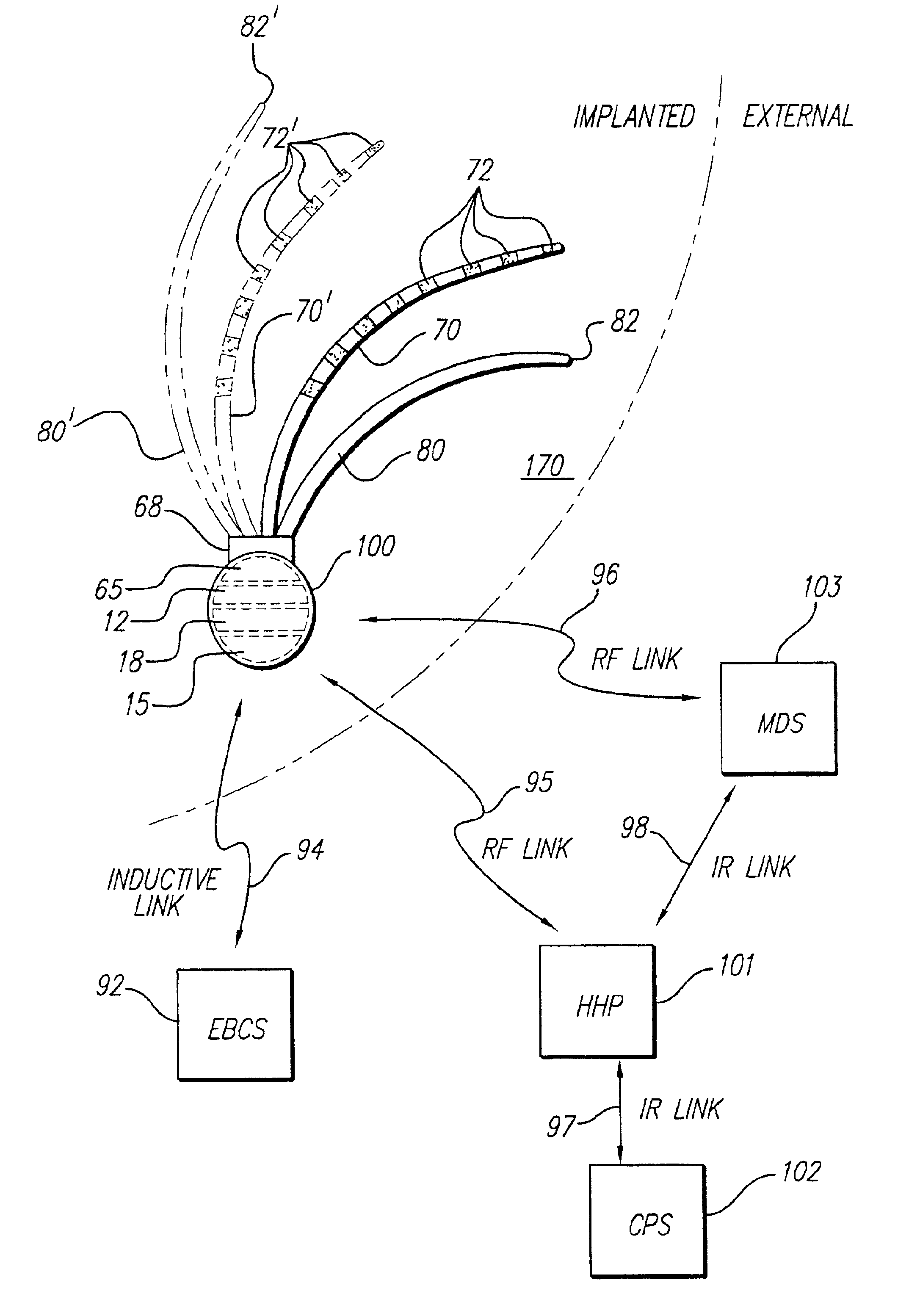
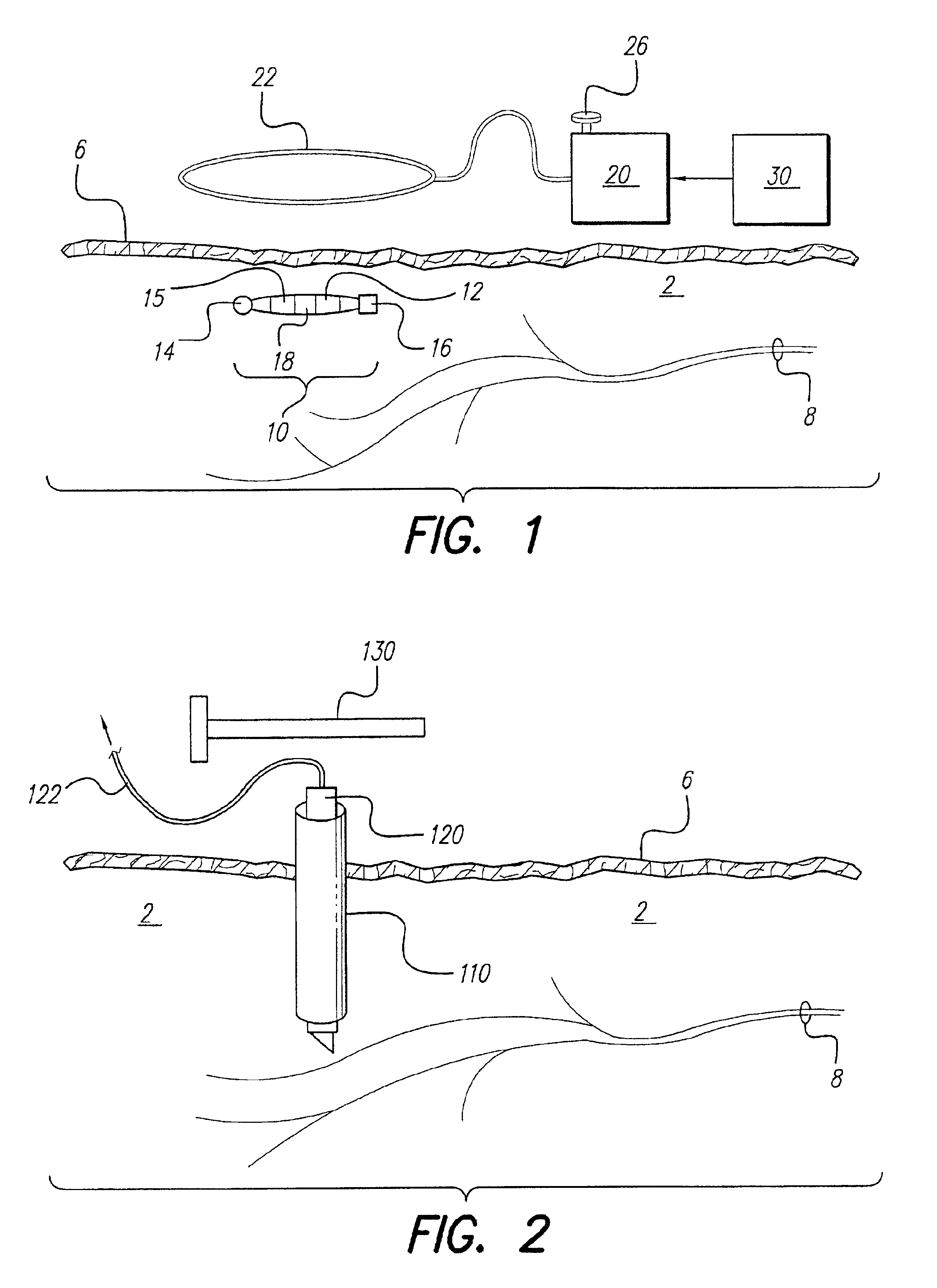
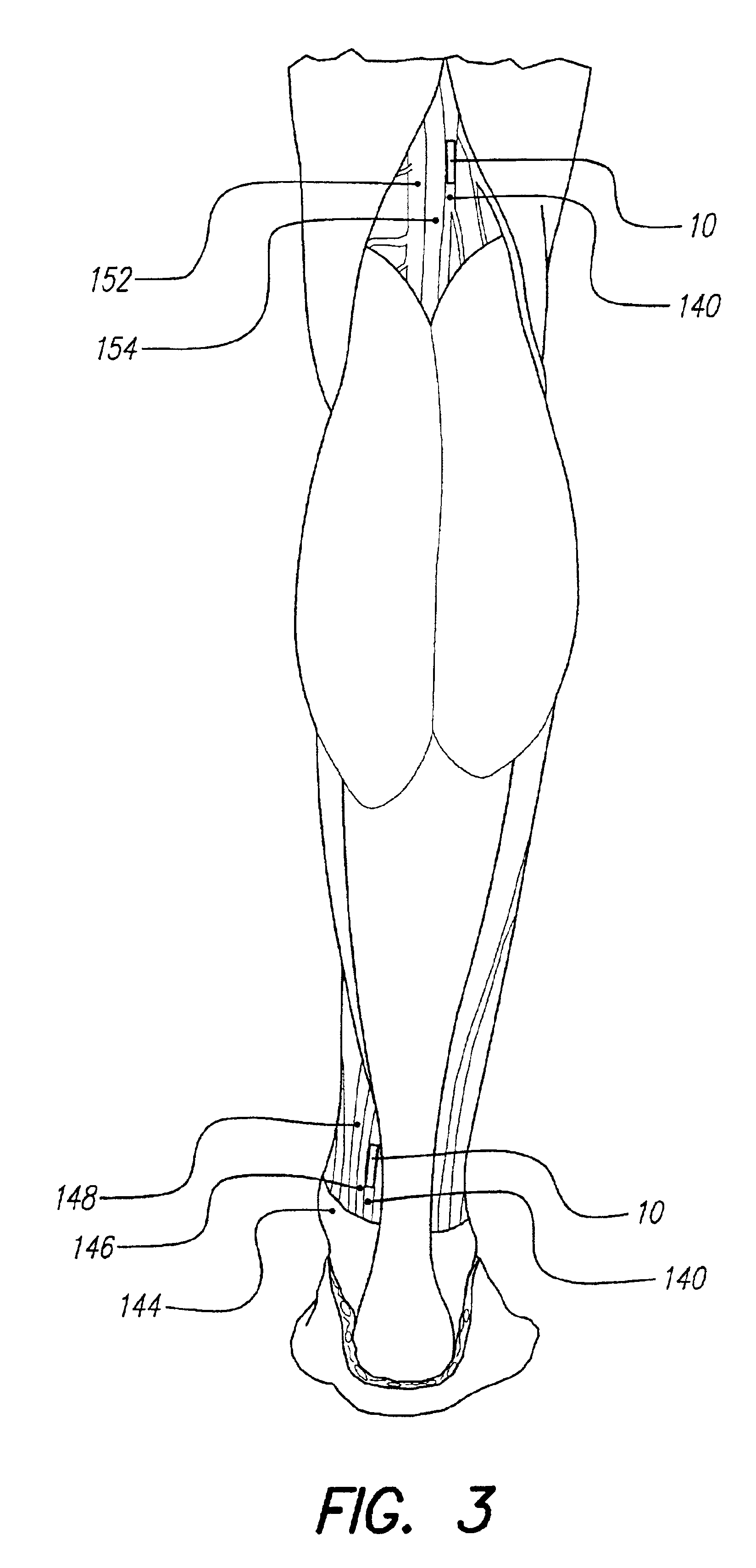
[0064]As indicated above, the present invention reduces or eliminates incontinence and / or pelvic pain by stimulating various nerve pathways with various stimulation systems and methods. For example, the present invention includes a system for using one or more stimulating drugs and / or electrical stimulation to directly activate the pudendal nerve and / or its branches by means of an implantable signal generator and electrode(s) and / or an implantable pump and catheter(s). One or more electrodes may be surgically attached to the nerve(s) or implanted adjacent to the nerve(s) to provide electrical stimulation, and / or one or more catheters may be surgically attach...


PUM


Abstract

A method and system for treatment of incontinence, urgency, frequency, and / or pelvic pain includes implantation of electrodes on a lead or the discharge portion of a catheter adjacent the perineal nerve(s) or tissue(s) to be stimulated. Stimulation pulses, either electrical or drug infusion pulses, are supplied by a stimulator implanted remotely, and through the lead or catheter, which is tunneled subcutaneously between the stimulator and stimulation site. For instance, the system and method reduce or eliminate the incidence of unintentional episodes of bladder emptying by stimulating nerve pathways that diminish involuntary bladder contractions, improve closure of the bladder outlet, and / or improve the long-term health of the urinary system by increasing bladder capacity and period between emptying. Moreover, the system and method allow a patient to be taught to receive one or more patterns of neural stimulation that can be prescribed by a physician and administered without continuous oversight by a clinical practitioner.

Description

[0001]This application is a continuation-in-part (CIP) of copending U.S. patent application Ser. No. 09 / 642,979, filed 18 Aug. 2000, now U.S. Pat. No. 6,735,474 which in turn is a CIP of PCT patent application Ser. No. PCT / US99 / 14775, filed 29 Jun. 1999, which in turn claims priority to, and the benefit of, prior U.S. patent application Ser. No. 60 / 091,762, filed 6 Jul. 1998. U.S. patent application Ser. No. 09 / 642,979 also claims the benefit of U.S. patent application Ser. No. 60 / 173,054, filed 24 Dec. 1999. The above-listed applications are incorporated herein by reference.FIELD OF THE INVENTION[0002]The present invention relates to implantable stimulator systems, and more particularly to an implantable stimulator system utilizing one or more implantable stimulators for treating incontinence and / or pain.BACKGROUND OF THE INVENTION[0003]Urinary incontinence is a clinical condition characterized by failure to hold urine in the bladder under normal conditions of pressure and filling....

Claims


Application Information

Patent Timeline
IPC IPC(8): A61N1/36A61N1/32A61N1/372A61N1/34A61N1/00
CPCA61N1/36007A61N1/36071A61N1/36017A61N1/37205
Inventor MANN, CARLA M.WHITEHURST, TODD K.MCGIVERN, JAMES P.LOEB, GERALD E.RICHMOND, FRANCES J. R.
Owner BOSTON SCI NEUROMODULATION CORP
Why Eureka
Eureka Blog
Learn More