
Biodegradable tissue retractor

Inactive Publication Date: 2005-08-30
FOCAL
View PDF64 Cites 37 Cited by
  • Summary
  • Abstract
  • Description
  • Claims
  • Application Information

AI Technical Summary

Benefits of technology

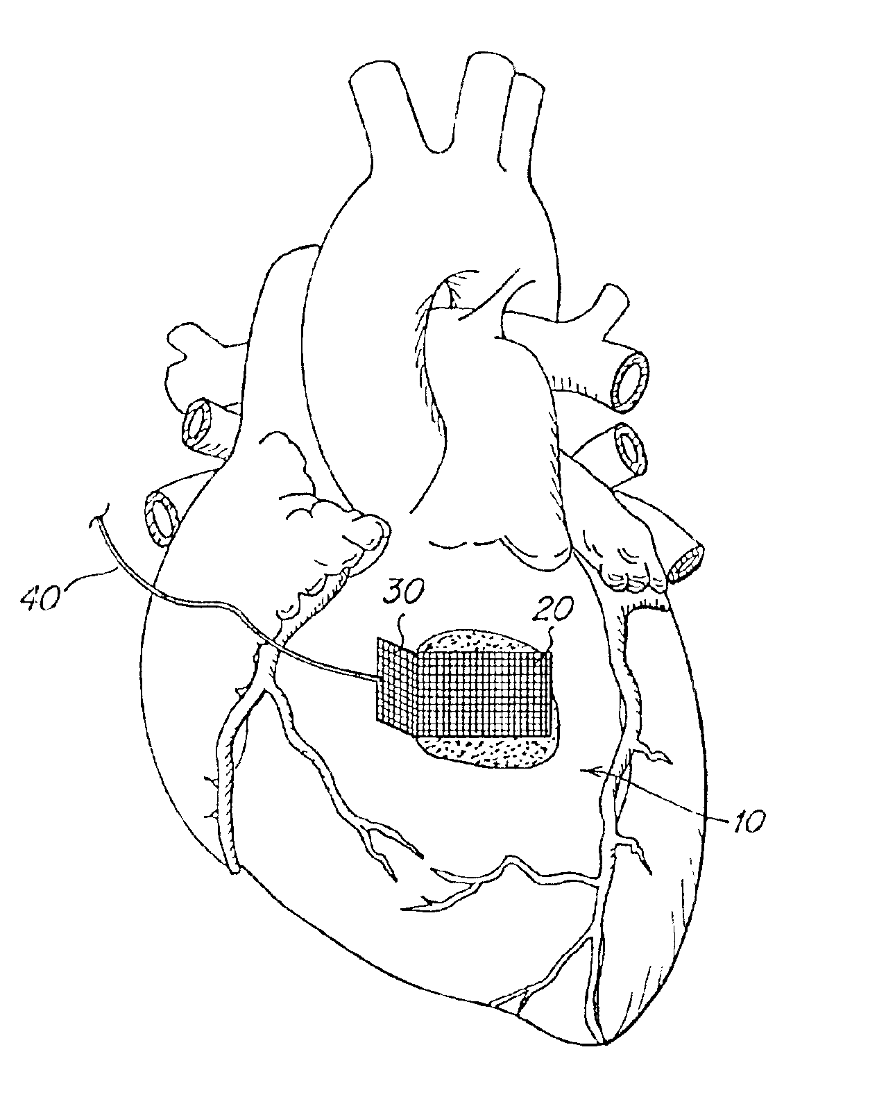
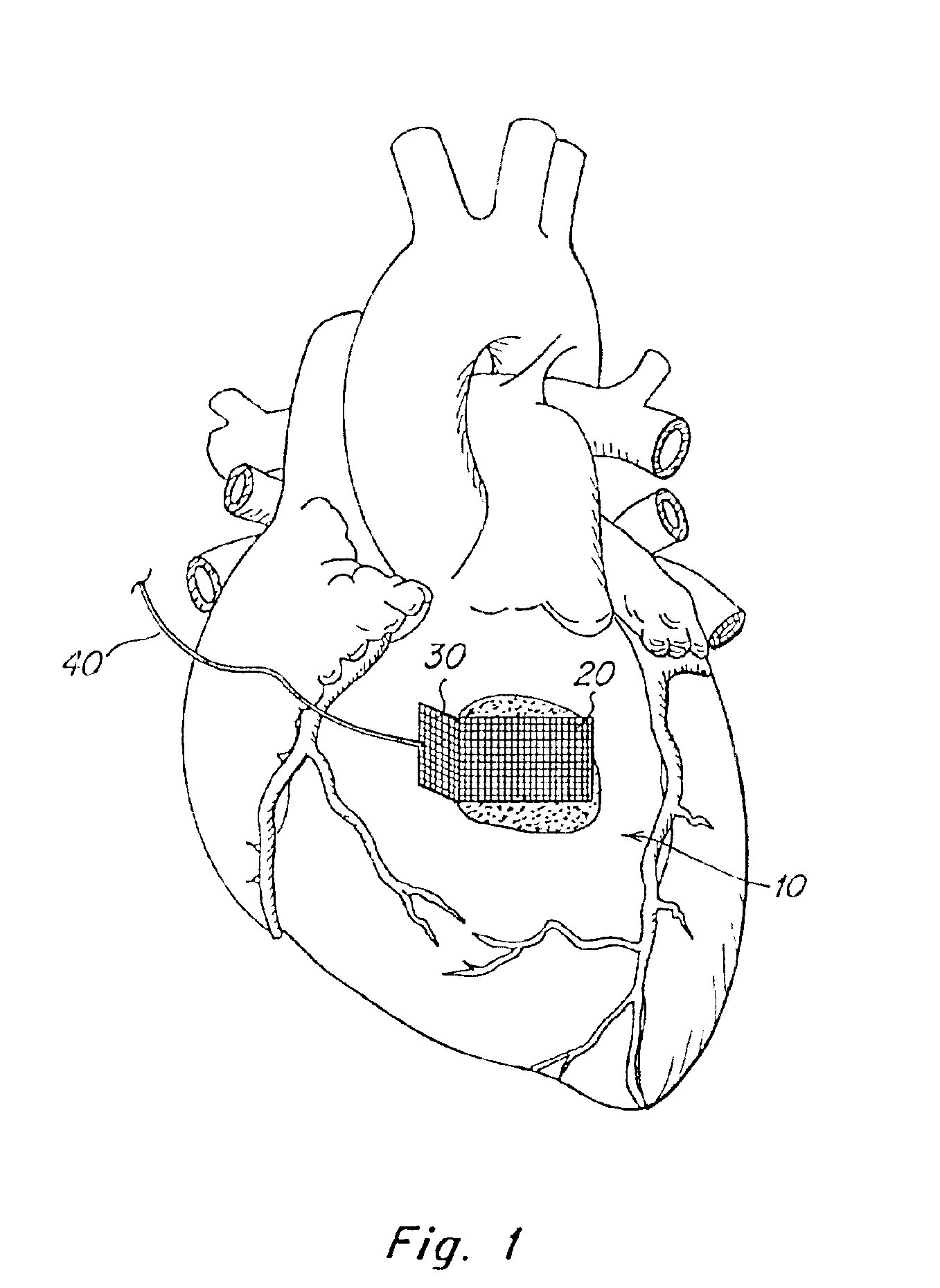
[0010]In one aspect, the invention provides a method that involves applying a tissue-affixing article to a surface of tissue internally of a patient. The tissue-affixing article is adhered to the surface of the tissue, and the tissue is retracted by exerting a manipulative force on the tissue-affixing article. In one embodiment the method is to affix an adhesive to tissue, internally of a patient, and to retract the tissue via the adhesive, that is, to physically manipulate the tissue by applying force directly or, preferably, indirectly, to the adhesive. This can be accomplished by affixing a patch of a material having sufficient tensile strength to the tissue, using a suitable adhesive. In one embodiment, an unadhered portion of the patch may then be easily grasped and moved or stabilized by suitable retraction means, such as a retracting instrument, or a suture that is passed through the unadhered portion of the patch, or which is permanently pre-attached to the patch. Alternatively, the patch may be completely adhered to the tissue, and a suture may then be passed through the adhered patch to provide tension without necessarily penetrating the tissue, or a retracting instrument may be applied to the adhesive or the patch. In each case, the patch can distribute the applied force over a larger surface area of the tissue than that which would be contacted directly by the retracting means, thus reducing potential tissue trauma, as might occur if a suture were passed directly through tissue and retracted. Moreover, the ability to achieve and maintain retraction of a tissue in a confined space using a means of retraction which minimizes obstruction of access ports is particularly important in minimally-invasive surgery, such as laparoscopy or other endoscopic surgery. This concept is particularly applicable to organs such as cardiac tissue, lung tissue, and GI (gastrointestinal) tissue in minimally invasive surgical procedures.

Problems solved by technology

Retraction or manipulation may also be accomplished by implanting a suture into the tissue, but there is a significant risk of tissue tearing even with strong, muscular tissue.

Method used


Image

  • Biodegradable tissue retractor
  • Biodegradable tissue retractor
  • Biodegradable tissue retractor

Examples


example

[0062]The nature of the invention is clearly demonstrated by an operational example. The technique was used to manipulate the beating heart of a live dog. A primer solution buffered to physiological pH and containing 50 ppm Eosin Y, 0.10 M neutral triethanolamine (TEA) buffer, and 10% of a polymerizable macromer (which contained a polyethylene glycol backbone (3500 MW) with about 5 lactate residues attached, and endcapped with acrylic acid esters) was brushed onto a tissue-affixing article, specifically a 2 cm by 4 cm area of a 2 cm×6 cm polyester (Mersilene™) mesh patch, and also onto the epicardial surface of the LV apex of a beating dog heart that was exposed via thoracotomy. A layer of sealant prepolymer (20% 35,000 MW PEG with trimethylene carbonate linkages and acrylate end caps) also containing Eosin Y and TEA buffer were applied over the primer on the heart. The mesh was placed onto the coated tissue, and a layer of sealant was brushed onto the mesh / tissue surface. Two 20-se...


PUM


Abstract

This invention describes novel methods and devices for stabilizing and retracting tissue during surgery, in particular internal tissue. Patches of material, preferably biodegradable, are adhered to tissue surfaces. By manipulation of the patches, for example directly with forceps, or via sutures attached to the patches, tissues can be retracted or otherwise manipulated with minimal trauma to the tissues. The method is especially useful in minimally-invasive surgery.

Description

RELATED APPLICATION[0001]This is application is a continuation application of U.S. patent application Ser. No. 09 / 634,366, filed Aug. 8, 2000 (now abandoned), which is a continuation application of U.S. patent application Ser. No. 09 / 045,401, filed Mar. 20, 1998 (now U.S. Pat. No. 6,123,667, issued Sep. 26, 2000, which claims the benefit under Title 35, U.S.C. §119(e) of U.S. provisional application Ser. No. 60 / 042,054, filed Mar. 20, 1997, incorporated herein by reference.FIELD OF THE INVENTION[0002]The invention relates generally to surgical devices, and more particularly to materials and methods for retraction and stabilization of tissues, using biodegradable patches adhered to tissue in a releasable manner.BACKGROUND OF THE INVENTION[0003]During surgery, it is often necessary to retract tissue, especially internal tissue, to operate upon it or upon adjacent tissue. This is conventionally accomplished using forceps, or other mechanical gripping devices adapted for the purpose. Re...

Claims


Application Information

Patent Timeline
IPC IPC(8): A61B17/03A61B17/02A61B17/08A61B17/00A61B17/06A61B17/04A61M31/00
CPCA61B17/0218A61B17/085A61B17/00491A61B17/06A61B2017/00004A61B2017/00495A61B2017/00951A61B2017/0237A61B2017/081
Inventor POFF, BRADLEY C.HERMAN, STEPHEN J.PICHON, DEAN M.SAWHNEY, AMARPREET S.
Owner FOCAL
Learn More