
Unidirectional presentation of membrane proteins in nanoparticle-supported liposomes

a technology of membrane proteins and nanoparticles, applied in the direction of viruses/bacteriophages, instruments, medical ingredients for detecting antibodies, etc., can solve the problems of difficult to make nanodisc samples in large quantities, difficult to present these proteins to immune systems to induce antibodies, and inability to incorporate transmembrane proteins

Pending Publication Date: 2022-03-31
PRESIDENT & FELLOWS OF HARVARD COLLEGE
View PDF0 Cites 0 Cited by
  • Summary
  • Abstract
  • Description
  • Claims
  • Application Information

AI Technical Summary

Benefits of technology

The patent describes a method for attaching a coating to a substrate and adding a functional moiety to the coating. The substrate can be a globular, solid, hollow, porous, multi-layer, or nanoparticle. The functional moiety can be a protein, DNA, RNA, PNA, LNA, or a biological molecule like a membrane bound protein or membrane transport protein. The method can also involve adding a tag to the substrate and detecting the tag using a coated substrate. The technical effects include improved detection and purification of proteins, improved functionalization of substrates, and improved detection of membrane-bound proteins.

Problems solved by technology

But presenting these proteins to immune systems to induce antibodies is a difficult problem, in particular, for those whose structural integrity can only be preserved on the membrane.
The unstable nature of the liposome, i.e., its tendency to fuse with other cellular vesicles, could be another source of risk in its application.
Greater stability in serum could be achieved using interbilayer-crosslinked multilamellar vesicle (ICMV)-coated particles, but the technique has not been demonstrated to incorporate transmembrane proteins.
But, nanodisc samples are generally difficult to make in large quantities.
Moreover, regular nanodiscs usually can only contain 1-2 copies of protein due to its small size (10-15 nm in diameter), and are thus not ideal for inducing strong immunogenic responses in vivo.
The success of this approach, however, depends on the efficiency of protein incorporation into VLP, which needs laborious optimization for each target and often cannot be controlled manually.

Method used


Image

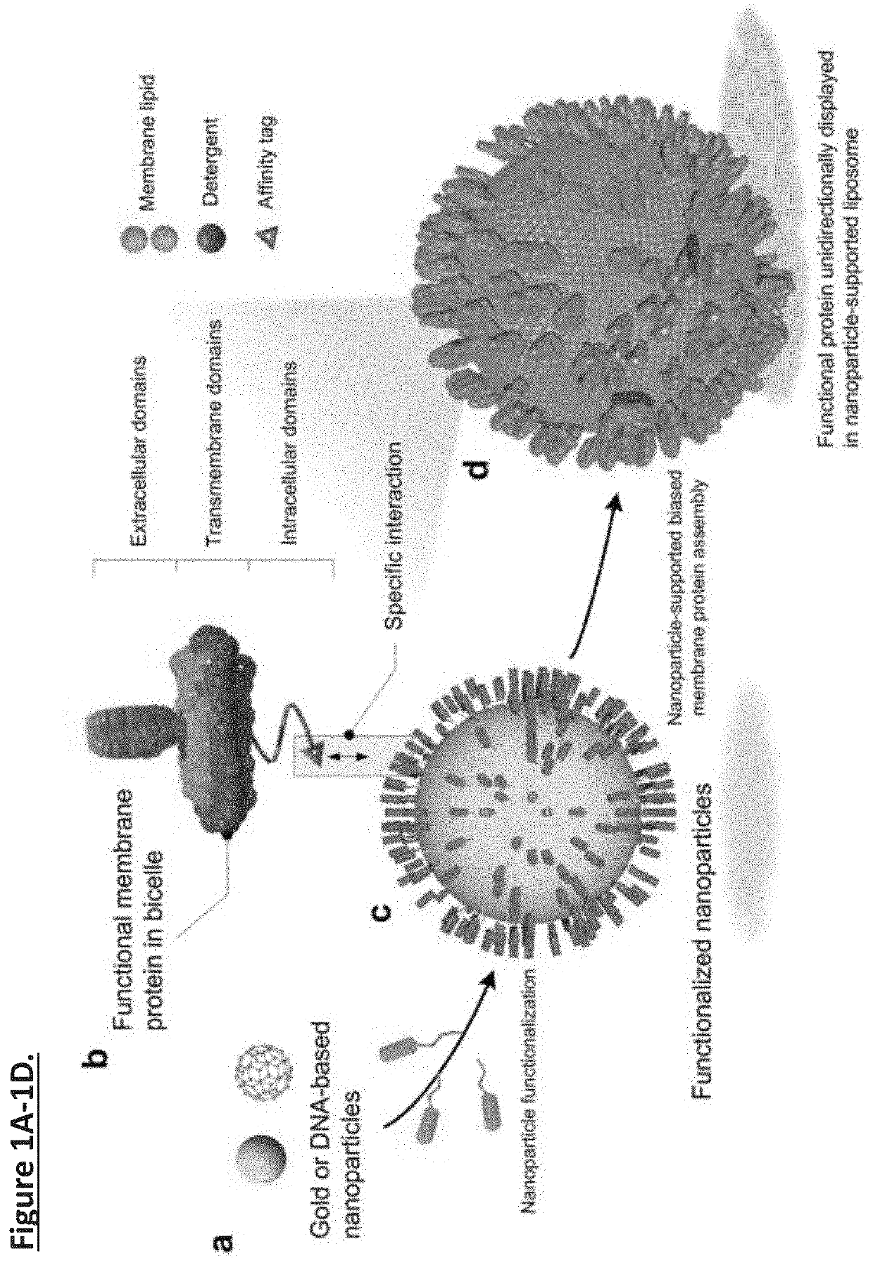
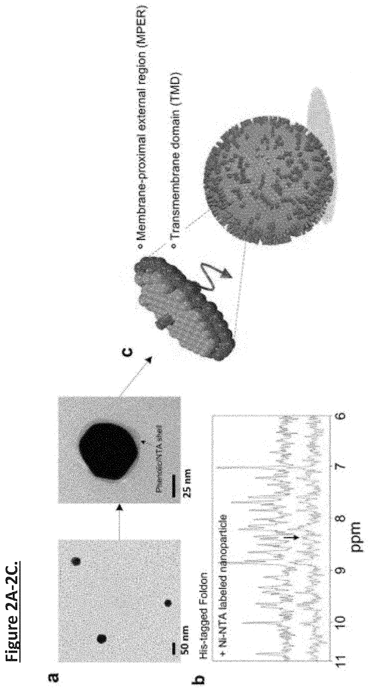
  • Unidirectional presentation of membrane proteins in nanoparticle-supported liposomes
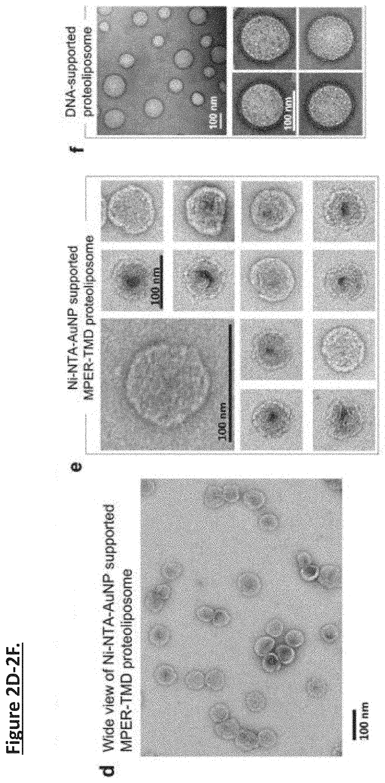
  • Unidirectional presentation of membrane proteins in nanoparticle-supported liposomes
  • Unidirectional presentation of membrane proteins in nanoparticle-supported liposomes

Examples


example 1

Nanoparticle Preparations

[0035]Gold-polyphenol nanoparticle production and functionalization. 0.8 mg / ml of tannic acid (Sigma Aldrich) was prepared in ddH2O. Chloroauric acid (Sigma Aldrich) was added to tannic acid solution drop by drop to the final concentration of 0.4 mM. The mixture was incubated at room temperature with stirring at 800 rpm for 20 minutes to form polyphenol-stabilized gold nanoparticles (AuNPs). AuNPs were spun down at 12,000 g for 10 minutes. Pellet was then washed with ddH2O. Resuspended AuNPs in ddH2O was thoroughly sonicated before centrifugation again. The centrifugation and washing steps were repeated twice. AuNPs were then mixed with 0.04% glutaraldehyde (Electron Microscopy Sciences) and 0.5 mg / ml Nα,Nα-Bis(carboxymethyl)-L-lysine (Lysine-NTA) (Sigma Aldrich) at 45° C. for 1 hour. The conjugated NTA-AuNPs were then spun down and washed with ddH2O for three times. 0.5 mM NiCl2 was added to the NTA-AuNP solution. Ni-NTA-AuNPs were then washed with ddH2O fo...

example 2

Zeta-Potential Measurement

[0036]The zeta-potentials were measured using a Zetasizer Nano-ZS (Malvern Instruments, UK) with a 633 nm He—Ne ion laser. The capsules were suspended in 10 mM phosphate buffer (pH 7.4) before adding different nanoparticle solutions. Measurements were repeated three times. The results were expressed as the mean and standard deviation obtained from the three measurements.

[0037]Interaction between Ni-NTA-AuNPs and His6-tag. Foldon is the C-terminal domain of T4 fibritin containing 27 residues and forms highly-stable trimer. Foldon with C-terminal His6-tag was cloned into the pET-15 vector and expressed in BL21(DE3) cells at 37° C. (induced with 1 mM isopropyl-β-d-thiogalactopyranoside (IPTG) for 6 hours). The protein was purified by Ni-NTA affinity (HisPur Ni-NTA resin, Thermo Fisher) and size exclusion chromatography (superdex 75 column, GE Healthcare). The NMR oneone echo experiment was used to record the 1D 1H spectrum of a 450 μl Foldon sample (30 μM Fold...

example 3

MPER-TMD Plasmid Construction, Expression and Purification

[0038]The MPER-TMD corresponds to a fragment of HIV-1 gp41 (clade D, isolate 92UG024.2) spanning residues 660-710; it contains the entire MPER (residues 660-683) and the TMD (residues 684-705). FLAG-tag and His6-tag sequences were added to the N- and C-termini of the MPER-TMD, respectively. The FLAG-MPER-TMD-His6 DNA was cloned into the pMM-LR6 vector as a fusion to the C-terminus of the trpLE sequence.

[0039]The MPER-TMD plasmid was transformed into E. coli BL21(DE3) for expression. Cell cultures were grown at 37° C. in LB media until OD600 reached 0.6, and cooled to 22° C. before induction with 100 μM isopropyl β-D-thiogalatopyranoside (IPTG) at 22° C. for overnight. The MPER-TMD was extracted from inclusion bodies, cleaved by cyanogen bromide, and purified by HPLC as described previously. The purified MPER-TMD were lyophilized and validated by SDS-PAGE and MALDI-TOF mass spectrometry.


PUM

PropertyMeasurementUnit
sizeaaaaaaaaaa
volumeaaaaaaaaaa
volumeaaaaaaaaaa

Abstract

Presentation of membrane proteins to host immune systems has been a challenging problem due to complexity arising from the poor in vivo stability of the membrane-mimetic media often used for solubilizing the membrane proteins. The Inventors report the use of functionalized, biocompatible nanoparticles as substrates to guide the formation of proteoliposomes that can present many copies of membrane proteins in a unidirectional manner. The approach was demonstrated to present the membrane-proximal region of the HIV-1 envelope glycoprotein. These nanoparticle-supported liposomes are broadly applicable as membrane antigen vehicles for inducing host immune responses. In some instances, the technology supports generation of antibodies that do not generate an immunogenic response in comparison to conventional protein presentation (i.e., liposome).

Description

CROSS-REFERENCE TO RELATED APPLICATIONS[0001]This application is a 35 U.S.C. § 371 National Phase Entry Application of International Patent Application No. PCT / US2019 / 068601 filed on Dec. 26, 2019, which designated the U.S., which claims benefit under 35 U.S.C. § 119(e) of U.S. Provisional Application No. 62 / 785,897 filed Dec. 28, 2018, the contents of which are incorporated herein by reference in their entireties.STATEMENT REGARDING FEDERALLY-SPONSORED RESEARCH[0002]This invention was made with government support under GM116898 awarded by the National Institutes of Health. The government has certain rights in the invention.SEQUENCE LISTING[0003]The instant application contains a Sequence Listing which has been submitted in ASCII format via EFS-Web and is hereby incorporated by reference in its entirety. Said ASCII copy, created on Dec. 26, 2019, is named 002806-094040WOPT_SL.txt and is 980 bytes in size.FIELD OF THE INVENTION[0004]Described herein are compositions and methods relat...

Claims


Application Information

Patent Timeline
Patent Type & Authority Applications(United States)
IPC IPC(8): A61K9/51A61K39/21A61K9/127G01N33/543A61K39/00
CPCA61K9/5115A61K39/21A61K2039/55555G01N33/54346C12N2740/16234A61K9/1271A61K39/12A61K9/0019G01N33/5432
Inventor CHOU, JAMESCHEN, WEN
Owner PRESIDENT & FELLOWS OF HARVARD COLLEGE
Learn More