Promoters and uses thereof
- Summary
- Abstract
- Description
- Claims
- Application Information
AI Technical Summary
Benefits of technology
Problems solved by technology
Method used
Image
Examples
example 1
[0212]Promoter Sequence of hSNCG
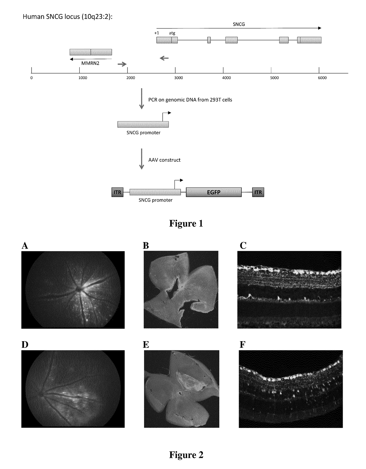
[0213]The SNCG gene is located on chromosome 10 (10q23.3) (FIG. 1). It contains 5 exons extending over 3.5 kb from +1 transcription site to the end of the 5th exon. The 1st exon of the multimerine 2 gene (MMRN2) is located 863 bp downstream of +1 SNCG transcription site. In order to extract the human sequence of the SNCG promoter, the −785 to +163 region (indicated by arrows in FIG. 1) was amplified from HEK 293T cells.
[0214]The amplification primers were the followings:
Forward primer:(SEQ ID NO: 2)5′-CACAAGCCAGTTCCTGTCC-3′,andReverse primer:(SEQ ID NO: 3)5′-GGGTGTGCAGGGTTGTG-3′.
[0215]The promoter sequence was amplified by 30 successive cycles consisting of a 30 second denaturation step at 94° C., followed by a one minute annealing step at 54° C., and an elongation step of one minute at 72° C.
[0216]The PCR product (SEQ ID NO: 1) was then subcloned into the pENTR-D / TOPO plasmid by the method using DNA topoisomerase I and sequenced. The sequence was con...
example 2
[0224]C57BL6 / J adult mice (6 weeks) received an intravitreal injection of AAV-2 / Y444F vectors comprising the promoter of SEQ ID NO: 1 and a nucleic acid encoding GFP protein (cf. example 1 and FIG. 1) diluted at a dose of 1011 vg (vector genome) in PBS. One month after injection, the fundus and the retina had a normal appearance, indicating the lack of toxicity of the vector or the surgical procedure (FIG. 2A).
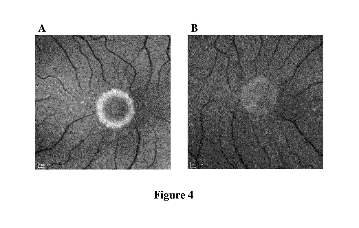
[0225]The expression of GFP reporter gene in the retina was observed by in vivo imaging of the fundus with direct fluorescence (FIG. 2A). Animals were anesthetized by intraperitoneal injection of a mixture of ketamine and xylazine, and the pupils were dilated by topical application of neosynephrine and mydriaticum. After total pupil dilation, the animals were examined by a fluorescence camera specially designed to view the rodent fundus (MicronIII, PhoenixLaboratories).
[0226]With the fluorescence, numerous cell bodies expressing GFP were detected, and nerve fibers converging t...
example 3
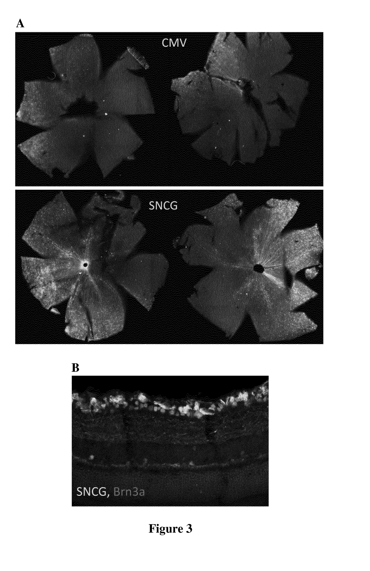
[0227]In order to characterize the expression pattern of the hSNCG promoter in the retina, the inventors have compared the hSNCG promoter (SEQ ID NO: 1) to the mPGK promoter, a ubiquitous promoter known to provide a high level of expression in the retina.
[0228]Two groups of C57BL6 / J mice were injected intravitreally with AAV2-Y444F vectors diluted in PBS at a dose of 1011 vg and encoding GFP under the control of mPGK or hSNCG promoter. One month after injection, the GFP expression was analyzed in vivo as described above. Animals were then euthanized and tissues fixed by intracardiac perfusion of a solution containing 4% paraformaldehyde in PBS. The eyeballs were subsequently extracted, post-fixed one hour in the same fixative, cryoprotected in a solution containing 25% sucrose in PBS, and then cut with a cryostat to a thickness of 16 μm. GFP expression was then analyzed under a microscope by visualization of direct fluorescence and after immunolabeling using Bnr3a as specific marker...
PUM
| Property | Measurement | Unit |
|---|---|---|
| Fraction | aaaaa | aaaaa |
| Digital information | aaaaa | aaaaa |
| Digital information | aaaaa | aaaaa |
Abstract
Description
Claims
Application Information
 - R&D
- Intellectual Property
- Life Sciences
- Materials
- Tech Scout
- Unparalleled Data Quality
- Higher Quality Content
- 60% Fewer Hallucinations
Browse by: Latest US Patents, China's latest patents, Technical Efficacy Thesaurus, Application Domain, Technology Topic, Popular Technical Reports.
© 2025 PatSnap. All rights reserved.Legal|Privacy policy|Modern Slavery Act Transparency Statement|Sitemap|About US| Contact US: help@patsnap.com


