
Flourescent exomarker probes for hydrogen sulfide detection

a fluorescent exomarker and probe technology, applied in the field of fluorescent exomarker probes for hydrogen sulfide detection, can solve the problems of limited diagnostic tools and no prior art report a potent probing device for mapping endogenous h2s formation in cancer cells over normal cells

Active Publication Date: 2018-10-25
AMRITA VISHWA VIDYAPEETHAM
View PDF0 Cites 3 Cited by
  • Summary
  • Abstract
  • Description
  • Claims
  • Application Information

AI Technical Summary

Benefits of technology

The present patent relates to hydrogen sulfide probe compounds, their methods of preparation, and their use as diagnostic markers for various diseases. The compound of formula I has been found to have high selectivity for detecting hydrogen sulfide over other molecules, making it useful for identifying the presence of this molecule in a sample. The compound can be used in a pharmaceutical composition to treat diseases such as neurodegenerative disorders, diabetes, hypertension, and cancer. The compound can also be used to detect hydrogen sulfide in a sample, such as cells from adipose, muscle, cartilage, bone, mucosa, lung, heart, brain, liver, kidney, pancreas, or vasculature. The compound can be visualized using microscopy or other imaging techniques. The patent also includes a process for preparing the compound of formula I.

Problems solved by technology

However, these have been limited as diagnostic tools as they are unable to provide spatial and temporal information of in vivo concentration of H2S and its distribution in living cells and organisms.
It's a challenging task to detect H2S in a microenvironment like mitochondria over competing thiols due to difficulty of monitoring H2S and thiol levels simultaneously in complex environment of sulfur redox homeostasis.
It remains a challenge so far detecting mitochondrial H2S specifically and accurately.
However, none of the prior arts reported a potent probing device to map endogenous H2S formation in cancer cells over normal cells.

Method used


Image

  • Flourescent exomarker probes for hydrogen sulfide detection
  • Flourescent exomarker probes for hydrogen sulfide detection
  • Flourescent exomarker probes for hydrogen sulfide detection

Examples


example 1

of Mito-HS

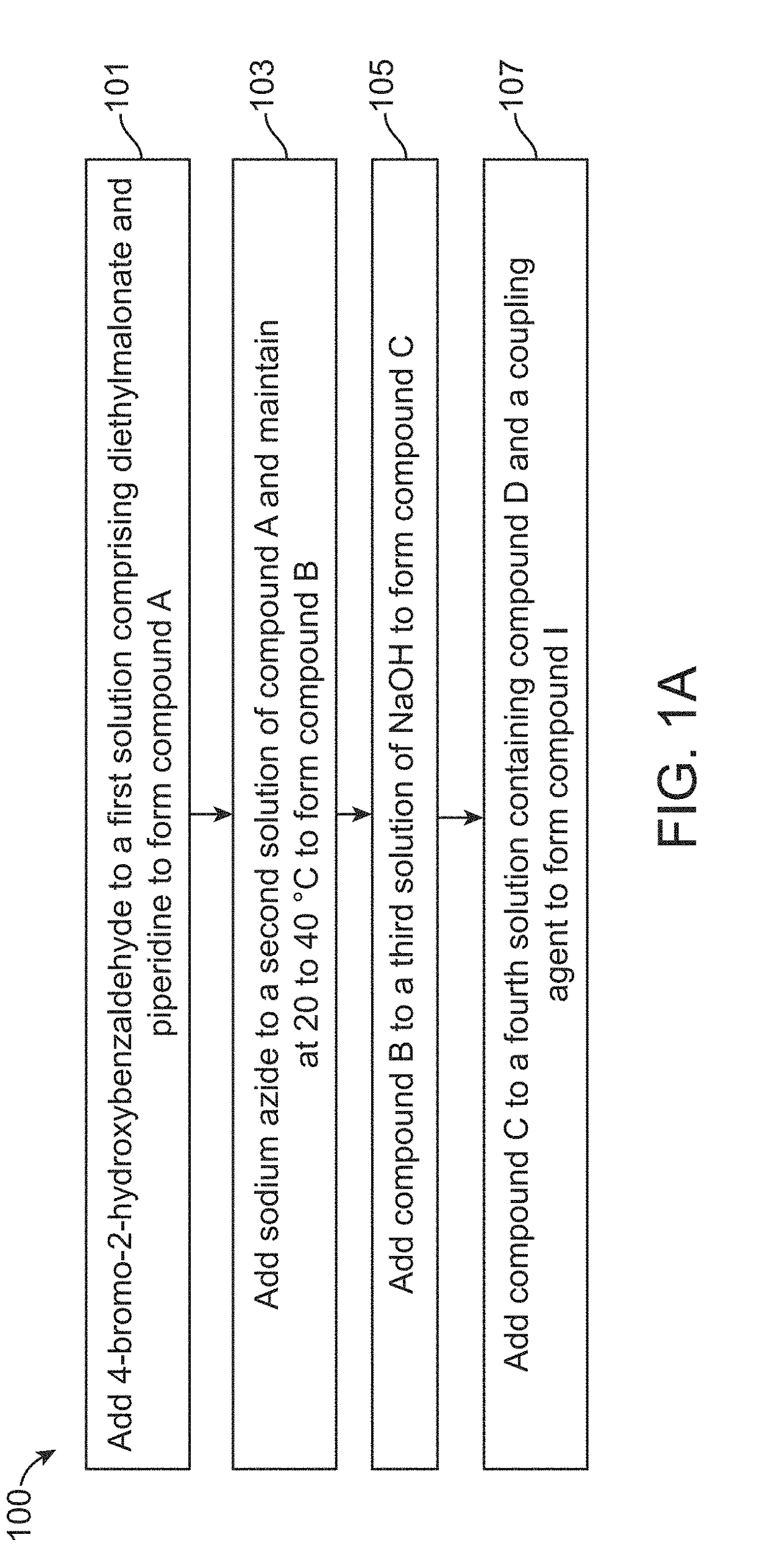
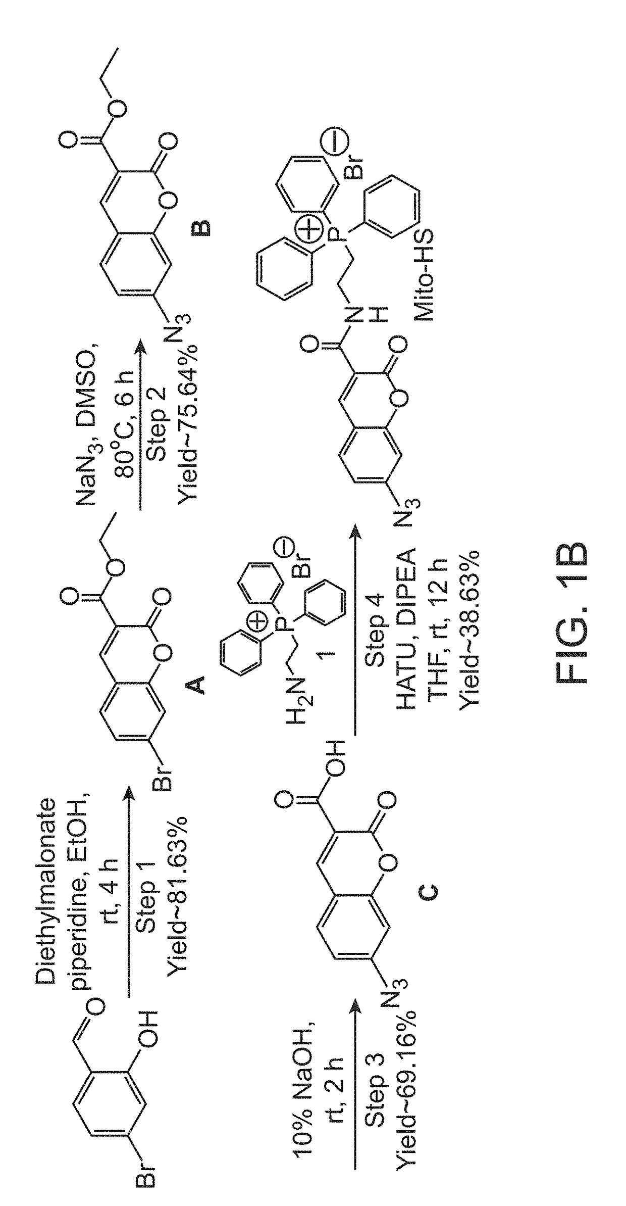
[0066]A solution of 4-bromo-2-hydroxybenzaldehyde (1.0 g, 4.97 mmol) was prepared by adding in 20 mL of ethanol. Diethylmalonate (955 mg, 5.97 mmol) and piperidine (1.27 g, 14.92 mmol) were added to it followed by continuous stirring for 3 hours at room temperature. Ethanol was evaporated after competition of the reaction. The residue obtained was dissolved in 2N HCl and extracted with ethyl acetate The extracted organic layer was washed with water and brine solution followed by drying over anhydrous sodium sulfate. The organic layer was kept in reduced pressure to get concentrated to obtain white colour solid product (1.20 g, 81.63%), named as compound A.

[0067]The yield for the above synthesis was 98.10%, determined by liquid chromatography-mass spectrometry (LCMS). ‘H and’3C NMR were performed for compound A. 1H-NMR (400 MHz, DMSO-d6): δ 8.76 (s, 1H); 7.86 (d, 1H, j=9.08 Hz); 7.78 (s, 1H); 7.62-7.60 (dd, 1H, j=6.88 Hz); 4.30 (q, 2H); 1.30 (q, 3H); 13C-NMR (100 MHz, DMSO-...

example 2

of Endogenous H2S Selectively in Cancer Cells

[0074]Human cervical cancer cells (HeLa), breast cancer cells (MDA-MB-231), prostate cancer cells (DU 145) and 3T3-L1 fibroblast cells were cultured in DMEM high glucose media supplemented with 10% fetal bovine serum, 1% Penstrep, 0.2% Amphotericin B. The cells were grown overnight at 37° C. incubator with 5% CO2. HeLa, MDA-MB-231, DU 145, and 3T3-L1 cells were seeded at a density of 0.3×10⁶ cells in 35 mm dish and kept overnight. The probe Mito-HS prepared in Example 1 was dissolved in 0.2% DMSO to make a stock concentration of 10 mM. The cells were treated with 5 μM of Mito-HS for 15 min. 300-550 nm excitation light was used to measure its fluorescence properties. Images were acquired using Zeiss Fluorescence Microscope (A1 Axiovert) with ×40 objective lens.

[0075]UV-Vis and fluorescence spectroscopy was studied and changes of Mito-HS was recorded in variable concentrations of Na2S (0-200 μM) in PBS buffer solution containing 0.2% of DMS...

example 3

ty Study of Mito-HS in Cellular Milieu

[0077]Fluorescence responses of Mito-HS (5 μM) in the presence of various biologically important analytes such as H2S, cysteine (Cys), H2O2, NaNO2, Cu(OAc)2, Zn(OAc)2, FeSO4, FeCl3, Na2CO3, GSH, and ascorbic acid (AA) NO, Na2S2O4 in aqueous solutions (in PBS, 0.2% DMSO, pH=7.4) at 37° C. were studied as shown in FIG. 5. Excitation wavelength was set at 380 nm and excitation and emission slit widths both set at 3 nm. Error bars were obtained from triplet experimental data. It was observed that none of the analytes showed considerable fluorescence change other than H2S. So, the experiment supported the proposed concept of using Mito-HS to track cellular H2S over other competing substances such as thiols.


PUM


Abstract

A fluorescence probe with mitochondrial targeting and two-photon property, its preparation method and application in detecting and tracking endogenous H2S in samples or living cells. The fluorescent probe is prepared by a four-step preparation method and demonstrates a UV-vis absorption increment λab=395 nm and ˜43 fold higher fluorescence intensity in the presence of H2S. The probe further demonstrates stability, selectivity for H2S over competing agents and sensitivity as low as 20 nm. A method of detecting endogenous H2S rapidly in the absence of any external stimulators is provided. Samples are contacted with the probe and the changes in fluorescence are monitored to detect H2S levels. The disclosed probe is non-toxic and suitable as a biomarker and therapeutic molecule in cancer and other diseases.

Description

CROSS-REFERENCES TO RELATED APPLICATIONS[0001]This application claims priority to Indian patent application No. 201741013739, filed on 18 Apr. 2017, the full disclosure of which is incorporated herein by reference.FIELD OF THE INVENTION[0002]The present application is related to synthesis of fluorescent exomarker probes and in particular to a fluorescent exomarker probe for hydrogen sulfide detection.DESCRIPTION OF THE RELATED ART[0003]Hydrogen sulfide (H2S) plays a key role in cellular signaling events such as a neurotransmitter in central nervous system of humans and mammals. H2S is generated in vivo enzymatically from sulfur containing amino acids such as cysteine and homocysteine in the presence of enzymes such as cystathionine-β-synthase (CBS), cystathionine-γ-lyase (CSE), and 3-mercaptopyruvate sulfurtransferase in a controlled manner in several organs such as heart, vasculature, brain, kidney, liver, lungs, pancreas, thoracic aorta, ileum, portal vein, and uterus. It can also...

Claims


Application Information

Patent Timeline
IPC IPC(8): G01N33/574C07F9/655G01N33/58
CPCG01N33/57411C07F9/65522G01N33/582G01N33/84
Inventor BHUNIYA, SANKARPRASADMISHRA, NANDITAVELUSAMY, NITHYABINOY, ANUPAMABOBBA, KONDAPA NAIDUNEDUNGADI, DIVYA
Owner AMRITA VISHWA VIDYAPEETHAM
Learn More