
Method for diagnosing APS using determination of anti-PAR1 antibodies

a technology of anti-par1 antibodies and aps, applied in the field of medicine, can solve the problems of high risk of death, laborious and time-consuming diagnosis, and rapid organ failur

Inactive Publication Date: 2018-10-04
CELLTREND
View PDF0 Cites 0 Cited by
  • Summary
  • Abstract
  • Description
  • Claims
  • Application Information

AI Technical Summary

Benefits of technology

The patent is about finding a way to diagnose a condition called antiphospholipid syndrome (APS) by checking for the presence of a specific antibody in a patient's sample. This method has been found to have better sensitivity and specificity than other methods for diagnosing autoimmune diseases. The patent also describes a way to remove the specific antibody from the patient's blood to help treat the condition.

Problems solved by technology

In some cases, APS leads to rapid organ failure due to generalised thrombosis; this is termed “catastrophic antiphospholipid syndrome” (CAPS) and is associated with a high risk of death.
For diagnosis of APS currently at least one clinical event, i.e. thrombosis or pregnancy complication, and two antibody blood tests spaced at least three months apart that confirm the presence of either lupus anticoagulant, or anti-β2-glycoprotein-I (since β2-glycoprotein-I antibodies are a subset of anti-cardiolipin antibodies, an anti-cardiolipin assay can be performed as a less specific proxy) are needed making the diagnosis laborious and time-consuming.
Further, the testing for antibodies to targets of aPL such as β2 glycoprotein 1 and antiphosphatidyl serine is currently under debate.
Even though different tools are used and / or discussed, the diagnosis currently is laborious and time-consuming.

Method used


Image

  • Method for diagnosing APS using determination of anti-PAR1 antibodies
  • Method for diagnosing APS using determination of anti-PAR1 antibodies
  • Method for diagnosing APS using determination of anti-PAR1 antibodies

Examples


example 1

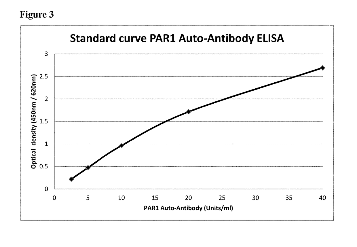
[0083]We measured the anti-PAR1 autoantibody in serum samples using a sandwich ELISA kit (CellTrend GmbH Luckenwalde, Germany). The microtiter 96-well polystyrene plates were coated with chemically synthesized human PAR1 isoform 1 (SEQ ID NO:1). To maintain the conformational epitopes of the receptor, 1 mM calcium chloride was added to every buffer. Duplicate samples of a 1:100 serum dilution were incubated at 4° C. for 2 hours. After washing steps, plates were incubated for 60 minutes with a 1:20.000 dilution of horseradish-peroxidase-labeled goat anti-human IgG (Jackson, USA) used for detection. In order to obtain a standard curve, plates were incubated with test sera from an anti-PAR1 autoantibody positive index patient. The ELISA was validated according to the FDA's “Guidance for industry: Bioanalytical method validation”.

[0084]To set a standard for the concentrations of the autoimmune antibodies, a standard curve was generated. In detail, a serum sample of a systemic sclerosis ...

example 2

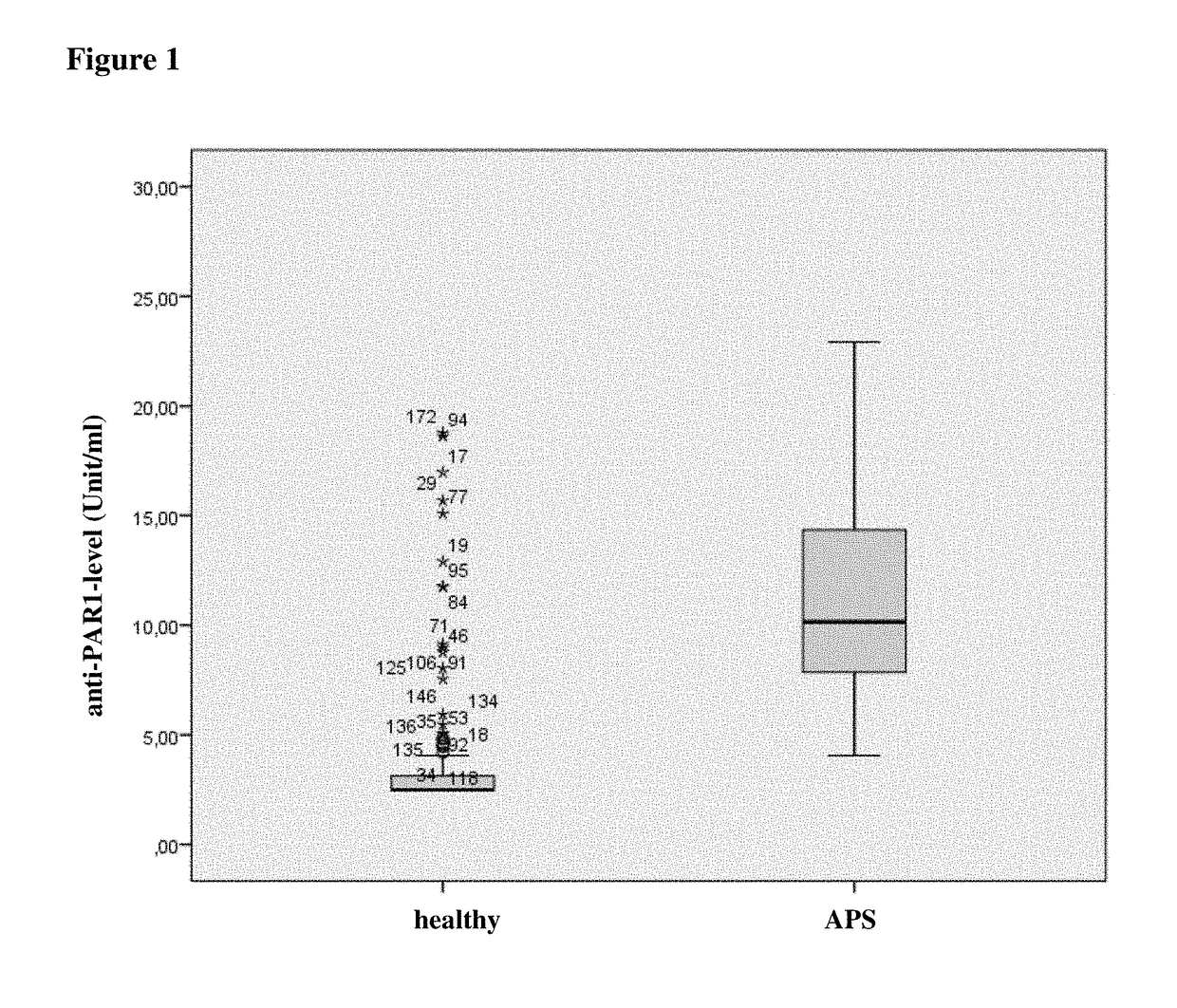
[0085]Anti-PAR1 antibody levels in serum samples from 198 healthy donors (“healthy”) and 83 patients with APS (“APS”) were measured using the kit and method of example 1. The levels were determined in units / mL. FIG. 1 shows the comparison of anti-PAR1 antibody level for case and control subjects. Patient suffering from APS had significantly increased levels (p<0.05) of anti-PAR1 antibodies as compared to healthy controls.

[0086]The analysis of percentiles revealed the following results:

TABLE 1Percentiles (weighted mean): Anti-PAR1-antibody levels-percentile in healthy donors (n = 198) and subjectssuffering from APS (n = 83).Percentile5102550759095healty subjects3.144.829.27(units / ml)Subjects with APS4.405.157.8210.1514.5020.2721.89(units / ml)

[0087]These data show that a threshold of e.g. 4 unit / ml lies within the 5th percentile of subjects with APS and about 90th of healthy subject, i.e. 95 percent of patients with APS have a higher level than 4 units / ml and 90 percent of healthy subj...


PUM

PropertyMeasurementUnit
optical densityaaaaaaaaaa
thresholdaaaaaaaaaa
fluorescence immunoassayaaaaaaaaaa

Abstract

The application relates to a method for diagnosis of an antiphospholipid syndrome (APS) in a subject, wherein presence or absence of an anti-protease-activated receptor 1 (PAR1) antibody is detected in a sample from the subject diagnosed, and wherein the presence of an anti-PAR1 antibody is indicative of the disease. Furthermore, it relates to the use of PAR1 for the diagnosis of APS, as well as to a method of removing anti-PAR1 antibodies from isolated blood of a subject upon detection of said anti-PAR1 antibodies in a sample of said patient.

Description

TECHNICAL FIELD OF THE INVENTION[0001]The present invention relates to the field of medicine, particular to the field of diagnosis of an autoimmune disease. Furthermore, the invention relates to the field of detection of PAR1 antibodies in a sample of a subject to be diagnosed for diagnosis of APS.BACKGROUND OF THE INVENTION[0002]Antiphospholipid syndrome or antiphospholipid antibody syndrome (APS or APLS), or often also Hughes syndrome, is an autoimmune, hypercoagulable state caused by antiphospholipid antibodies. APS provokes blood clots (thrombosis) in both, arteries and veins, as well as pregnancy-related complications such as miscarriage, stillbirth, preterm delivery, and severe preeclampsia. Antiphospholipid syndrome can be primary or secondary. In some cases, APS leads to rapid organ failure due to generalised thrombosis; this is termed “catastrophic antiphospholipid syndrome” (CAPS) and is associated with a high risk of death.[0003]Treatment of antiphospholipid syndrome is u...

Claims


Application Information

Patent Timeline
Patent Type & Authority Applications(United States)
IPC IPC(8): G01N33/564
CPCG01N33/564G01N2800/226G01N33/6893
Inventor HEIDECKE, HARALDSCHULZE-FORSTER, KAI
Owner CELLTREND
Learn More