
Intracellular organelle peptide targeted enzyme substrates

Active Publication Date: 2015-08-06
MARKER GENE TECH
View PDF3 Cites 10 Cited by
  • Summary
  • Abstract
  • Description
  • Claims
  • Application Information

AI Technical Summary

Benefits of technology

This patent describes a method for analyzing the activities of enzymes in cells or in isolated organelles. The method involves using fluorescent dyes that produce a visible signal when acted upon by specific enzymes. The dyes are prepared as a labeling solution and are used to incubate the cells or organelles with the dye solution for a certain amount of time. The resulting fluorescence is then detected using fluorescence detection systems. This method allows for the simultaneous measurement of two or more independent enzyme activities.

Problems solved by technology

Prior stains, methods and assays for visualizing acidic organelles are not useful for monitoring enzyme activities in living cells.
The fluorescent dyes neutral red and acridine orange are also commonly used for staining acidic organelles, but they lack specificity and are not well retained in the organelles, particularly after fixing and permeabilization.
In particular, methods to analyze Golgi aid ER enzyme functional activity within living cells have been hampered by the inability to monitor such activity inside individual organelles.
While such targeting peptides or proteins have been used as research tools for staining specific organelles their potential for real time analysis of enzyme activity within living cells or tissues remains unexplored.

Method used


Image

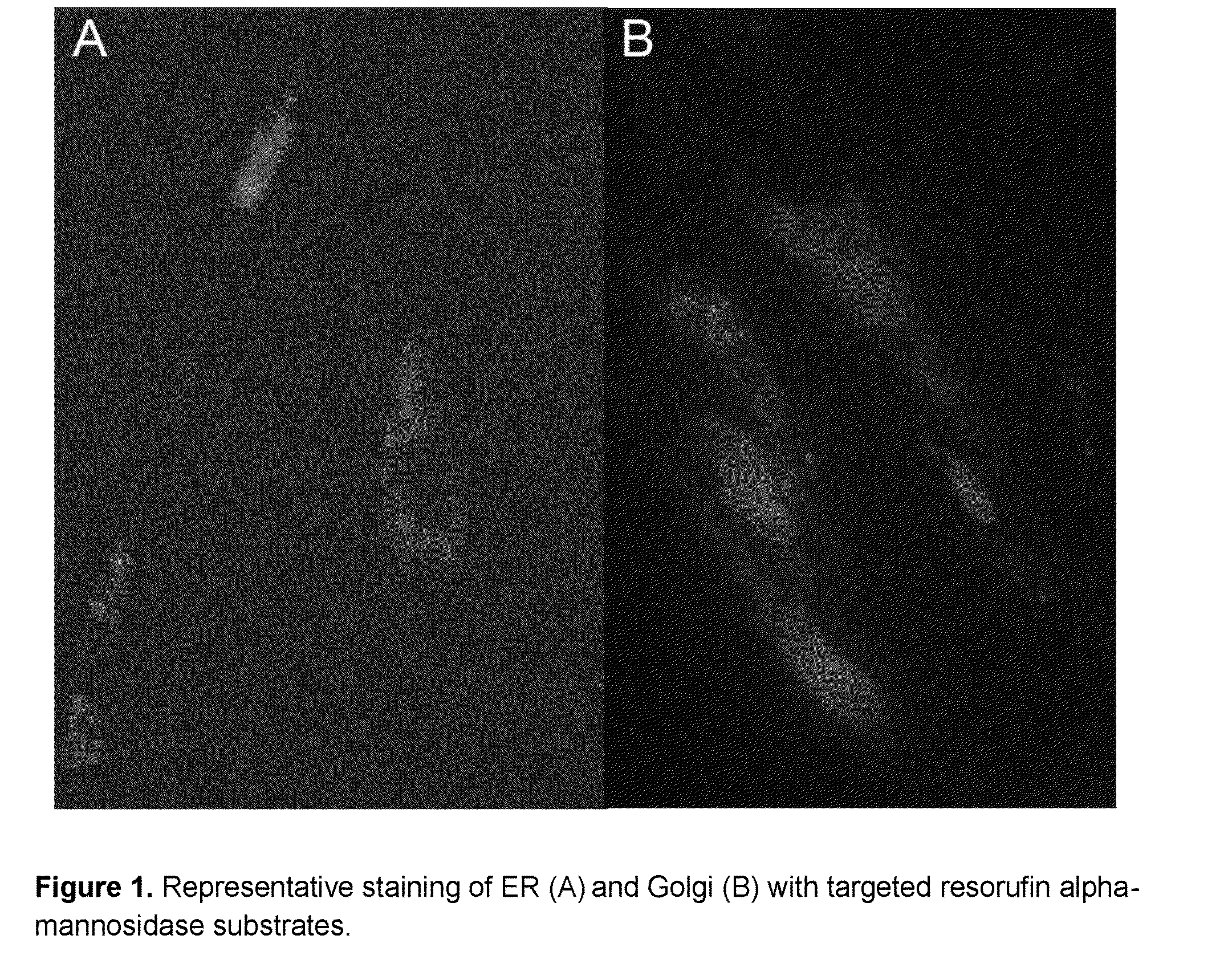
  • Intracellular organelle peptide targeted enzyme substrates
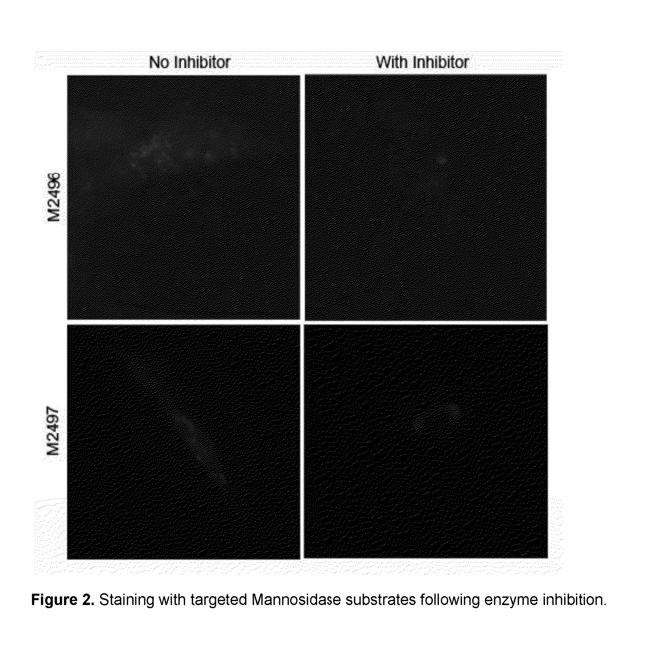
  • Intracellular organelle peptide targeted enzyme substrates
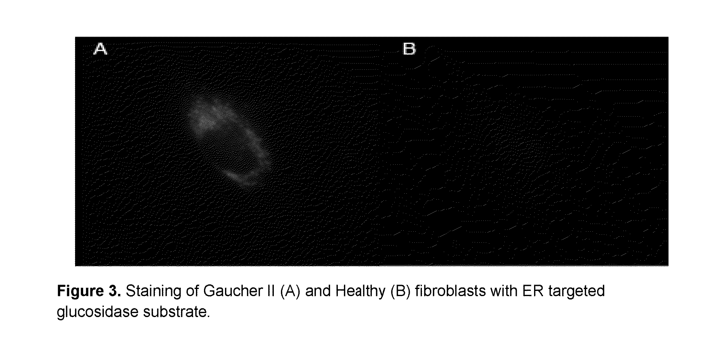
  • Intracellular organelle peptide targeted enzyme substrates

Examples


example 1

Preparation of a 7-Nitrobenzofurazan-Based Endoplasmic Reticulum-Targeted Dye

4-maleimido-1-butanammonium trifluoroacetate (M1959)

[0096]To a 250 mL round-bottom flask was added 4-amino-1-butanol (1.56 mL, 16.9 mmol) and water (35.0 mL). The solution was cooled to 0° C., and sodium bicarbonate (2.83 g, 33.7 mmol) was added. After 5 min, t-Boc anhydride (5.52 g, 25.3 mmol) in THF (5.0 mL) was added. After 17.5 h, the reaction solution was extracted with dichloromethane (3×50 mL). The organic layer was washed with water (2×50 mL), dried over MgSO4 and concentrated in vacuo. The resulting crude t-Boc carbamate (3.99 g) was added to a 100 mL round-bottom flask containing triphenylphosphine (4.88 g, 18.6 mmol) and maleimide (1.81 g, 18.6 mmol) in THF (50 mL). After 15 min, diisopropyl azodicarboxylate (4.00 mL, 20.3 mmol) was added. This reaction solution was allowed to stir for 16 h, the solution was concentrated in vacuo and the product purified via flash chromatography over SiO2, elutin...

example 2

Preparation of a 7-Nitrobenzofurazan-Based Golgi Apparatus-Targeted Probe

4-(4-maleimidobutanamino)-7-nitrobenzofurazan, conjugated to Golgi-targeting peptide (M2423)

[0099]To a 1.5 mL Eppendorf tube was added a solution of M2419 (100 μL, 2.0 mM in acetone, 0.2 μmol, a solution of H2N-G-A-S-D-Y-Q-R-L-C-COOH (SEQ ID NO:7) (100 μL, 1.0 mM in water, 0.1 μmol) and a solution of triethylamine (100 μL, 0.10 mM in water, 0.010 μmol). After gently agitating the vial by inversion for 18 h, the solution was diluted into water (500 μL) and extracted with chloroform (3×500 μL). The aqueous layer was lyophilized to give M2423, homogeneous by TLC (10% acetic acid / methanol) Rf=0.11.

example 3

Preparation of a Resorufin-Based α-Mannosidase Substrate Targeted to the Endoplasmic Reticulum Exhibiting Red Fluorescence after Enzyme Activity

N-Hydroxysuccinimide trifluoroacetate (M1027)

[0100]To a flame-dried 25 mL round-bottom flask was added N-hydroxysuccinimide (1.15 g, 10.0 mmol) and trifluoroacetic anhydride (3.80 mL, 27.5 mmol). After 2 h, the solution was concentrated in vacuo, co-evaporated with toluene (2×25 mL) and dried in vacuo to give M1027 (2.03 g, 9.62 mmol, 96%) as a white solid.

2-chloro-6-carboxyresorufin (M2458)

[0101]To a flame-dried 250-mL round-bottom flask was added 4-nitroso-6-chlororesorcinol (6.94 g, 40.0 mmol) and 2,6-dihydroxybenzoic acid (6.16 g, 40.0 mmol), and the solids dissolved slowly into conc. sulfuric acid (40.0 mL). The stirred solution was heated to 107° C. for 35 min., and the resulting red / brown solution was allowed to cool to room temperature, poured into ice-cold water (300 mL). After stirring for 15 min, the red precipitate was collected ...


PUM

PropertyMeasurementUnit
Magnetic fieldaaaaaaaaaa
Capacitanceaaaaaaaaaa
Fluorescenceaaaaaaaaaa

Abstract

This invention relates to substrates and methods for the visualization of intracellular organelles, such as the lysosome, peroxiosome, nucleus, Endoplasmic Reticulum and Golgi Apparatus, based upon organelle enzyme activity. Such compounds represent a novel combination of chemically distinct enzyme substrates with targeting and detection substrates which are activated by enzyme activity inside target organelles to produce a detectable signal. The organelle targeted enzyme substrates of this invention are designed to provide high fluorescence at lower pH values found in some organelles and can be used for monitoring enzyme activity inside cells at very low concentrations.

Description

CROSS-REFERENCE TO RELATED APPLICATIONS[0001]The present application is continuation-in-part of co-pending application U.S. Ser. No. 13 / 986,535 filed May 14, 2013 which is a divisional of application U.S. Ser. No. 12 / 381,560, filed Mar. 11, 2009 which issued as U.S. Pat. No. 8,460,862. The present application also claims priority in co-pending Provisional Application U.S. Ser. No. 62 / 176,131 filed Feb. 9, 2015.[0002]This invention was made with Government support under grant 1R43GM108137-01 and grant 5R43MH079542-02 awarded by the National Institutes of Health. The Government has certain rights in this invention.REFERENCE TO SEQUENCE LISTING[0003]The present invention includes a sequence listing submitted in Computer Readable Form (CRF) which is hereby incorporated by reference. The CRF includes a single file of 5 KB denoted “MGT-2-C1 sequences_ST25.txt” recorded on Apr. 10, 2015.FIELD OF THE INVENTION[0004]This invention relates substrates and methods for the fluorescent staining o...

Claims


Application Information

Patent Timeline
IPC IPC(8): G01N33/573C07K7/08G01N33/58C07K7/06
CPCG01N33/573C07K7/06C07K7/08G01N33/581G01N33/531G01N2333/924C12Q1/34C12Q1/44G01N33/5076G01N33/582A61K31/445A61K47/64A61K47/645Y02A50/30
Inventor NALEWAY, JOHN JOSEPHHARLAN, FIONA KARENLUSK, JASON SCOTT
Owner MARKER GENE TECH
Learn More