
Method of Preparation of Polysaccharide Fibres, Wound Covers that Contain Them, Method of Manufacturing of Wound Covers, and Apparatus for Preparation of Polysaccharide Fibres

Inactive Publication Date: 2015-04-30
CONTIPRO BIOTECH
View PDF0 Cites 20 Cited by
  • Summary
  • Abstract
  • Description
  • Claims
  • Application Information

AI Technical Summary

Benefits of technology

The patent text describes a method for making a nonwoven textile that is compact, tough, and elastic. This is achieved by creating strong bonds between the fibers through a wet compression process. The method also avoids using large amounts of inorganic salts in the coagulation process, which could negatively affect the microfibers. The result is a textile that has good blood clotting and antibacterial properties.

Problems solved by technology

Wound healing is a very complicated process which is nowadays studied intensively.
Even though the partial steps of the healing process are known, the whole complex is still undiscovered.
This leads to the fibrillation and microfibers formation.
However, there is a disadvantage of the fibers prepared by this process being very short, according to the said document their length is maximum 800 μm, so they form a hardly processable pulp.
The prepared microfibers are also very short.
Many biopolymers have the disadvantage of non-fusibility (their melting point is above their decomposition temperature).
For spinning, especially for the purpose of nonwoven textiles production, it is not possible to use progressive technologies of fiber production, such as “spunbond” (i.e. direct application of a fiber formed form a melt by a nozzle onto an endless belt where a web layer—the base of the nonwoven textile—is formed), or “meltblown” (i.e. the polymer melt leaving the nozzle is entrained with a hot air flow, which helps to form a characteristic type of the fiber as a base of a nonwoven textile).
The problem of this patent is that the procedure with chitosan comprises dissolving chitosan (40 g) in formic acid, then adding the ice cold acetic acid, and anhydride (500 ml).
The mixture is agitated for 12 hours at 0° C. At these given conditions, the rate of acetylation of chitosan amide groups, and therefore the conversion of chitosan to chitin, is questionable.
Another problem, naturally, is that the amine group protonizes by acid treatment to give the respective salts (—NH2+CH3COOH→—NH3+−OOCCH3).
However, the problem is that chitosan in its neutral form (amine groups are not protonized) does not have haemostatic and antimicrobial effects.
The essential problem of pure chitosan consists in that, according to our experiences, these fibers have a small strength and are fragile.
Another problem is that chitosan acidic aqueous solutions are logically spun into an alkaline solution thereby the chitosan has no protonized amino groups and therefore it loses its haemostatic and antibacterial effect (Fouda, M. M. G. et al., Use of chitosan / polyamine biopolymers based cotton as a model system to prepare antimicrobial wound dressing.
We can also conclude that neither wound covers based on staple microfibers longer than 800 μm made from pure schizophylan, chitin, chitin / chitosan-glucane complex, internal mixture of chitin and chitosan, or water soluble chitosan salt, are known, even if we take into account the fact that chitin and also chitosan fibers exist and it is not possible to distinguish exactly between chitin and chitosan.

Method used


Image

  • Method of Preparation of Polysaccharide Fibres, Wound Covers that Contain Them, Method of Manufacturing of Wound Covers, and Apparatus for Preparation of Polysaccharide Fibres
  • Method of Preparation of Polysaccharide Fibres, Wound Covers that Contain Them, Method of Manufacturing of Wound Covers, and Apparatus for Preparation of Polysaccharide Fibres
  • Method of Preparation of Polysaccharide Fibres, Wound Covers that Contain Them, Method of Manufacturing of Wound Covers, and Apparatus for Preparation of Polysaccharide Fibres

Examples


example 1

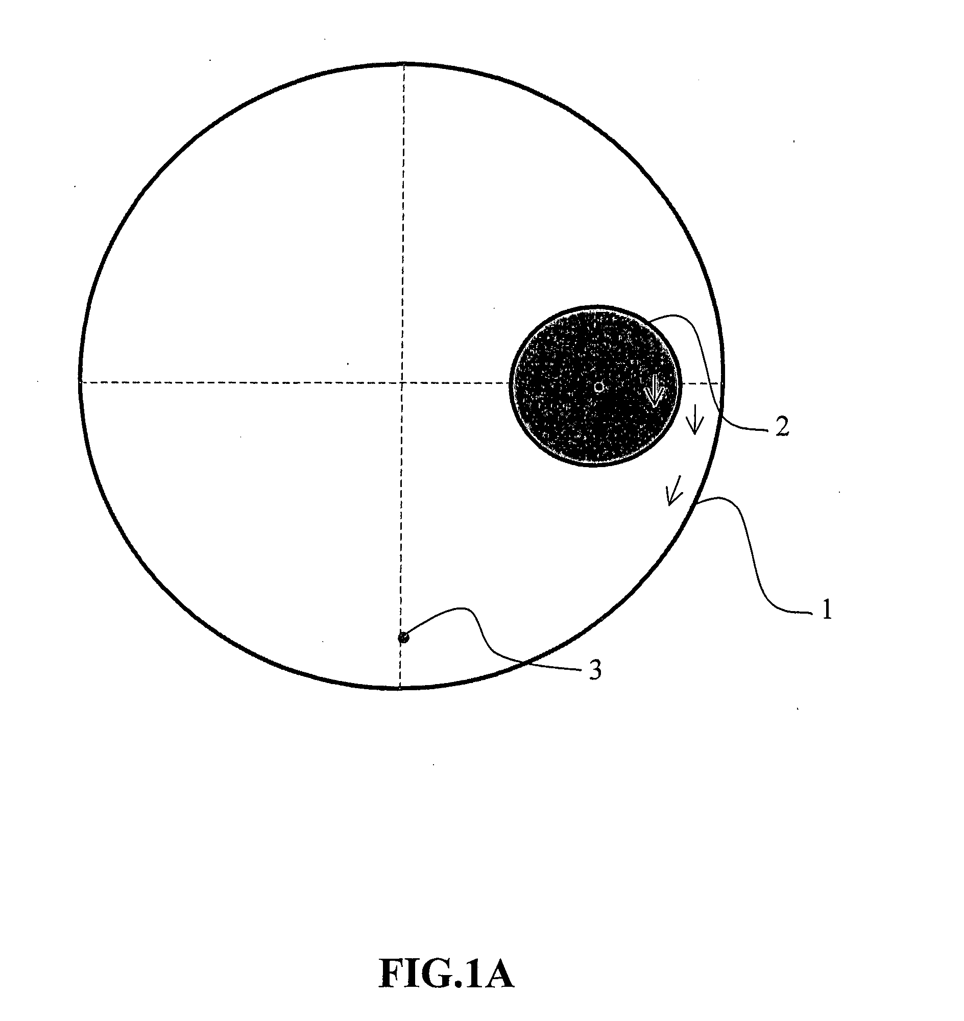
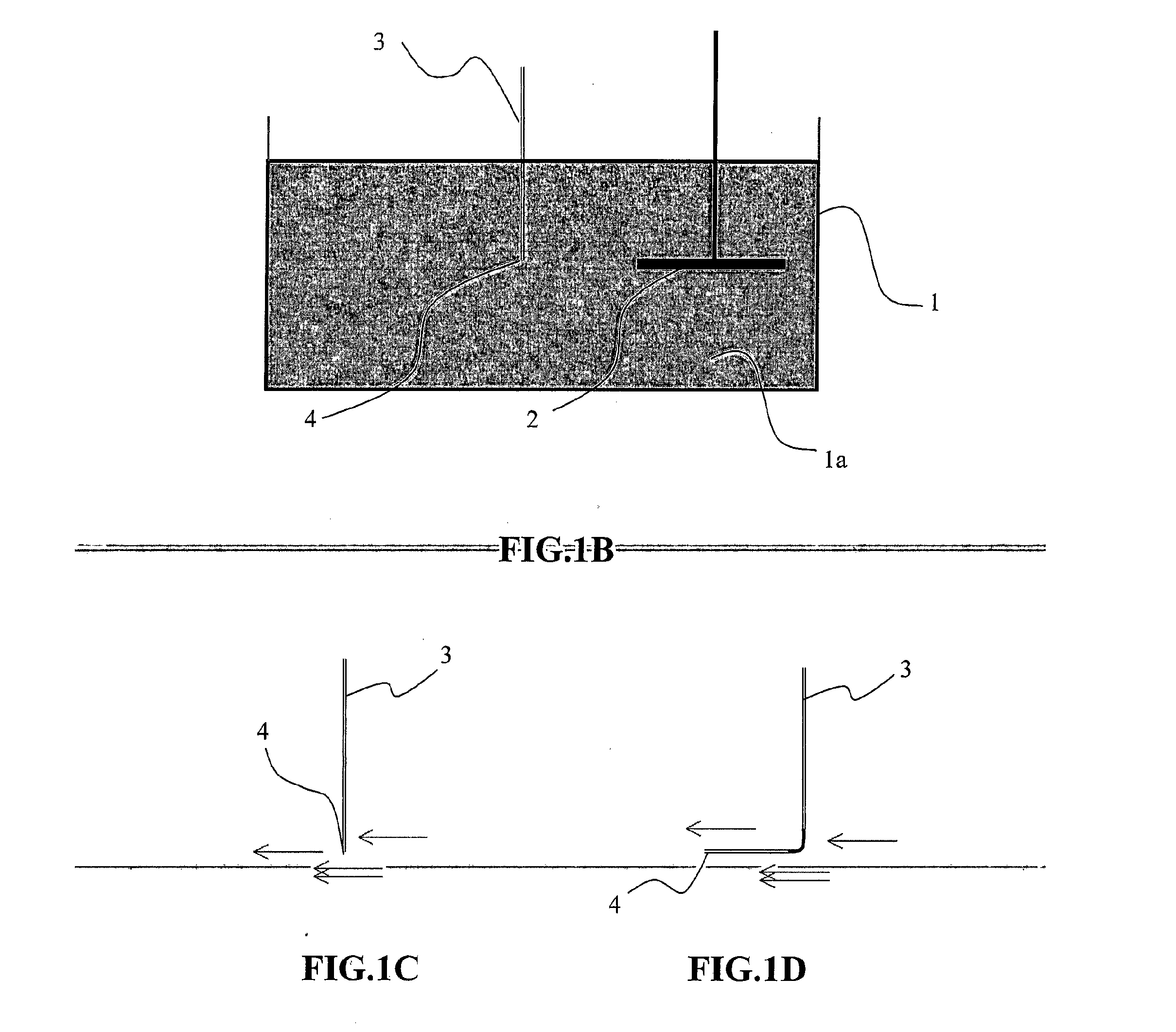
[0139]In the air atmosphere, at intense stirring, 6 grams of sodium hyaluronan with molecular weight of 1.7 MDa (determined by the SEC-MALLS method) was dissolved in 94 g of water with adding of 0.51 g NaOH to obtain a homogenous, well flowing, viscous solution suitable for spinning. This solution was spun by the wet method with a nozzle having the diameter of 0.4 mm into the spinning bath comprising propane-2-ol, stirred with an agitator, more specifically with the Heidolph DIAX 900 dispergator at 2500 rpm. The forming microfiber was collected from the “moving (non-stationary) bath” and transferred to the “maturation bath” consisting of propane-2-ol, where the coagulation of the fiber was completed. After 60 minutes, the microfiber was removed from the maturation bath and dried at the temperature of 60° C. The fiber character is shown in FIG. 3. The length of the microfibers obtained by this method is approximately 5-30 mm. This type of fibers is suitable for the construction of ba...

example 2

[0140]In the air atmosphere, at intense stirring, 6 grams of sodium hyaluronan with the molecular weight of 1.7 MDa (determined by the SEC-MALLS method) was dissolved in 94 g of water with adding of 0.51 g NaOH to obtain a homogenous, well flowing, viscous solution suitable for spinning. This solution was spun by the wet method with a nozzle having the diameter of 0.4 mm into the spinning bath comprising propane-2-ol, stirred with the Heidolph DIAX 900 dispergator at 2500 rpm. The forming microfiber was collected from the “moving bath” and transferred to the “maturation bath” consisting of propane-2-ol, where the coagulation of the fiber was completed. After 60 minutes the maturation bath containing the microfibers was poured into the Eta-Ergo knife mixer, where the fibers were shortened. Then the shortened fiber was filtrated off and the obtained filtration cake was compressed to the form of a “paper” sheet, and the produced product was dried at the room temperature (approx. 25° C....

example 3

[0142]In the air atmosphere, at intense stirring, 6 grams of sodium hyaluronan with the molecular weight of 1.7 MDa (determined by the SEC-MALLS method) was dissolved in 94 g of water with adding of 0.51 g NaOH to obtain a homogenous, well flowing, viscous solution suitable for spinning. This solution was spun by the wet method with a nozzle having the diameter of 0.4 mm into the spinning bath comprising propane-2-ol, stirred with the Heidolph DIAX 900 dispergator at 2500 rpm. The forming microfiber was collected from the “moving bath” and transferred to the “maturation bath” consisting of propane-2-ol, where the coagulation of the fiber was completed. After 60 minutes the maturation bath containing the microfibers was poured into the Eta-Ergo knife mixer, where the fibers were shortened. Then the shortened fiber was filtrated off and the filtration cake was dried without compression at 20° C. A voluminous layer of microfibers—a cotton wool showed in FIG. 5—was thereby obtained as a...


PUM

PropertyMeasurementUnit
Temperatureaaaaaaaaaa

Abstract

The invention relates to the method of the preparation of polysaccharide fibers based on hyaluronic acid, a compound comprising hyaluronic acid and metal ions, schizophylan, chitin / chitosan-glucan complex, a compound comprising chitin / chitosan-glucan complex and metal ions, internal mixture of chitin and chitosan, a compound comprising internal mixture of chitin and metal ions, sodium alginate, potassium alginate, ammonium alginate, xanthane, xanthane sodium salt, xanthane potassium salt, oxycellulose, oxycellulose sodium salt, oxycellulose potassium salt, carboxymethyl cellulose, carboxymethyl cellulose sodium salt, and carboxymethyl cellulose potassium salt, or a mixture of polysaccharides, in a non-stationary coagulation bath. Further the invention relates to covers of internal and external wounds and skin defects based on these fibers, and a method of production thereof, and the apparatus for the preparation of polysaccharide fibers.

Description

FIELD OF THE INVENTION[0001]This invention relates to a method and an apparatus for preparing polysaccharide staple microfibers, wound covers based on the microfibers, and the method of their preparation.BACKGROUND OF THE INVENTION[0002]Treatment of skin defects, and acute, or chronic wounds is a very important part of the modern medicine. This also relates to the increasing age of population being treated with the use of more and more modern procedures. Wound healing is a very complicated process which is nowadays studied intensively. Even though the partial steps of the healing process are known, the whole complex is still undiscovered. Nowadays the wet healing is preferred, based on supplying the humidity from the dressing, and using the regulated drainage of the tissue fluid. Use of a vacuum pump with the low pressure is also modern. During the last 10 years, the system based on the utilization of biological and physical properties of the hyaluronic acid has been prepared. Espec...

Claims


Application Information

Patent Timeline
IPC IPC(8): A61L15/28A61F13/00
CPCA61L15/28A61L2300/60A61L2300/802A61F13/00995D06M11/83D01D1/02D01D1/06D01D5/06D01D5/40D01F2/24D01F9/00D01F9/04D06M2101/02C08L5/08C08L1/04C08L1/286C08L5/00C08L5/04A61F13/00063Y02E60/10
Inventor BURGERT, LADISLAVHRDINA, RADIMVELEBNY, VLADIMIRABDEL-LATTIF, ABDEL MOHSENSULAKOVA, ROMANASOBOTKA, LUBOSBETAK, JIRISMIRNOU, DZIANIS
Owner CONTIPRO BIOTECH
Learn More