
Detection of bacterial (mollicutes) contamination

a technology of bacterial contamination and detection method, which is applied in the field of detection of bacterial contamination within biological samples, can solve the problems of difficult detection with a conventional microscope, small physical size of cells, and cell culture contamination, and achieve the enhancement of detection sensitivity and specificity of contaminant, and improve the pcr-based amplification of target sequences.

Inactive Publication Date: 2015-03-26
ROCHE DIAGNOSTICS OPERATIONS INC
View PDF0 Cites 0 Cited by
  • Summary
  • Abstract
  • Description
  • Claims
  • Application Information

AI Technical Summary

Benefits of technology

[0013]The present disclosure provides an improved PCR-based amplification of a target sequence by suppressing non-specific amplification products. This improvement concerns the use of a primer pair optimized to amplify a nucleic acid of a contaminant in the background of genomic DNA of a first organism. When DNA from a second organism, suspected as comprising the contaminant, is subjected to the same polymerase chain reaction (PCR) based amplification reaction, detection sensitivity and specificity of the contaminant is enhanced when an amount of genomic DNA of the first organism is present in the amplification reaction.
[0015]According to some embodiments, relative to the volume of the sample, a predetermined amount of DNA from CHO cells is added to at least one of the sample, the processed sample, the purified nucleic acids, or the reaction mixture. In such embodiments, the added DNA from CHO cells reduces unspecific amplification in the step of detecting.

Problems solved by technology

Mycoplasma cell culture contamination can occur due to contamination from individuals or contaminated cell culture medium ingredients, for example.
Mycoplasma cells are physically small—less than 1 μm—and are difficult to detect with a conventional microscope.
Severe Mycoplasma infections have the potential to destroy a cell line.
However, such linkage is considered controversial (see, Alexeeva, I., et al., J Clin Microbiol 44 (2006) 91-97, were a blinded study of rRNA species failed to detect any footprint of Spiroplasma in scrapie-infected hamster brain).
Moreover, the possibility of Mycoplasma contamination during biopharmaceutical production, cell therapy, and tissue engineering is also a concern and potential major problem.
These culture-based techniques require a long time to achieve results.
Furthermore, cultivation of certain Mycoplasma species may not be possible or reliable.

Method used


Image

  • Detection of bacterial (mollicutes) contamination
  • Detection of bacterial (mollicutes) contamination
  • Detection of bacterial (mollicutes) contamination

Examples


example 1

The MYCOTOOL Kit and Assay

[0075]The MYCOTOOL PCR Mycoplasma Detection Kit is an in vitro nucleic acid amplification test optimized for the detection of bacteria belonging to the Mollicutes. These include Mycoplasma hyorhinis, M. arginini, M. pneumoniae, M. fermentans, M. orale, M. pirium, M. salivarum, M. hominis, M. synoviae, Spiroplasma mirium, S. citri, and Acholeplasma laidlawii.

[0076]The MycoTool Kit comprises two subkits: Subkit 1 (“Detection Prep Kit”; Roche Applied Science Catalog No. 05184592001) and Subkit 2 (“Detection Amplification Kit”; Roche Applied Science Catalog No. 05184240001).

[0077]The kit was used exactly according to the instructions of the manufacturer.

[0078]If not stated otherwise, a liquid sample selected from (i) cell culture supernatant, (ii) a suspension of cultured cells and (iii) amniotic fluid was lysed by adding an aqueous buffer containing a guanidinium salt and proteinase K, mixing the buffer with the sample, and incubating the mixture to effect ly...

example 2

DNA from Mollicutes Species for Spiking of Samples

[0085]All samples used in the present Examples were from cultures free of prokaryotic contaminants.

[0086]Instead of sample material infected with a Mollicutes species, DNA prepared from Acholeplasma laidlawii or Mycoplasma orale was spiked to either sample material prior to lysis, or to isolated DNA prepared from sample material (if not indicated otherwise). For the purpose of spiking DNA was prepared from reference cultures (standard conditions) of Acholeplasma laidlawii (ATCC 27556) and Mycoplasma orale (ATCC 23714). For each culture the cell titer expressed as colony forming units (cfu) was determined. Quantities of spiked DNA which were applied reflected the number of cfu determined for the respective culture from which the DNA was prepared.

[0087]Mollicutes DNA as indicated above was used in the spiking experiments described below. The size of a typical specific PCR fragment amplified from Mollicutes DNA (specific amplification p...

example 3

Detection of GAPDH Genomic Target Sequences in Samples from MDCK Cell Culture (Suspended Cells)

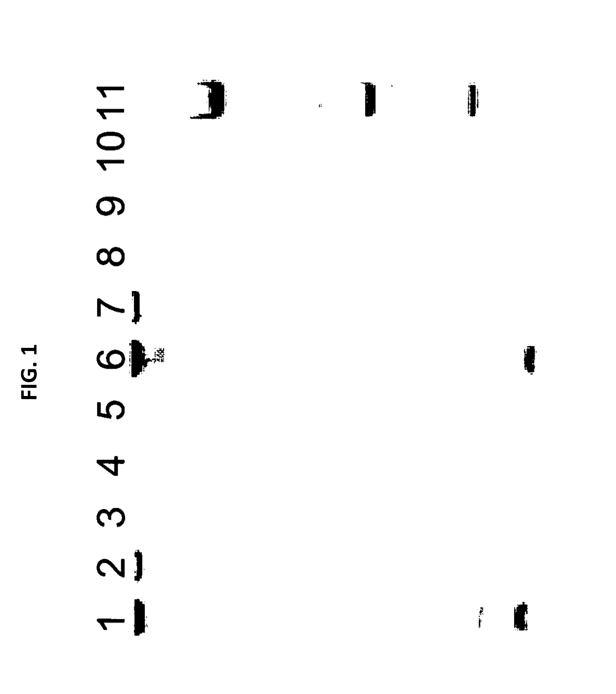
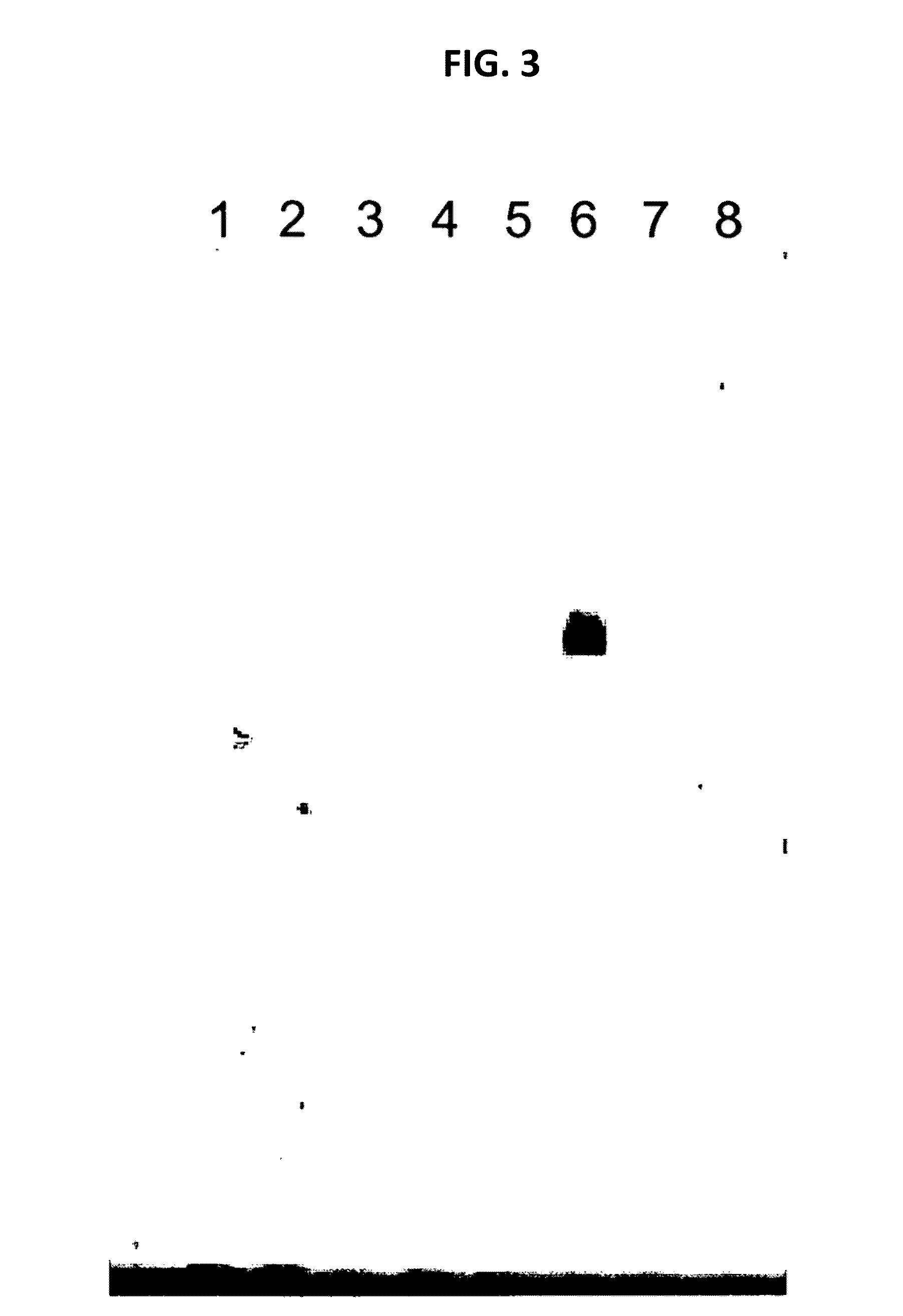
[0088]A culture of MDCK cells free of any prokaryotic organisms and having a cell titer of 0.79×10⁶ cells / ml was used. Acholeplasma laidlawii DNA was added to the sample at a concentration of 3 colony forming units (cfu) per 1 ml sample. Nucleic acids were isolated from the spiked sample material as specified in the instruction manual of the MYCOTOOL kit (see also Example 1). Two different nucleic acid preparations were made, the first without addition of CHO cell DNA, the second with the addition of CHO cell DNA (50 mg per 1 ml sample) to the sample material.

[0089]The preparation was repeated, however without spiking Acholeplasma laidlawii DNA to the sample. Again, nucleic acids were prepared with or without CHO cell DNA.

[0090]Several dilutions of the DNA preparations were made in TE buffer. An aliquot of each dilution was subjected to GAPDH-specific PCR according to the MYCOTOOL instruct...


PUM

PropertyMeasurementUnit
temperatureaaaaaaaaaa
pHaaaaaaaaaa
volumeaaaaaaaaaa

Abstract

The present disclosure provides a method and system for the PCR amplification of a target sequence which suppresses non-specific amplification products. The disclosure concerns the use of a primer pair optimized to amplify a nucleic acid of a contaminant in the background of genomic DNA of a first organism. When DNA from a second organism suspected for comprising the contaminant is subjected to the same PCR-based amplification reaction, detection sensitivity and specificity of the contaminant is enhanced when an amount of genomic DNA of the first organism is present in the amplification reaction.

Description

RELATED APPLICATIONS[0001]This application is a continuation of U.S. application Ser. No. 13 / 364,050 filed Feb. 1, 2012, which is a continuation of International Application No. PCT / EP2010 / 004655, filed Jul. 29, 2010, which claims the benefit of European Patent Application No. 09009965.6, filed Aug. 1, 2009, the disclosures of which are hereby incorporated by reference in their entirety.SEQUENCE LISTING[0002]The instant application contains a Sequence Listing which has been submitted in ASCII format via EFS-Web and is hereby incorporated by reference in its entirety. Said ASCII copy, created on Jan. 31, 2012, is named Sequence_Listing—26237_US.txt, and is 1,128 bytes in size.BACKGROUND[0003]1. Field of the Disclosure[0004]The present disclosure relates to contamination within biological samples. More specifically, the instant disclosure relates to a system and method for detecting bacterial contamination within biological samples.[0005]2. Description of the Related Art[0006]Mycoplas...

Claims


Application Information

Patent Timeline
Patent Type & Authority Applications(United States)
IPC IPC(8): C12Q1/68
CPCC12Q1/689C12Q1/6848C12Q2527/125C12Q2549/125C12Q1/686C12Q2527/137C12Q2600/158
Inventor BIRKNER, CHRISTIAN
Owner ROCHE DIAGNOSTICS OPERATIONS INC
Learn More