
Mr imaging with b1 mapping

a b1 mapping and magnetic resonance technology, applied in the field of magnetic resonance (mr) imaging, can solve the problems of difficult integration into clinical workflow, difficult bsub>1 /sub>mapping techniques, and become more difficult, so as to minimize t2* relaxation and fast readout

Active Publication Date: 2015-02-12
KONINKLJIJKE PHILIPS NV
View PDF11 Cites 14 Cited by
  • Summary
  • Abstract
  • Description
  • Claims
  • Application Information

AI Technical Summary

Benefits of technology

The patent describes a method for improving the efficiency of B1 mapping in magnetic resonance imaging (MRI) by combining a DREAM B1 mapping approach with a suppression sequence for MR signals emanating from the blood pool in the heart. This allows for dynamic RF shimming, where the B1 maps are automatically adapted to the motion of the patient. The method also includes a threshold-based masking of the B1 maps to avoid degradation due to flow artifacts and a plurality of FID and stimulated echo signals generated by a train of reading RF pulses with small flip angles. The suppression sequence may include a black-blood preparation pulse and other magnetization preparation pulses to further improve B1 mapping quality. Overall, the method enables faster and more robust B1 mapping in MRI, leading to improved image quality and reduced motion-induced artifacts.

Problems solved by technology

However, as the MR frequency increases, with increasing main magnetic field strength, this becomes more difficult due to conductive losses and wavelength effects within the body of the patient.
However, most B1 mapping techniques are relatively slow, making integration into the clinical workflow difficult.
This is in particular the case for MRA and, most notably, cardiac B1 mapping, where ECG triggering and breath-holding used to suppress physiological motion further reduces scan efficiency.

Method used


Image

  • Mr imaging with b1 mapping
  • Mr imaging with b1 mapping
  • Mr imaging with b1 mapping

Examples


Embodiment Construction

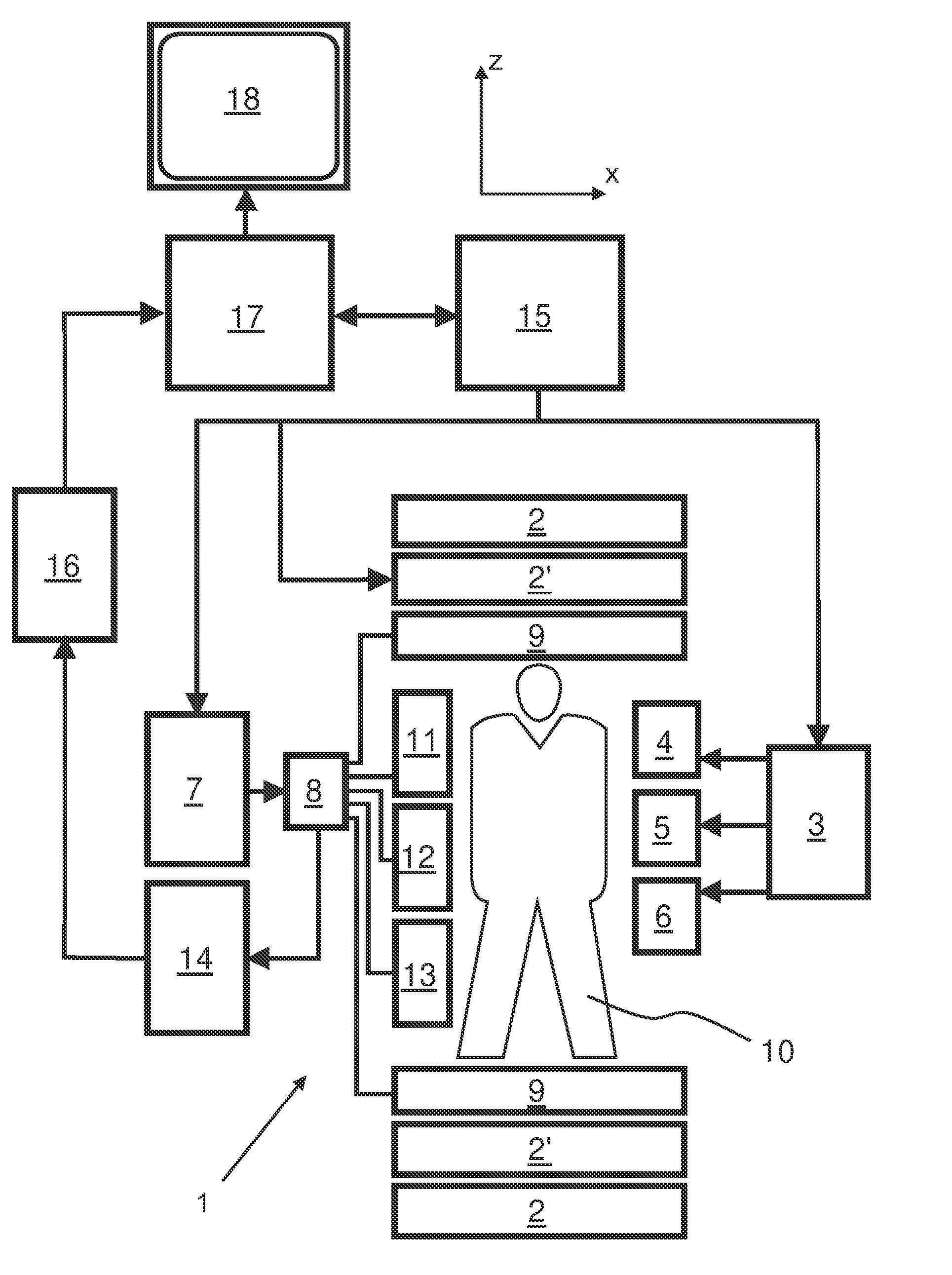
[0033]With reference to FIG. 1, a MR device 1 is shown. The device comprises superconducting or resistive main magnet coils 2 such that a substantially uniform, temporally constant main magnetic field B0 is created along a z-axis through an examination volume. The device further comprises a set of (1st, 2nd, and—where applicable—3rd order) shimming coils 2′, wherein the current flow through the individual shimming coils of the set 2′ is controllable for the purpose of minimizing B0 deviations within the examination volume.

[0034]A magnetic resonance generation and manipulation system applies a series of RF pulses and switched magnetic field gradients to invert or excite nuclear magnetic spins, induce magnetic resonance, refocus magnetic resonance, manipulate magnetic resonance, spatially and otherwise encode the magnetic resonance, saturate spins, and the like to perform MR imaging.

[0035]Most specifically, a gradient pulse amplifier 3 applies current pulses to selected ones of whole-...


PUM


Abstract

The invention relates to a method of MR imaging, wherein a portion of a body (10) placed in the examination volume of a MR device (1) is subjected to an imaging sequence (IMG) of RF pulses and switched magnetic field gradients. The imaging sequence (IMG) is a stimulated echo sequence including i) at least two preparation RF pulses (α) radiated toward the portion of the body (10) during a preparation period (21), and ii) one or more reading RF pulses (β) radiated toward the portion of the body (10) during an acquisition period (22) temporally subsequent to the preparation period (21). One or more FID signals (I1) and one or more stimulated echo signals (I2) are acquired during the acquisition period (22). A B1 map indicating the spatial distribution of the RF field of the RF pulses within the portion of the body (10) is derived from the acquired FID (I1) and stimulated echo (I2) signals. It is an object of the invention to provide an improved B1 mapping method which can be applied in magnetic resonance angiography (MRA) and, in particular, in cardiac imaging, where the acquisition of a complete B1 map needs to be performed within the diastolic phase of the heartbeat. The invention proposes that the portion of the body (10) is subjected to a suppression sequence (BB) for suppression of MR signals emanating from blood prior to the imaging sequence (IMG). Moreover, the invention relates to a MR device (1) and to a computer program for a MR device (1).

Description

FIELD OF THE INVENTION[0001]The invention relates to the field of magnetic resonance (MR) imaging. It concerns methods of MR imaging of at least a portion of a body. The invention also relates to a MR device and to a computer program to be run on a MR device.BACKGROUND OF THE INVENTION[0002]Image-forming MR methods which utilize the interaction between magnetic fields and nuclear spins in order to form two-dimensional or three-dimensional images are widely used nowadays, notably in the field of medical diagnostics, because for the imaging of soft tissue they are superior to other imaging methods in many respects, do not require ionizing radiation and are usually not invasive.[0003]According to the MR method in general, the body of the patient to be examined is arranged in a strong, uniform magnetic field (B0 field) whose direction at the same time defines an axis (normally the z-axis) of the co-ordinate system on which the measurement is based. The magnetic field produces different ...

Claims


Application Information

Patent Timeline
IPC IPC(8): G01R33/565A61B5/026A61B5/055G01R33/24G01R33/561
CPCG01R33/5659G01R33/246A61B5/0263G01R33/56509A61B5/055G01R33/5612
Inventor NEHRKE, KAYBOERNERT, PETER
Owner KONINKLJIJKE PHILIPS NV
Learn More