
Bioartificial proximal tubule systems and methods of use

Inactive Publication Date: 2012-11-29
DEPUY SYNTHES PROD INC
View PDF2 Cites 10 Cited by
  • Summary
  • Abstract
  • Description
  • Claims
  • Application Information

AI Technical Summary

Benefits of technology

[0025]Another embodiment of the invention is a method of differentiating one or more cells differentiable into renal cells into renal proximal tubule epithelial cells under conditions sufficient to allow the differentiation, whereby the differentiated cells form an epithelial monolayer and whereby tension is applied to the cells. The method may further comprise the step of seeding the cells on a decellularized scaffold prior to differentiating the cells. When the method comprises this step, the method can be used to produce a bioartificial device of the invention. The decellularized biological matrix scaffold may be derived from mammalian (such as e.g. porcine) tissue such as mucosal or submucosal tissue. In one embodiment, the decellularized biological matrix scaffold is derived from a mammalian alimentary canal. In another embodiment, the decellularized biological matrix scaffold is derived from the stomach, duodenum, jejunum, ileum or colon of a mammal. The one or more cells differentiable into renal cells is selected from the group consisting of primary renal tubule epithelial cells, inducible pluripotent stem cells or progenitor cells differentiated into renal cells or renal progenitor cells, stem cells isolated from the kidney or progenitor cells isolated from the kidney and mixtures thereof. In one embodiment, the one or more cells differentiable into renal cells are mammalian kidney-derived cells (such as e.g. human kidney-derived cells). The mammalian kidney-derived cells may be obtained from the kidney cortex, kidney medulla, kidney subcapsular region and mixtures thereof. In one embodiment, the kidney-derived cells are capable of self-renewal and expansion in culture, positive for the expression of one or more of Oct-4, Pax-2, and Rex-1 and negative for the expression of one or more of Sox2, FGF4, hTert and Wnt-4. In an alternate embodiment, the kidney-derived cells are capable of self-renewal and expansion in culture, positive for expression of at least one of Eya1, Pax2, WT1, FoxD1, BMP7, BMP2, GDF5, EpoR or Rex-1, and negative for expression of at least one of Sox2, FGF4, hTert or Wnt-4. In yet another embodiment, the kidney-derived cells are also positive for at least one of cell-surface markers HLA I, CD24, CD29, CD44, CD49c, CD73, CD166, or SSEA-4, and negative for at least one of cell-surface markers HLA II, CD31, CD34, CD45, CD56, CD80, CD86, CD104, CD105, CD117, CD133, CD138, CD141, or E-cadherin.

Problems solved by technology

This results in an inability of the kidney to excrete toxic metabolic wastes produced by the body.
In the United States, CKD is becoming increasingly common and is associated with poor health outcomes and high medical costs.
As a consequence of carrying out this important function, renal proximal tubule cells are often damaged by the toxic effect of these compounds.
However, the use of these cell lines has several disadvantages.
Furthermore, many of these cell lines are not human-derived.
The use of primary cells is cumbersome as the cells are typically freshly isolated and minimally expanded prior to being used for experiments.
The isolation process can be laborious with contamination of unwanted cell populations.
In addition, there can be significant variability of the donor source material.
Another common issue is the limited duration in which the primary cells will form an intact monolayer; this limits the cells utility for in vitro studies.
Porous filters (such as e.g. TRANSWELL® filters), while somewhat effective, are made from synthetic materials and, therefore, do not accurately represent the underlying matrix that renal tubule cells are typically exposed to in vivo.
The latter is problematic due to the laborious isolation technique and species differences that need to be considered.
Another disadvantage of these alternatives is their ability to only assess transport of xenobiotic compounds, which are applied to the culture media, into the lumen of the renal tubules.
The effect of such compounds on the normal functions of the tubules, such as e.g. glucose reabsorption or albumin uptake, cannot be assessed since there is no reliable way to introduce labeled test substances into the lumen or take samples of the luminal fluid of the tubules for assay.
In addition, the effects of changes in flow and physiological dynamic conditions that may occur under various in vivo scenarios cannot be assessed.

Method used


Image

  • Bioartificial proximal tubule systems and methods of use
  • Bioartificial proximal tubule systems and methods of use
  • Bioartificial proximal tubule systems and methods of use

Examples


example 1

Seeding and Differentiation of hKDC on Extracellular Matrix Scaffolds

[0095]This experiment tests the attachment, growth and differentiation of human kidney-derived cells (“hKDC”) on various configurations of extracellular (i.e decellularized) matrix scaffolds as well as to the traditional culture on collagen-coated transwells.

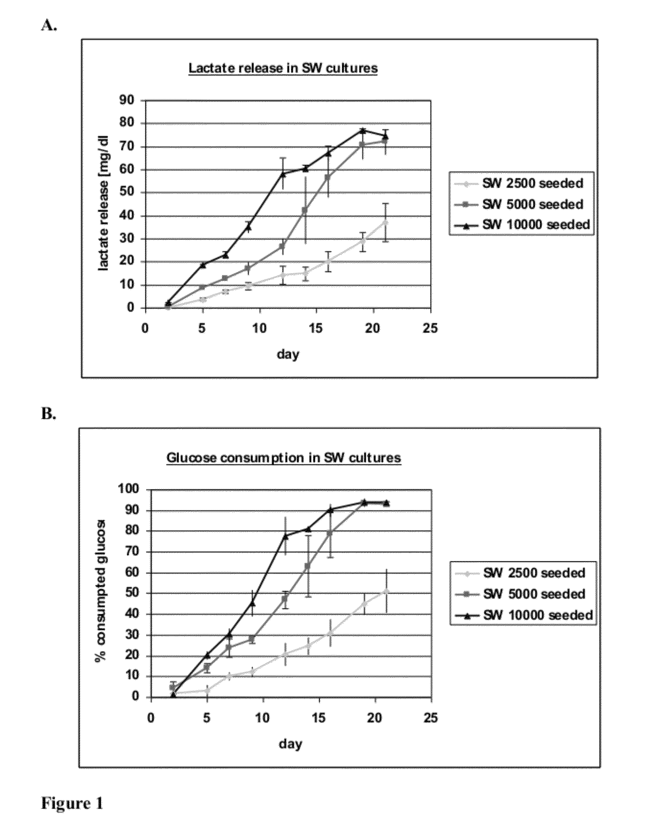
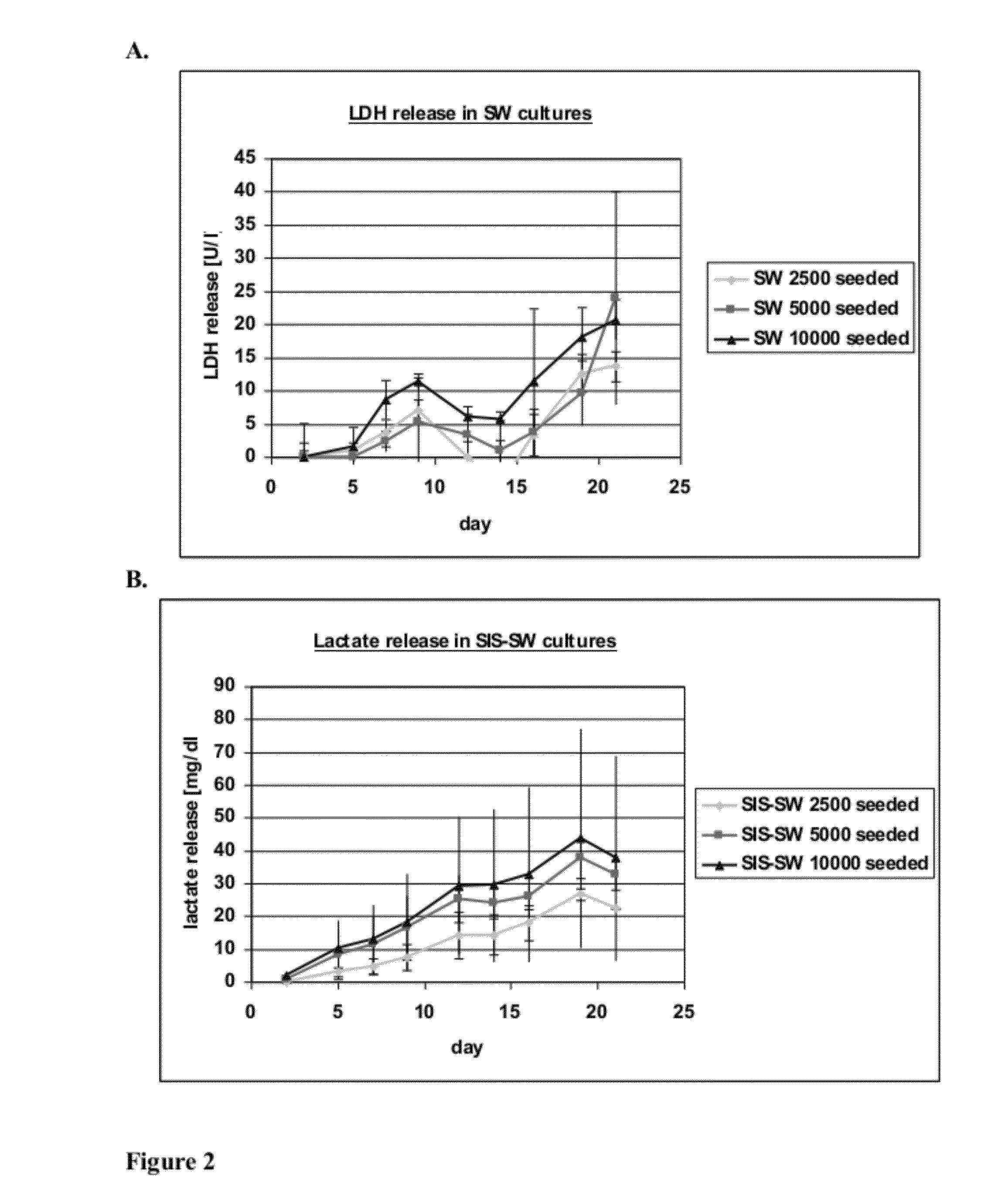
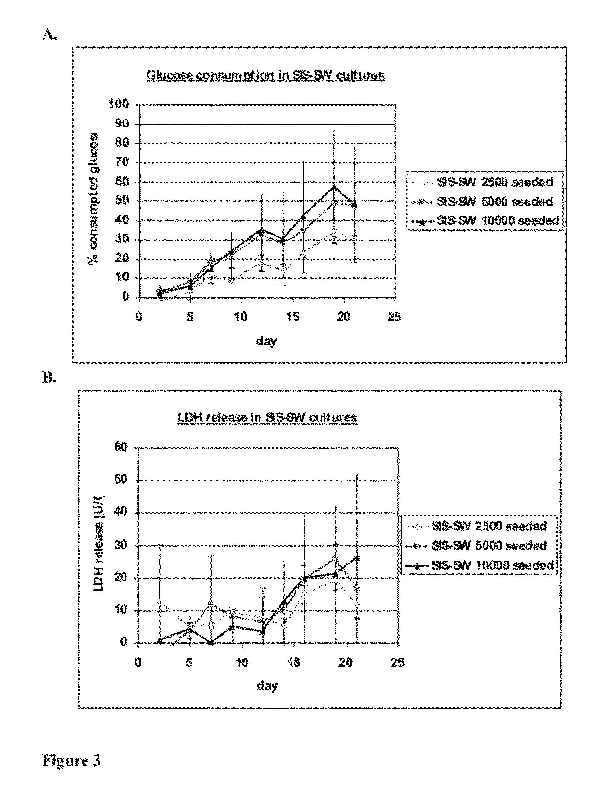
[0096]hKDC at passage 4 were seeded onto three different scaffold configurations and on transwells (Corning, Corning N.Y.). The cells were cultivated over a time period of three weeks with REGM™ renal epithelial growth medium (Lonza, Walkersville Md.). Each scaffold configuration was tested with three different cell concentrations: 2.5×10³, 5×10³, and 1×10⁴ cells. The configurations tested were: 1) collagen sandwich culture; 2) collagen-SIS sandwich culture; 3) SIS monolayer culture; and 4) collagen-coated transwells.

[0097]Collagen Sandwich Cultures

[0098]hKDC were cultivated between two collagen gel layers in 24-well plates. The bottom gels were cast by first m...

example 2

Optimization of Cell Seeding Concentration of hKDC on Decellularized Scaffolds

[0110]Example 1 demonstrated that cells seeded onto two-dimensional decellularized scaffolds without collagen formed a confluent epithelial monolayer expressing proximal tubule markers after three weeks of culture. The following experiments were conducted to optimize the cell seeding density and attempt to reduce the culture period necessary for formation of the monolayer.

[0111]Scaffolds were prepared by decellularizing segments of small intestine submucosa (SIS) as described in Example 1. hKDC at passage 4 were seeded onto the SIS scaffolds at three different concentrations (1×10⁴, 5×10⁴, and 1×10⁵ cells / well) and cultivated for three weeks with REGM™ renal epithelial growth medium (Lonza, Walkersville). Samples were removed for histological analysis after two and three weeks and were fixed with Bouin's fixative for 1 hour. Thereafter they were washed in water for at least 4 hours and embedded into paraff...

example 3

Immunohistochemistry of p-glycoprotein-1

[0118]Scaffolds were prepared by decellularizing segments of small intestine submucosa (SIS) as described in Example 1. hKDC at passage 4 were seeded onto the SIS scaffolds at 5×10⁴ cells / scaffold and cultivated for three weeks with REGM™ renal epithelial growth medium (Lonza, Walkersville). Samples were removed for histological analysis after three weeks and fixed with Bouin's fixative for 1 hour. Afterwards, they were washed in water for at least 4 hours and embedded into paraffin. Subsequently, 3 μm thick slices were prepared. Immunohistochemistry (IHC) was performed to confirm the expression of p-glycoprotein-1 (pgp-1 aka MDR1), which is an efflux transporter expressed by proximal tubule cells. Target retrieval of deparaffinized sections was achieved by enzymatic treatment with proteinase K, by heating in citrate buffer pH 6 (Dako, #S2369), or by heating in Tris / EDTA buffer pH 9 (Dako, #S2367). A blocking step with 3% hydrogen peroxidase w...


PUM


Abstract

This application invention discloses bioartificial proximal tubule device, constructed by preparing a decellularized biological matrix, seeding the biological matrix with mammalian kidney-derived cells and optionally mammalian endothelial cells. The device may then be cultured statically or matured using bioreactor culture to allow differentiation of the kidney cells into functioning proximal tubule cells. The device is capable of carrying out proximal tubule functions. The application also describes various methods of making the proximal tubule devices. The application also further describes methods of use of bioartificial proximal tubule devices for e.g. in vitro studies of tubule cell transport, toxicity effects of various compounds or pharmaceutical compound screening.

Description

CROSS-REFERENCE TO RELATED APPLICATIONS[0001]This application claims the benefit of U.S. Provisional Application No. 61 / 490,890, filed May 27, 2011, the content of which is incorporated by reference in its entirety.FIELD OF THE INVENTION[0002]The invention generally relates to a bioartificial proximal tubule device comprising a biological scaffold and one or more progenitor cells (such as a e.g. mammalian kidney-derived cells) that are differentiated into a renal proximal tubule cell monolayer on the scaffold. The invention further relates to the methods of preparing and culturing the device in a bioreactor. Also provided are methods of use of the device for in vitro nephrotoxicity or pharmaceutical compound screening.BACKGROUND OF THE INVENTION[0003]Chronic kidney disease (CKD) and end-stage renal disease (ESRD) are defined by a decline in renal function, primarily the glomerular filtration rate. This results in an inability of the kidney to excrete toxic metabolic wastes produced ...

Claims


Application Information

Patent Timeline
IPC IPC(8): C12N5/071
CPCC12N5/0686C12N2533/92C12N2533/54C12N2503/00C12N5/0602A61L27/38
Inventor KAZANECKI, CHRISTIANCOLTER, DAVID C.SCHANZ, JOHANNAHOPPENSACK, ANKEHANSMANN, JANWALLES, HEIKE
Owner DEPUY SYNTHES PROD INC
Learn More