Methods and devices for high throughput crystal structure analysis by electron diffraction

a crystal structure and electron diffraction technology, applied in the direction of material analysis using wave/particle radiation, instruments, nuclear engineering, etc., can solve the problems of limiting the contribution of x-ray crystallography and consequently to any attempt at high throughput analysis to nanoscience, and the traditional method of atomic structure determination, x-ray crystallography, etc., to achieve improved identification/fingerprinting of individual grains/crystallites, high throughput rates, and improved quality orientation phase map

Active Publication Date: 2011-09-15
NANOMEGAS SPRL +2
View PDF3 Cites 46 Cited by
  • Summary
  • Abstract
  • Description
  • Claims
  • Application Information

AI Technical Summary

Benefits of technology

[0037]Another embodiment of the invention is a method and a device as described above where enhanced identification/fingerprinting of individual grains/crystallites and better quality orientation phase maps are obtained by combining beam scanning according to the method described above with ED energy filtering.
[0038]Another embodiment of the inventio...

Problems solved by technology

Unfortunately, the traditional method for atomic structure determination, X-ray crystallography, cannot be used for single crystals with sizes in the nanometer range, either because the limit size for single crystal structure determination with modern Synchrotron sources is above 5 microns, or because powder X-Ray patterns show extremely poor resolution (e.g. peak broadening and overlapping) at decreasing crystallite size (nm).
This simple fact limits the contribution of X-ray crystallography and consequently to any attempt of high throughput analysis to nanoscience, a scientific area crucial in many fields, from semiconductors to pharmaceuticals and proteins.
Therefore, compounds that only exist at nanocrystalline state are usually out of reach for the X-ray diffraction methods of structure determination and as a result, such nanostructures, despite their given importance, are unknown.
The resulting lack of knowledge on the underlying structure-property relationships often prohibits breakthrough developments in a whole research sector or may cause fatal delays in the cycle of further product development.
The resulting lack of knowledge of the underlying structure-property relationships often prohibits breakthrough developments in a whole research sector, or may cause fatal delays in the cycle of further product development.
Transmission electron microscopy (TEM) which uses an electron beam in transmission mode, gives a direct image of the structure, but suffers from lens distortions and limited resolution.
This is contrary to the use of an electron beam in electron diffraction techniques which can go to very high resolution e.g. 0.5 Ångström or even higher, but suffer from dynamic scattering.
However, it is currently not possible to use ED intensities directly (unlike X-Ray techniques) to solve crystal structures; these drawbacks currently limit general use of ED as reliable technique for nanocrystal structure analysis.
Conventional TEM used in diffraction mode do not always allow high-tilt angles due to restrictions caused by the geometr...

Method used


Image

  • Methods and devices for high throughput crystal structure analysis by electron diffraction
  • Methods and devices for high throughput crystal structure analysis by electron diffraction
  • Methods and devices for high throughput crystal structure analysis by electron diffraction

Examples


example 1

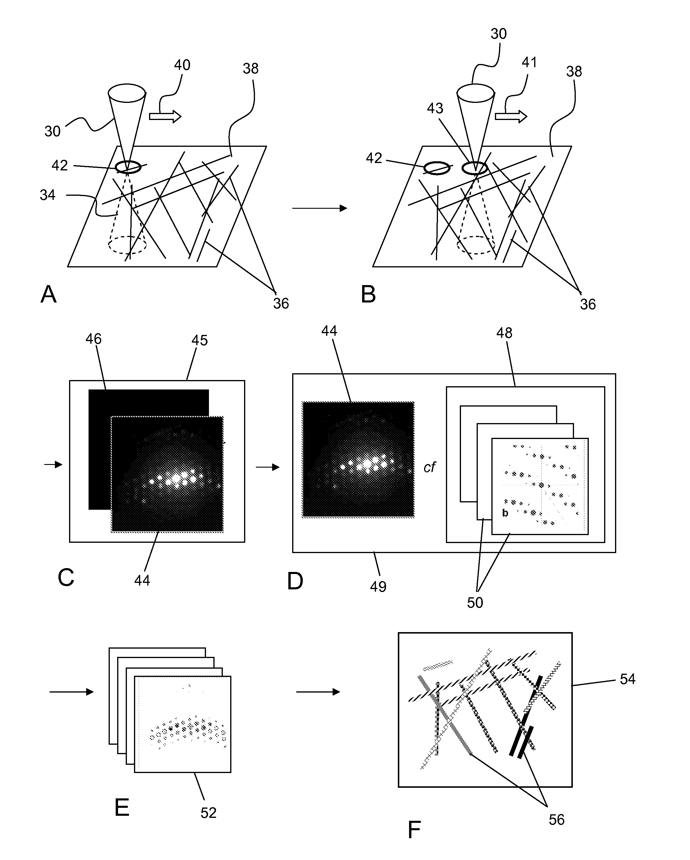
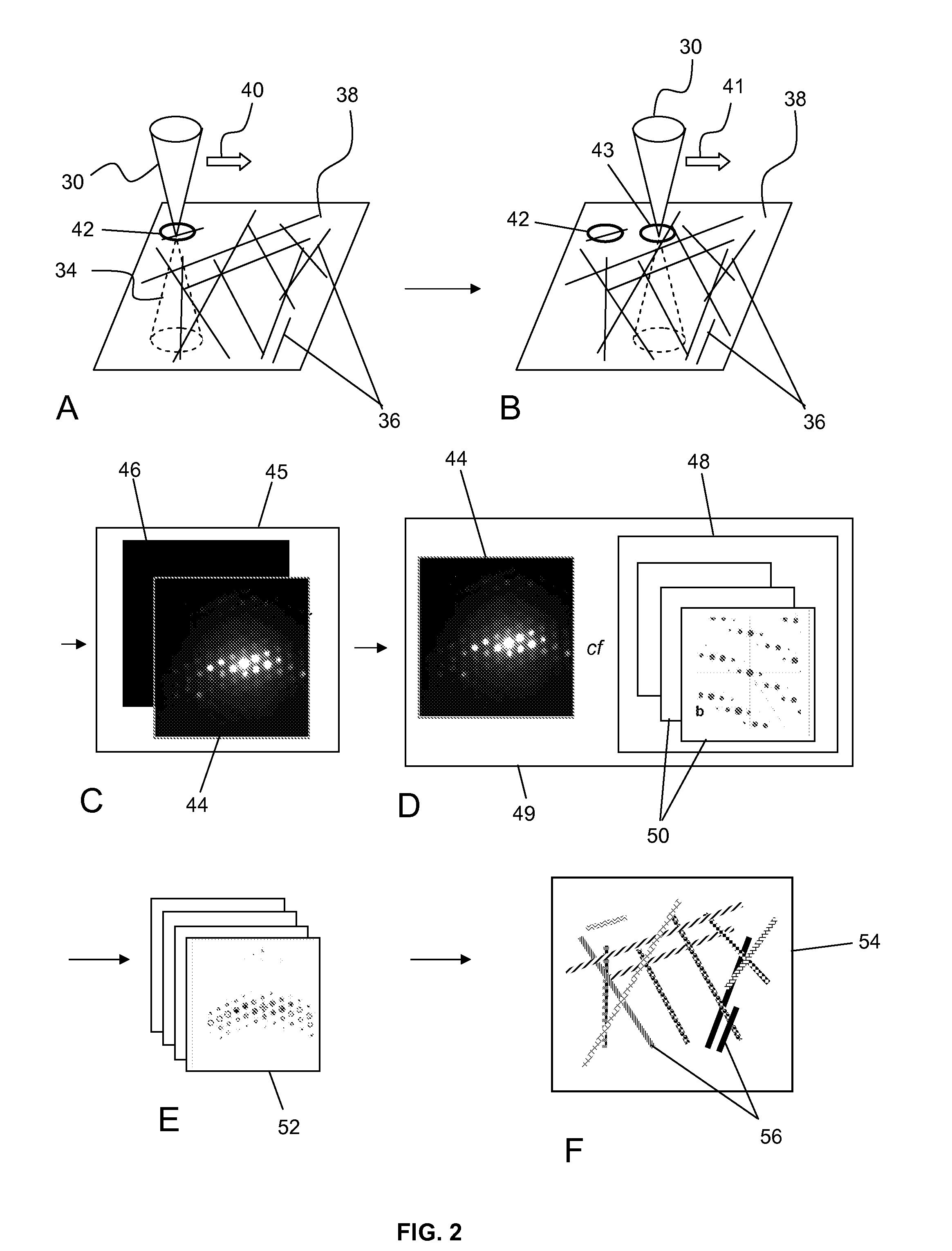
[0141]An example of template matching to determine the orientation of crystals in a sample of nanocrystalline copper is depicted in FIG. 5. FIGS. 5A and 5B show simulated templates generated for all orientations over the stereographic triangle. Shown in FIG. 5C is the experimental ED pattern acquired using a TEM equipped with a CCD camera of one location in the sample of nanocrystalline copper. The ED pattern was compared to every simulated template; shown in FIG. 5D is a matching template superimposed with circles from the experimental ED pattern. A correlation indexes map was generated as shown FIG. 5E showing the best match, i.e. the most probable crystal orientation at the point of acquisition.

example 2

[0142]An example of template matching to determine the orientation of crystals in a sample of asbestos fiber crystals is depicted in FIG. 6 when a scanning protocol (precession) is not used compared with when it is used. Beam scanning is used in both cases. An experimental ED pattern was first acquired using a TEM at a location in the sample of asbestos fiber crystals with precession not used, resulting in an ED pattern shown in FIG. 6A captured by a CCD camera. The ED pattern was compared to every simulated template; shown in FIG. 6B is a matching template superimposed with circles from the experimental ED pattern. A corresponding correlation index map was generated, with the orientation indicated with a white spot (S), as shown in FIG. 6C. According to matching, ED pattern is 0.65° misoriented in relation to the nearest ZA [1 01]. An experiments ED pattern was then acquired at the same location in the sample of asbestos fiber crystals with precession used, resulting in an ED patte...

example 3

[0143]A further example of template matching to determine the orientation of crystals in a sample of asbestos fiber crystals is depicted in FIG. 7 employing beam scanning, when a scanning protocol (precession) is not used compared with when it is used. Beam scanning is used in both cases. An experimental ED pattern was first acquired using a TEM at a location in the sample of asbestos fiber crystals with precession not used, resulting in an ED pattern shown in FIG. 7A captured by a CCD camera. The ED pattern was compared to every simulated template; shown in FIG. 7B is a matching template superimposed with circles from the experimental ED pattern. A corresponding correlation index map was generated, with the orientation indicated with a white spot (S), as shown in FIG. 7C. According to matching ED pattern is −2° misoriented in relation to the nearest ZA [1-10]. An experiment ED pattern was then acquired at the same location in the sample of asbestos fiber crystals with precession us...


PUM


Abstract

A method and device for electron diffraction tomography of a crystal sample, which employs scanning of the electron beam over a plurality of discrete locations of the sample, in combination with a beam scanning protocol as the beam converges at every discrete location (42, 43) of the sample (38) to obtain a series of electron diffraction patterns, use of template matching to determine crystal orientations and thickness maps to obtain a common intensity scaling factor.

Description

FIELD OF THE INVENTION[0001]The present invention is in the field of determination of crystal structure characteristics, such as internal three dimensional structure, crystal orientation, phase, and thickness by an electron diffraction analyzer, using techniques that are suitable for high-throughput structural analysis, and which can be applied to crystals having dimensions in the nanometer range.BACKGROUND[0002]High throughput X-Ray powder screening of materials for useful properties reduce development times by up to orders of magnitudes, at a fraction of costs. For example, ability to unequivocally differentiate one crystallographic phase from another has proven to be the greatest asset in the pharmaceutical field: powder X-Ray diffraction patterns of each crystalline form contain characteristic sets of peaks unique to that form, allowing the patterns to be used as fingerprints. Fully automatic powder crystal data evaluation may allow speedy (pattern analyzed in minutes) and cost ...

Claims


Application Information

Patent Timeline
IPC IPC(8): H01J37/26
CPCG01N23/20058H01J37/05H01J37/222H01J37/256H01J2237/2802H01J37/2955H01J2237/226H01J2237/25H01J37/28
Inventor NICOLOPOULOS, STAVROSBULTREYS, DANIELRAUCH, EDGARD
Owner NANOMEGAS SPRL
Why Eureka
Try Eureka