
Fluorescent emulsion of indocyanine green

a technology of indocyanine green and fluorescence, which is applied in the field of fluorescence emulsion of indocyanine green, can solve the problems of poor soluble in water, difficult use of indocyanine green fluorescent labels, and some properties of indocyanine green, so as to improve imaging performance, improve optical characteristics, and improve performan

Inactive Publication Date: 2011-08-18
COMMISSARIAT A LENERGIE ATOMIQUE ET AUX ENERGIES ALTERNATIVES
View PDF6 Cites 5 Cited by
  • Summary
  • Abstract
  • Description
  • Claims
  • Application Information

AI Technical Summary

Benefits of technology

The invention proposes a formulation of indocyanine green (ICG) in the form of a nanoemulsion, which has improved optical characteristics and can be used as a diagnostic agent. The nanoemulsion is stable and can be prepared in an isotonic medium, making it suitable for injection in humans. The formulation is easy to prepare, inexpensive, and can be adapted to different pharmaco-kinetics. It can also be functionalised and used for fluorescence imaging in clinical applications.

Problems solved by technology

However, ICG has some properties which render its use as a fluorescent label problematic.
First of all, ICG is an amphiphilic compound with a solubility of from 5 to 10 mg / ml and is therefore poorly soluble in water.
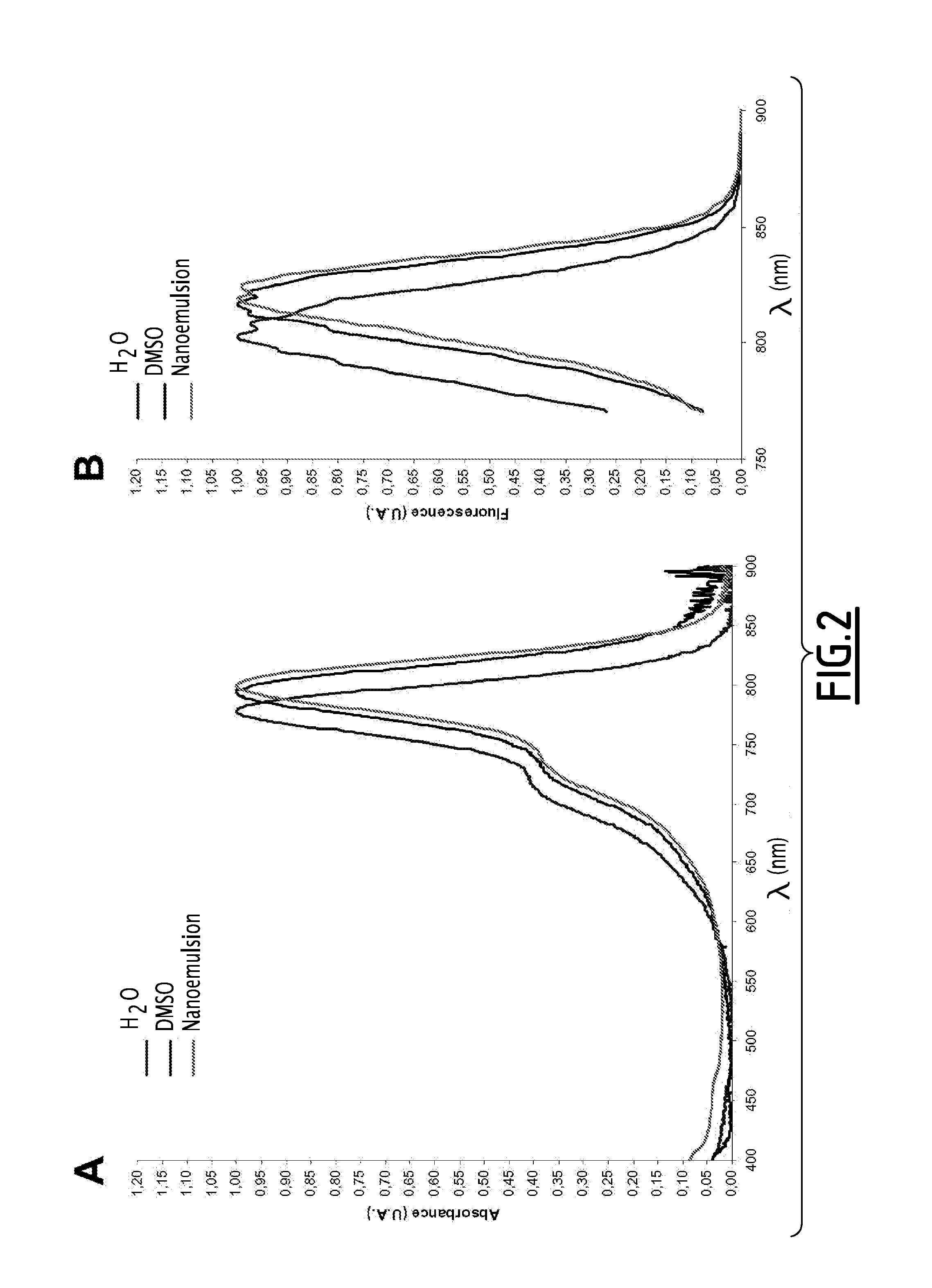
Moreover, ICG has poor stability and a low fluorescence quantum yield in aqueous solution, especially because of the formation of those poorly emissive dimers.
In addition, ICG is adsorbed on plasma proteins to a considerable degree when injected by the intravenous route, which alters the absorption and emission spectrum.
It is therefore difficult to distinguish the ICG fluorescence from the tissue autofluorescence using a fluorescence device based on pulsed luminous excitation.
Finally, liposomes are vesicles with a double-layer shell and generally have particle sizes greater than 100 nm in diameter; solutions of particles of that size diffuse light and do not permit satisfactory extravasation of the blood circulation to tumour tissues and internalisation in the cells.
However, formulations based on non-covalent adsorption bonds have low chemical stability and limit the choice of biological targeting ligands which can be used.
However, such synthetic liposomes are complicated to synthesise, require the use of synthetic polymers and do not directly yield nanoparticles suitable for permitting successful extravasation of the blood circulation to tumour tissues and internalisation in the cells, namely less than 100 nm and even less than 50 nm.
However, that type of formulation leads in most cases to emulsions in which the droplet size is too large to limit the diffusion of light and ensure satisfactory colloidal stability and furtivity after injection in vivo.

Method used


Image

  • Fluorescent emulsion of indocyanine green
  • Fluorescent emulsion of indocyanine green
  • Fluorescent emulsion of indocyanine green

Examples


example 1

Formulation of ICG in Emulsion Form

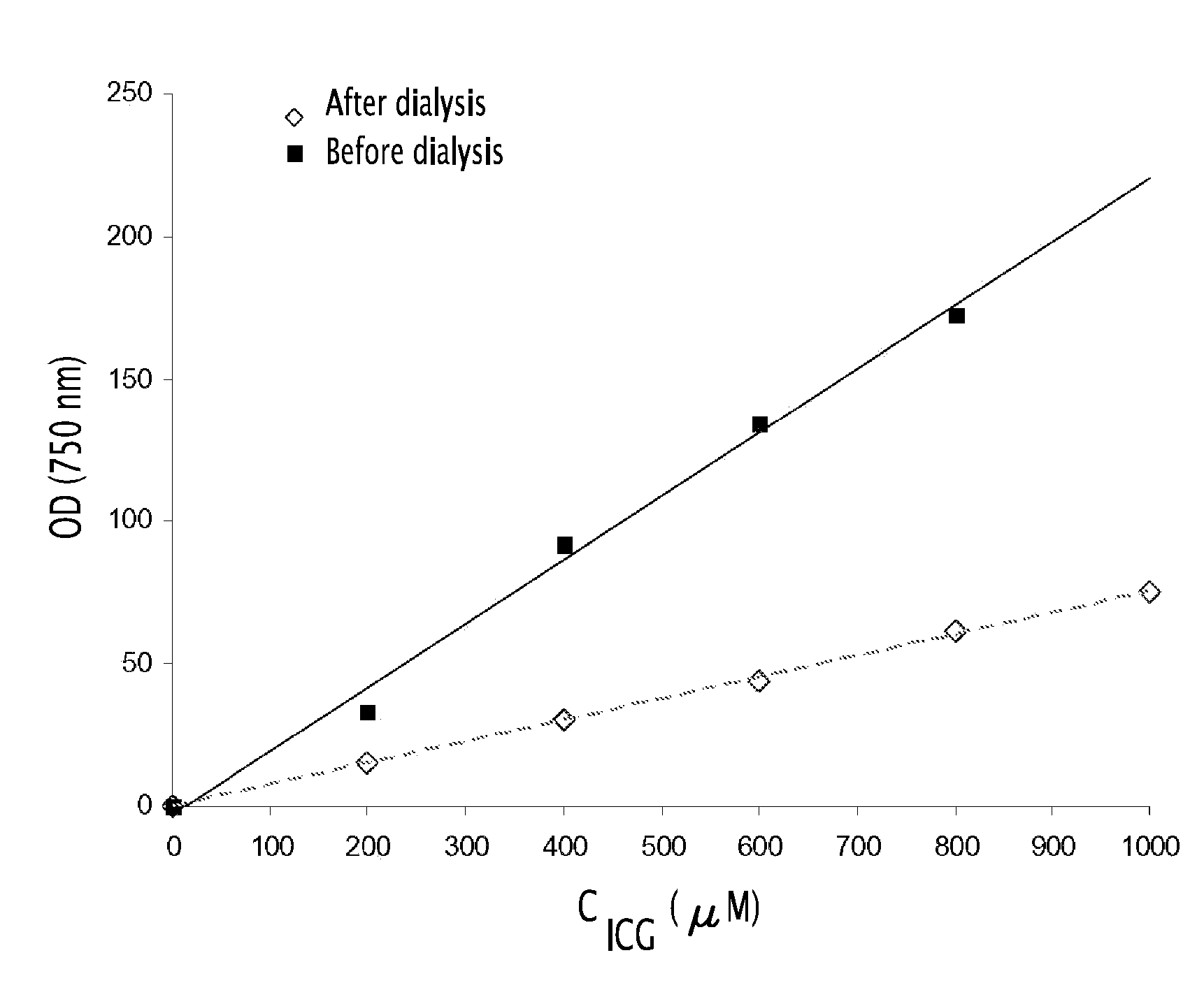
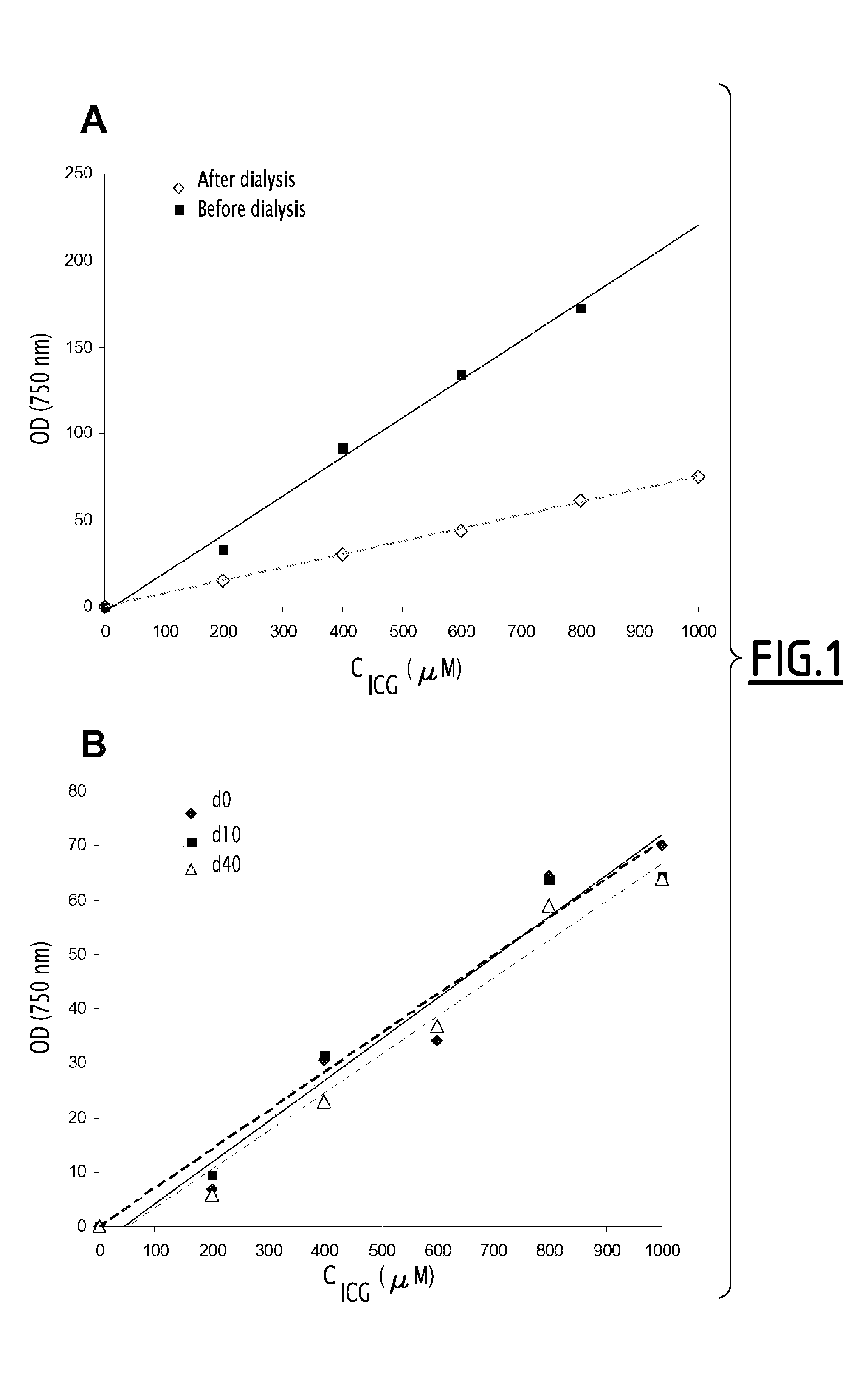
[0134]In a suitable container there was prepared a premixture constituted by 0.05 g of soybean oil (Sigma-Aldrich), 0.150 g of semi-synthetic glycerides sold under the trade name Suppocire® NC (Gattefossé) and 0.310 mg and 9.30 mg of ICG (Cardiogreen, Sigma-Aldrich or Infracyanine Serb laboratoires) in solution in dimethylsulfoxide (DMSO) as well as 0.100 g of soybean lecithin (enriched with 45% phosphatidylcholine) sold by Lipoïd under the trade name Lipoïd® S45.

[0135]After evaporation of the DMSO in vacuo, the residue is heated to 50-60° C. and the liquid mixture is maintained at that temperature for the emulsification (at ambient temperature the mixture becomes waxy).

[0136]The continuous phase was prepared by mixing 0.05 g of glycerol, 0.331 g of polyoxyethylene stearate having 50 ethylene oxide units, sold under the trade name Myrj® 53 by ICI Americas Inc., and 154 mM sodium chloride solution to make the mixture up to 1.7 g. The solution was th...

example 2

Imaging of Vascularisation

[0149]Male rats of the strain Sprague Dawley (Harlan France) are anaesthetised by isoflurane inhalation (4% for induction and 2% for maintenance) and then placed beneath an imaging device. The device is constituted by: 1) a light source emitting in the excitation band of ICG and the excitation power of which is of the order of several mW / cm2 and 2) a CCD camera coupled to a lens adapted to the observed sample. The assembly is equipped with filters which allow the excitation light and flare to be avoided and only the fluorescence light to be collected. Dissection of the region of the neck is then carried out in order to isolate the carotid and the jugular vein. An ICG formulation prepared as described in Example 1 with a load rate of 350 μM after dialysis is then injected intravenously. Fluorescence imaging of the vascular system in that region is then carried out with visualisation firstly of the carotid and then of the jugular vein as shown in FIG. 7. This...

example 3

Imaging of Sentinel Nodes

[0150]Female mice of the strain Nude (Janvier) are anaesthetised by isoflurane inhalation (4% for induction and 2% for maintenance) and then placed beneath the fluorescence imaging device described above.

[0151]10 μA of an ICG formulation (0.5 nmole injected) prepared as described in Example 1 or of ICG dissolved in glucose water (1 nmole injected) are injected intradermally into the right rear paw.

[0152]Temporal monitoring of the mice which had received the injections was then carried out by fluorescence imaging. It is noted that the ICG tracer formulated as nanoemulsions accumulates rapidly (from the first 5 minutes) and preferentially in the lymph nodes close to the injection site. In addition, the use of the ICG tracer formulated as nanoemulsions permits access to a more sensitive detection of the lymph nodes as compared with the results obtained using ICG formulated in 5% glucose water, as shown in FIGS. 8A and 8B.


PUM


Abstract

The present invention relates to a formulation of indocyanine green in the form of a nanoemulsion, comprising a continuous aqueous phase and at least one dispersed oily phase, in which the oily phase comprises indocyanine green, at least one amphiphilic lipid and at least one solubilising lipid.It relates also to a process for the preparation and to the use of the formulation.

Description

[0001]The present invention relates to a novel formulation of indocyanine green which can be used as a diagnostic agent, in particular for fluorescence imaging, and to a process for the preparation thereof and uses thereof.PRIOR ART[0002]Fluorescence imaging is an imaging technique which is based on the injection of a fluorescent label into an animal or human and detection of the localisation of the fluorescent label. The instrumentation accordingly comprises an excitation source for the fluorescent label and a detector for the fluorescence emitted by the label.[0003]Nowadays, fluorescence imaging appears as a complementary imaging technique to other modalities such as MRI (magnetic resonance imaging), PET (positron emission tomography), SPECT (single photon emission computed tomography), ultrasonic echography, radiography or X-ray tomography.[0004]Fluorescence imaging has a number of advantages over the other imaging techniques:[0005]it does not use ionising rays and therefore does...

Claims


Application Information

Patent Timeline
Patent Type & Authority Applications(United States)
IPC IPC(8): A61K49/00
CPCA61K49/0078A61K49/0034A61P43/00
Inventor GOUTAYER, MATHIEUNAVARRO Y GARCIA, FABRICETEXIER-NOGUES, ISABELLE
Owner COMMISSARIAT A LENERGIE ATOMIQUE ET AUX ENERGIES ALTERNATIVES
Learn More