[0014]To improve the regenerative capacity of biomaterials scaffolds, biomolecules have been incorporated to present biochemical cues that direct
cellular functions. This approach requires that the biomolecules are precisely tailored to the surface of the
biomaterial to ensure that the appropriate cellular binding domains are presented for maximum bioactivity. To improve the compatibility and regenerative potential of biomaterials scaffolds, FN is a
protein of interest to adsorb to the surfaces, based on its role in
cell adhesions, migration, and differentiation. However, several studies indicate that when FN is passively adsorbed to the surface of biomaterials, its conformation is effected by surface properties, which modulate cellular
binding site presentation as well as
biological activity. This invention relates, in part, to the effect of passive adsorption of FN on epithelialization of a
basal lamina analog. Additionally the presentation sites of the central cellular
binding domain of FN were evaluated based on the preparation technique of the
basal lamina analog and the conjugation strategy. Overall it was determined that EDC conjugation of FN to the surface of self-assembled CI membranes improved
binding site availability.
[0016]In unwounded epidermis, between 10 and 20% of basal keratinocytes are proliferative, based on the location of the skin. In an
acute wound environment,
keratinocyte proliferation is increased. Within hours after injury, keratinocytes at the
wound edge become activated and undergo a phenotypic change which facilitates migration over the wound
bed. A proliferative burst occurs 24 to 72 hours
post injury and after
wound closure, the
proliferative capacity of the basal layer returns to normal status. This invention includes FN treated scaffolds that closely mimic the wound environment, and provide the appropriate signals for proliferation to occur. Once the cells sense that a
monolayer is formed, proliferation returns to normal and the cells begin to undergo differentiation and migrate upward to create a
stratum corneum that provides protection from the environment.
[0019]Various investigations have evaluated covalent conjugation strategies to improve the presentation and bioactivity of FN over passive adsorption on various surfaces. The use of a
carbodiimide conjugation strategy was evaluated to crosslink the membranes as well as to covalently bind FN. This crosslinking agent has been highly successful in crosslinking collagen and improving its degradation resistance and mechanical properties, as well as
coupling chondroitin sulfate,
heparin sulfate, and
heparin to the surface of collagen scaffolds. The current invention relates, in part, to a method to covalently conjugate FN to the surface of both collagen based scaffolds resulting in a significant increase in cellular
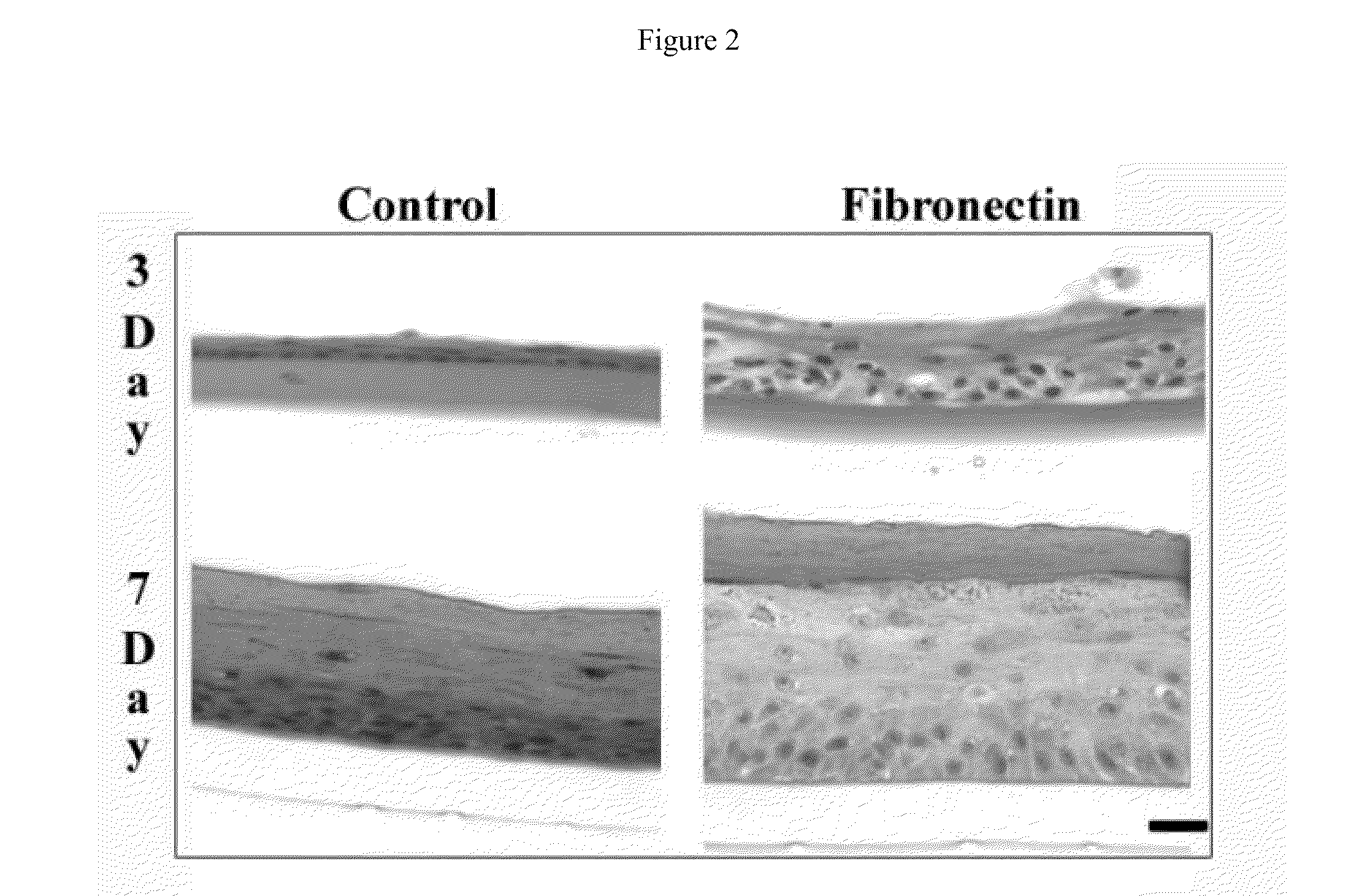
binding site availability of FN when compared to that of using passive adsorption. When keratinocytes were cultured at 3 days at the A / L interface on self-assembled basal lamina analogs with no FN, passively adsorbed FN, and EDC conjugated FN, an increase in epithelial thickness was found between all surfaces. This data also corresponds with the data from the FN cellular
binding domain availability analysis. Overall the results from these studies indicate that the cellular
binding domain of FN can be enhanced on collagen-based biomaterials and directly influences functions important for epithelialization. The information gained from this invention can be applied to other model systems where the enhancement of cellular binding sites of FN on collagenous biomaterials would enhance tissue functionality. Additionally this information can be used in the design of engineered tissues where the incorporation of a basal lamina analog is necessary to direct epithelial polarity and functions as well as to separate
cell types and act as a selectively permeable barrier, such as in the glomerulus of the
kidney or the
small intestine.
[0021]To create a microfabricated basal lamina analog produced from self-assembled CI,
photolithography was used. A master pattern was created on a
silicon wafer to produce channels with specified features of 200 μm depth and 50 μm, 100 μm, 200 μm, and 400 μm widths. A negative replicate was produced using PDMS and acid soluble
type I collagen was self-assembled on the surface of the negative replicate PDMS pattern. Previously, a similar strategy was used to create basal lamina analogs using a collagen-GAG coprecipitate with different
processing techniques to create a basal lamina analog laminated to a dermal
scaffold. When comparing the two strategies to produce microfabricated basal lamina analogs, it was found that the features of the microfabricated basal laminas when composed of collagen-GAG had a greater error associated with both the widths and depths (mean width error varied from 13-30% and mean depth varied from 7.4-16.2%), than the features on the self-assembled CI lamina analogs (mean width errors varied from 2-9% and mean depths varied from 0.9-2.5%). Although the depths and widths of the self-assembled CI membranes varied from the design specifications, the method of the invention using self-assembled CI demonstrates improved fidelity for recapitulating topographical features as well as a defined starting
biochemistry for enhanced FN EDC conjugation.
[0032]Overall this invention has focused on developing a bioengineered skin substitute that recapitulates biochemical and microtopographical features found at the DEJ to enhance epithelialization and interfollicular ESC localization. It was found that 50 and 100 μm width channels with approximate depths of 150 μm contain a full epithelial layer after 7 days at A / L interface culture. When comparing these values to epithelialized DED or native skin, it was found that the epithelial thicknesses were not statistically different from one another and also contain similar values of proliferating basal keratinocytes. Additionally, the bioengineered skin of the invention substitute containing a microtopographical basal lamina analog provides an excellent
model system to evaluate the proper ESC niche through both surface markers and
label-retaining studies in order to enhance the regenerative capacity of bioengineered
skin substitutes.


