Magnetic resonance angiography with flow-compensated and flow-sensitive imaging

a technology flow-sensitive imaging, which is applied in the field of magnetic resonance angiography with flow-sensitive and flow-compensated and flow-sensitive imaging, can solve the problems of difficult cardiac phase finding for many patients, high signal obliteration in the arteries of angiography, and difficult to find cardiac phases, etc., to achieve the effect of reducing or increasing flow sensitivity

Active Publication Date: 2010-11-04
SIEMENS HEALTHCARE GMBH
View PDF4 Cites 29 Cited by
  • Summary
  • Abstract
  • Description
  • Claims
  • Application Information

AI Technical Summary

Benefits of technology

[0011]An object of the present invention is to improve angiography that is not contrast agent-intensified and to increase the signal difference in the arteries given the acquisition of the two data sets in order to obtain high-contrast MR angiography images.
[0012]According to the invention, a method is provided to generate an MR angiography image in which a first MR data set of an examination region is acquired in which the vessels in the examination region are shown with high signal intensity. Furthermore, a second MR data set of the examination region is acquired in which the vessels in the examination region are shown with low signal intensity. An angiographic magnetic resonance image is calculated via mathematical combination of the first and second data set. According to the invention, the first data set is now acquired with an imaging sequence with reduced flow sensitivity and the second data set is acquired with an imaging sequence with a flow sensitivity increased relative to the first data set. According to the invention, the MR data set in which the vessels in the examination region are shown with high signal is thereby improved. In the acquisition of this first MR data set, the flow sensitivity is reduced, so an MR data set is generated that leads to an optimized bright signal in the arteries. The optimization of the first MR data set with light vessel depiction is of particular important area given fast blood flow or in patients with high or varying heart rate. The improvement of the bright signal portions in the vessels in the acquisition of the first MR data set leads to a higher stability of the angiography method. In particular, it is no longer necessary (as in the prior art) to detect the time period without flow in the arteries since the present method shows vessels with high signal even at low flow speeds. The problem of the signal obliteration in the first MR data set is therefore prevented. By the use of an essentially flow-insensitive imaging sequence and a flow-sensitive imaging sequence it is possible to show vessels with medium or fast flow speeds. The signal attenuation occurring in the center of the vessel (where the flow is highest) in conventional methods is avoided in the acquisition of the first data set. By the method according to the invention, this effect is avoided and the signal homogeneity in the vessels is improved in the first data set. An additional advantage of the method according to the invention is that the maximum speed and the minimum speed of the blood flow have a lesser importance to the acquisition of the two MR data sets than in previous methods. The image quality of the MR angiography image thus can be improved, even in the case of irregular heart rates in EKG-triggered acquisitions.
[0017]The first MR data set is advantageously acquired during the diastole of the cardiac cycle in which the blood flow in the vessels in the examination region is slow, and the second MR data set is acquired during the systole of the cardiac cycle in which the blood flow in the vessels in the examination region is fast. However, if the imaging sequence with reduced flow sensitivity and the more flow-sensitive imaging sequence operate reliably, it is also possible to modify the data acquisition so that both the first data set and the second data set are acquired in the same cardiac phase, for example during the systole with fast arterial flow. In this embodiment the third disadvantage described above—that errors occur in the subtraction of the two MR images due to the pulsing state of the artery and the size difference during the systole and the diastole—can be prevented. If both data sets are acquired during the systole, the errors induced in the vessel disposition by pulsation can be reduced or, respectively, avoided in the vessel.
[0020]The increased flow sensitivity of the second MR data set can also be achieved by activating a flow-dephased preparation before the actual imaging sequence, or before sub-segments of the actual imaging sequence. It is likewise possible to acquire one of the two data sets using a series of RF pulses with variable flip angles, with the difference of the flow sensitivities of the first MR data set and of the second MR data set being achieved or additionally emphasized by the use of different flip angle curves.

Problems solved by technology

First, it has been shown in practice that it is very difficult for many patients to find a cardiac phase in which absolutely no flow occurs, such that the MR image with the bright flow signal often has regions with less signal or without signal in the arteries. This particularly applies for patients with rapid heart rates.
Second, this type of angiography imaging is problematical, in particular in regions with fast flow. For example, with the aforementioned technique good results (i.e. good MR angiography images) can be achieved in the lower feet while it is more difficult to achieve the same result quality in the upper feet or the pelvis. The problem of signal obliterations in the arteries with high signal also occurs for patients with irregular heart beat since it is difficult to precisely determine the phase of the rapid flow during the systole and the phase of the low flow during the diastole.
A third disadvantage is that the strong pulsing of the vessels leads to changes in the vessel diameter, which can lead to false results in taking the difference of the images of the systole and the diastole.
A fourth disadvantage of the method described above is that EKG triggering is necessary during the image acquisition in order to correlate the image acquisition with the heart beat for the acquisition of the MR images during the systole and the diastole.
However, in this case it is more difficult to achieve a sufficient flow sensitivity in the data set in which the vessels should be shown dark.

Method used


Image

  • Magnetic resonance angiography with flow-compensated and flow-sensitive imaging
  • Magnetic resonance angiography with flow-compensated and flow-sensitive imaging
  • Magnetic resonance angiography with flow-compensated and flow-sensitive imaging

Examples


Embodiment Construction

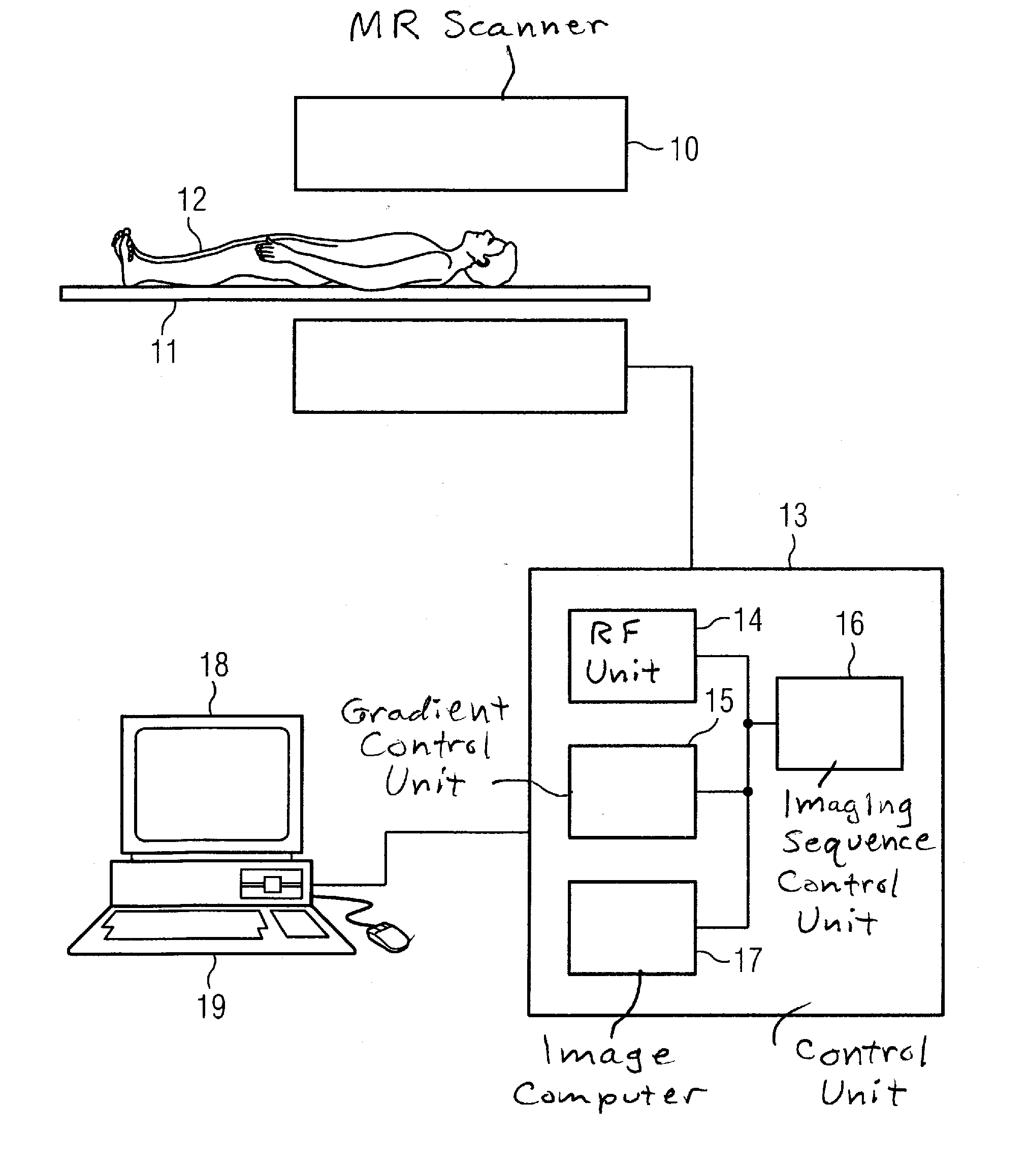
[0029]Shown in FIG. 1 is an MR system with which MR angiography images according to the invention can be generated with which a better image quality is achieved via improvement of the MR acquisition with bright vessel depiction. The MR system has scanner 10 that contains a basic field magnet to generate a polarization field BO. An examination subject 12 arranged on a bed 11 is driven into the middle of the magnet where the acquisition of the MR signals from an examination region is implemented by radiation of RF pulses and switching (activation / deactivation) of gradients in the scanner 10. The manner by which MR images can be generated in a pulse sequence with a progression of RF pulses and switching of gradients is known to those skilled in the art and need not be described in detail herein. The MR system is connected with a central control unit 13 with which the MR system is controlled. The central control unit moreover has an RF control unit 14 that controls the switching of the ...


PUM


Abstract

In a magnetic resonance angiography method with flow-compensated and flow-sensitive imaging and a magnetic resonance apparatus for implementing such a method, a first MR data set of the examination region is acquired with an imaging sequence in which vessels in the examination region are shown with high signal intensity, a second MR data set of the examination region with an imaging sequence in which the vessels in the examination region are shown with low signal intensity, and the angiographic magnetic resonance image is calculated in a processor by taking the difference of the first and second data set. The first data set is acquired with an imaging sequence with reduced flow sensitivity and the second data set is acquired with an imaging sequence with an increased flow sensitivity compared to the initial imaging sequence.

Description

BACKGROUND OF THE INVENTION[0001]1. Field of the Invention[0002]The present invention concerns a method to generate an angiographic magnetic resonance image of an examination region and a magnetic resonance system for implementing such a method.[0003]2. Description of the Prior Art[0004]In magnetic resonance tomography there are multiple possibilities to show the blood vessels in angiograms that can either be based on a contrast agent-intensified signal acquisition, or acquired without contrast agent by using the effect of flowing magnetization during the image acquisition. Since not all examined persons tolerate the administration of contrast agent, magnetic resonance angiography that is not contrast agent-intensified is gaining importance.[0005]In magnetic resonance angiography it is typically desirable to show only the arteries and to suppress the MR signal from the veins. In MR angiography that is not contrast agent-intensified, the method that is used depends on the desired exa...

Claims


Application Information

Patent Timeline
IPC IPC(8): A61B5/055
CPCA61B5/0263A61B5/055A61B5/7285G01R33/56509G01R33/56316G01R33/5635G01R33/5614
Inventor BI, XIAOMINGSCHMIDT, MICHAELASCHMITT, PETERWEALE, PETER
Owner SIEMENS HEALTHCARE GMBH
Why Eureka
Try Eureka